3185
MR Molecular Imaging of Pancreatic Ductal Adenocarcinoma during a Therapeutic Vaccine Regimen1Biomedical Engineering, Case Western Reserve University, Cleveland, OH, United States, 2Case Western Reserve University, Cleveland, OH, United States, 3Cleveland Clinic, Cleveland, OH, United States
Synopsis
Keywords: Molecular Imaging, Cancer
Motivation: Pancreatic ductal adenocarcinoma (PDAC) is a highly aggressive form of cancer with a low survival rate. Immunotherapies have been able to overcome some of the barriers of traditional therapy with PDAC, resulting in increased survival. However, immunotherapy results in responsive and non-responsive tumors.
Goal(s): This study aims to use MR molecular imaging with a contrast agent that's specific to the tumor microenvironment to determine responders and monitor therapy.
Approach: MR molecular imaging T1w images at discrete timepoints with contrast injection and during therapy.
Results: CNR changes were detected using MR molecular imaging on mice bearing PDAC tumors during a vaccine regimen.
Impact: There is an absence of biomarkers/predictive tools for pancreatic cancer response to immunotherapy. There’s a critical need for therapeutic strategies that improve patient outcomes in tandem with imaging methods that can monitor disease progression and accurately guide clinical decision making.
Introduction
Pancreatic ductal adenocarcinoma (PDAC) is a highly metastatic form of cancer, the disease is characterized by desmoplasia and rapid metastasis into distant organs through the ducts (1). Chemotherapy is the standard care for advanced PDAC but yields limited efficacy and fails to improve the overall survival of patients. Immunotherapies using therapeutic vaccines have become breakthrough therapies for aggressive cancers (2-4). Cancer vaccines can stimulate anti-tumor immunity through the use of tumor specific or associated antigens. These vaccines provoke cell specific immunity and induce humoral immunity to inhibit tumor growth and increase immunogenic tumor cell death (5). While some of these cancer vaccines have shown promising results in both preclinical and clinical settings, we still lack imaging tools to adequately assess the responses of patients (6-9). We hypothesize that targeting the tumor immune microenvironment through neoantigens will allow for T-cell response and concurrent stromal remodeling that can be imaged through MR molecular imaging. We previously developed and characterized a contrast agent specific to an alternative splice of fibronectin, EDB-FN (10-12). This fibronectin splice is upregulated in aggressive cancers and its expression has been linked to tumor invasion and inflammation in the tumor microenvironment. Based on T1w and DCE imaging we were able to noninvasively characterize changes in the stroma of the tumor in vaccine treated mice.Methods
In preparation for mouse therapy, murine KPC PDAC cells were orthotopically implanted in C57BL/6 mice for in vivo imaging. Following implantation mice were imaged at three distinct timepoints, day 10 for baseline, day 28 following therapy administration and day 46 after the active immunogenic window. T1-weighted MR images were obtained using 2D fast spin-echo, 3D-FLASH and DCE FLASH sequences pre- and post-i.v.-injection of 0.1 mmol/kg MT218 using a 3T small animal scanner (MRSolutions, Surrey UK). Mice then received 2 weekly doses of vaccine and were imaged at two post-vaccine timepoints.Contrast-to-noise ratio (CNR) were calculated using muscle as the control tissue. Differences in signal intensity were calculated by normalizing post-contrast images to pre-contrast images. All animal studies were conducted in accordance with CWRU’s Institutional Animal Care and Use Committee. The targeted contrast agent, MT218, was provided by Molecular Theranostics, LLC. Mice were sacrificed and tumors were excised and embedded in formalin. Fixed samples were subsequently stained for H&E for cellular context and morphology, immunochemistry using anti-G4 for EDB-FN expression and Sirius red for stroma.Results
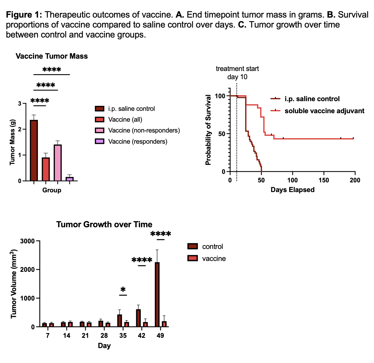
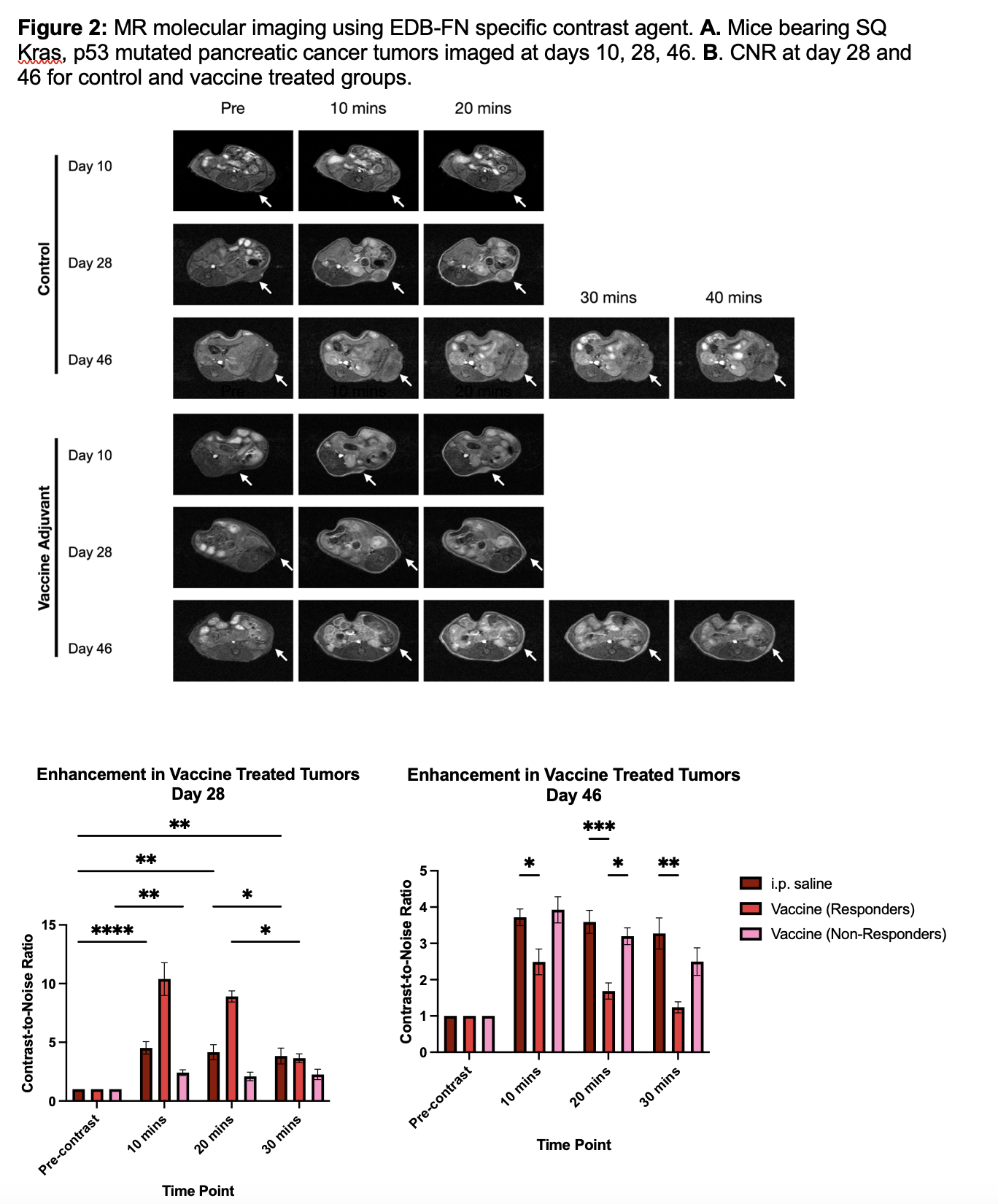
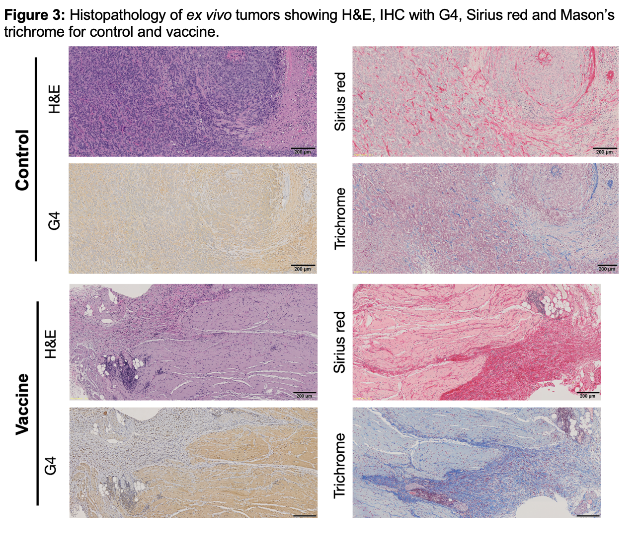
Therapy with vaccine resulted in decreased tumor mass compared to control tumors (Fig 1 A). Mice treated with tumors also resulted in prolonged survival, with median survival increasing by 27 days (Fig 1 B). Additionally, vaccine treated mice had substantially attenuated tumor growth compared to control, with significant differences in tumor growth occurring after day 35 (Fig 1 C). Based on tumor growth from MRMI and caliper measurements we were able to stratify tumors into responders, partial and non-responders based on the RECIST criteria. MRMI showed differences to growth over time as well as signal enhancement. Quantification of CNR using ROI measurements showed differences in vaccine and control at day 28. Once tumors were classified, responders had higher CNR of 10.4-fold enhancement in comparison to partial-responders which had 2.4 and 4.5-fold for controls (at 10 mins post-contrast). At day 46, vaccine responders with detectable tumors had 2.5-fold enhancement whereas partial-responders had a normalized CNR of 3.9 and controls has a nCNR of 3.7. Histopathology confirmed the patterns of enhancement and further elucidated vaccine responder EDB-FN expression through the presence of fibrotic scars. Through magnetic resonance molecular imaging (MRMI) with a contrast agent targeted to the fibronectin in the tumor microenvironment we were able to noninvasively characterize changes in the stroma of the tumor between treatment groups and responses. We found increased enhancement corresponded to ex vivo fibronectin expression which contributes to desmoplastic and aggressive nature of PDAC. Thus, we were able to assess tumor progression more effectively and establish a criterion for MRMI monitoring of ICT.Conclusion
Anti-cancer vaccine therapy resulted in a transient increase in fibronectin production in responders, followed by a stark decrease thus allowing for a positive correlation between MRMI signal and therapeutic efficacy. Thus, we were able to utilize a fibronectin-based contrast agent to monitor the progression of therapy in a preclinical setting.Acknowledgements
This work was funded by the NCI R01 CA235152 and R44CA265626References
- Siegel RL, Miller KD, Fuchs HE, Jemal A. Cancer Statistics, 2021. CA Cancer J Clin. 2021;71(1):7-33. Epub 2021/01/13. doi: 10.3322/caac.21654. PubMed PMID: 33433946.
- Zou W, Chen L. Inhibitory B7-family molecules in the tumour microenvironment. Nat Rev Immunol. 2008;8(6):467-77. Epub 2008/05/27. doi: nri232610.1038/nri2326. PubMed PMID: 18500231.
- Kraehenbuehl L, Weng CH, Eghbali S, Wolchok JD, Merghoub T. Enhancing immunotherapy in cancer by targeting emerging immunomodulatory pathways. Nat Rev Clin Oncol. 2022;19(1):37-50. Epub 2021/09/29. doi: 10.1038/s41571-021-00552-7. PubMed PMID: 34580473.
- Saxena, M., van der Burg, S.H., Melief, C.J.M. et al. Therapeutic cancer vaccines. Nat Rev Cancer 21, 360–378 (2021). https://doi.org/10.1038/s41568-021-00346-0
- Liu, J., Fu, M., Wang, M. et al. Cancer vaccines as promising immuno-therapeutics: platforms and current progress. J Hematol Oncol 15, 28 (2022). https://doi.org/10.1186/s13045-022-01247-x
- Rojas, L.A., Sethna, Z., Soares, K.C. et al. Personalized RNA neoantigen vaccines stimulate T cells in pancreatic cancer. Nature 618, 144–150 (2023). https://doi.org/10.1038/s41586-023-06063-y
- Khong, H., & Overwijk, W. W. (2016). Adjuvants for peptide-based cancer vaccines. Journal for immunotherapy of Cancer, 4, 1-11.
- Neri, D., & Bicknell, R. (2005). Tumour vascular targeting. Nature Reviews Cancer, 5(6), 436–446. https://doi.org/10.1038/nrc1627
- Niemi, J. V. L., Sokolov, A. V., & Schiöth, H. B. (2022). Neoantigen Vaccines; Clinical Trials, Classes, Indications, Adjuvants and Combinatorial Treatments. Cancers, 14(20), 5163. https://doi.org/10.3390/cancers14205163
- Qiao, P., Ayat, N. R., Vaidya, A., Gao, S., Sun, W., Chou, S., … Lu, Z. (2020). Magnetic Resonance Molecular Imaging of Extradomain B Fibronectin Improves Imaging of Pancreatic Cancer Tumor Xenografts, 10(October), 1–13. https://doi.org/10.3389/fonc.2020.586727
- Schilb, A. L., Ayat, N. R., Vaidya, A. M., Hertz, L. M., Hall, R. C., Scheidt, J. H., … Lu, Z. (2021). Efficacy of Targeted ECO / miR-200c Nanoparticles for Modulating Tumor Microenvironment and Treating Triple Negative Breast Cancer as Non-invasively Monitored by MR Molecular Imaging, 1405–1418.
- Han, Z., Li, Y., Roelle, S., Zhou, Z., Liu, Y., Sabatelle, R., … Lu, Z.-R. (2017). Targeted Contrast Agent Specific to an Oncoprotein in Tumor Microenvironment with the Potential for Detection and Risk Stratification of Prostate Cancer with MRI. Bioconjugate Chemistry, 28(4), 1031–1040. https://doi.org/10.1021/acs.bioconjchem.6b00719
Figures