3184
Accuracy, repeatability, and sensitivity of breath-hold calibrated fMRI for the mapping of absolute CMRO21Cardiff University Brain Research Imaging Centre (CUBRIC), School of Physics & Astronomy, Cardiff University, Cardiff, United Kingdom, 2Institute for Advanced Biomedical Technologies and Department of Neurosciences, Imaging, and Clinical Sciences, University G. D’Annunzio of Chieti-Pescara, Chieti, Italy
Synopsis
Keywords: Oxygenation, Metabolism, CMRO2 & oxygen extraction fraction & OEF & calibrated fMRI
Motivation: Gas-calibrated fMRI provides a sensitive measurement of cerebral oxygen consumption, but its applicability is limited to young, healthy participants who can tolerate the gas challenges.
Goal(s): Our goal was to establish the feasibility of breath-hold calibrated fMRI (bhc-fMRI) to replace gas challenges.
Approach: We compared bhc-fMRI to a global oxygen extraction fraction (OEF) measurement, measured within session repeatability and tested the sensitivity of the method to a visual stimulus.
Results: We found similar agreement to global OEF measurements as for gas-calibrated fMRI and moderate within session repeatability. Robust responses to continuous visual stimulation of CBF/CMRO2/OEF (+15%/+11%/-3%) demonstrate good sensitivity of the bhc-fMRI method.
Impact: We establish breath-hold calibrated fMRI as a viable alternative to gas-calibrated fMRI for mapping cerebral oxygen consumption. Breath-hold calibrated fMRI is simple to implement and is tolerable for ageing cohorts and patients with neurodegenerative pathologies.
Background
Dual-calibrated fMRI measurement of cerebral metabolism 1 is a sensitive method for mapping multi-parametric physiological parameters in the brain including cerebral oxygen consumption. However, the method requires specialized equipment, long acquisition times, experienced operators, and can be uncomfortable for participants. We recently introduced a single-gas (CO2) alternative 2 that constrains the Davis Model of the BOLD signal with a diffusion model of oxygen transport from capillaries to mitochondria to infer OEF and cerebral blood volume. Here, we replace the CO2 stimulus with a repeated breath-holding paradigm, increasing subject comfort and removing the need for ancillary equipment. The performance of the method was assessed in healthy volunteers at rest and during a visual task.Methods
We studied 35 healthy volunteers (24±5.6yrs, 16 female). MRI data were collected on a Siemens Prisma 3T MRI scanner with a 32-channel receive-only head coil. An MPRAGE T1-weighted scan was used for registration and brain segmentation purposes (matrix 165x203x197, 1 mm isotropic resolution, TR/TE = 2100/3.24ms). A dual-excitation (DEXI) pCASL sequence with background suppression was used to collect simultaneous BOLD and ASL data (TR = 4400ms; TE1/TE2 = 10ms/30ms; slices = 15; Slice thickness = 6mm; PLD = 1500ms; tag duration = 1500ms). The breath-holding protocol had 10 repeats of 20 seconds of breath-holding at end expiration. This was repeated at rest and during visual stimulation (reversing radial checkerboard at a frequency of 8Hz). A T2-Relaxation-Under-Spin-Tagging (TRUST) MRI sequence was acquired to estimate global OEF 3. We further collected a T1 inversion recovery sequence to calculate Hb (ΔTR/TE = 150ms/22ms, flip angle = 90 degrees). OEF estimates were made from the DEXI-pCASL data using an exhaustive search as in 2. However, the end-tidal regressor was replaced with the average grey matter response to breath-holding. The arterial oxygen content at rest was 127±8 mmHg, and 104±14 mmHg at the end of 20 seconds of breath-holding. These average values were used in the analysis of individual subjects.Results and Discussion
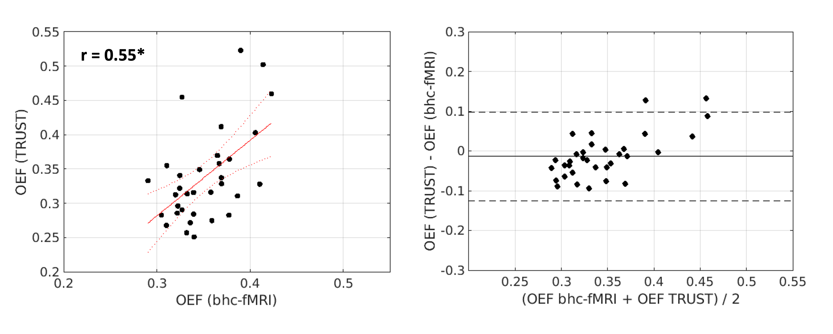
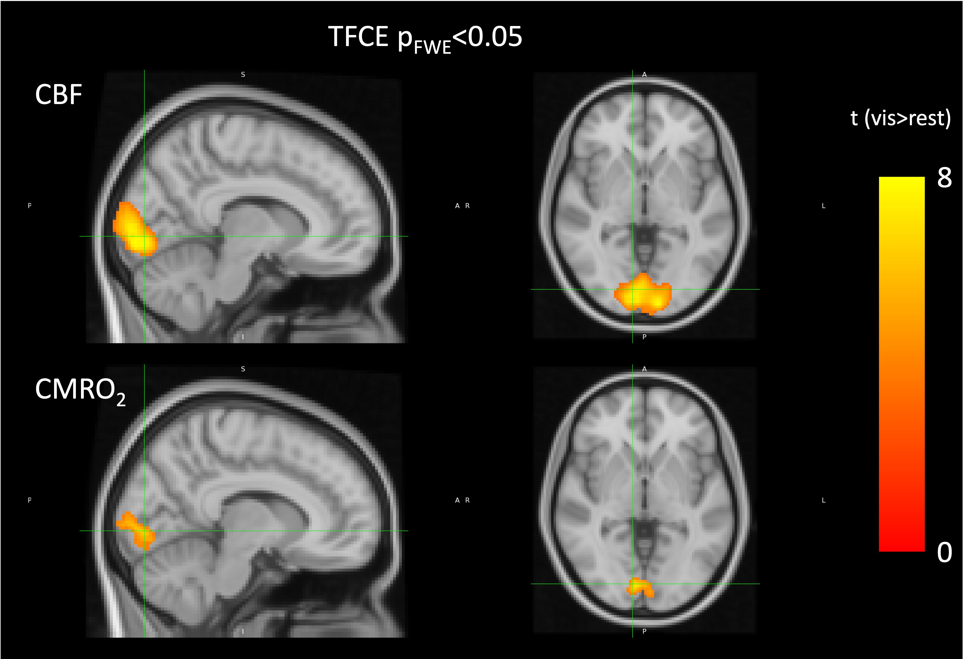
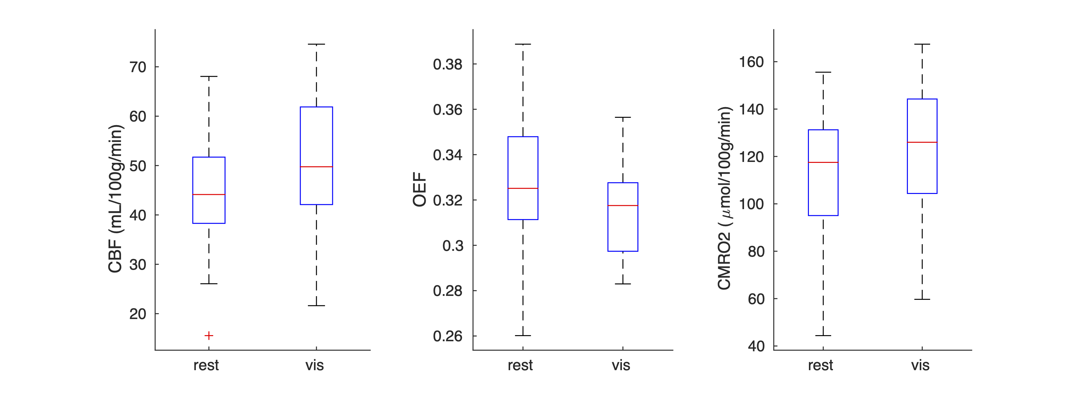
Figure 1 shows a scatterplot and Bland-Altman analysis of the mean grey matter OEF and the global TRUST estimate for each subject. The Pearson’s correlation coefficient between TRUST and bhc-fMRI is 0.55 (p<0.05) and is similar to that found for dual-calibrated fMRI (0.58) 2. The Bland-Altman analysis shows minimal bias of the mean. Within session repeatability, assessed over all GM between the rest and visual stimulation session, had a correlation coefficient of 0.76 (p<0.05). Figure 2 shows the regions of significant average CBF and CMRO2 change between the rest and visual stimulation. Within the ROI defined by significant CBF increase there is a 15±2% CBF increase, a 11±2% CMRO2 increase, and a 3±1% OEF decrease. Figure 3 shows a boxplot for parameter values in the visual ROI. The average neurovascular coupling constant (1.44) is in good agreement with the 1.4 coupling ratio found with dual-calibrated fMRI 4 and the 3.6% OEF decrease observed with a 5-minute R2’ measurement 5. This in-vivo study demonstrates that breath-hold calibrated fMRI has similar accuracy to dual-calibrated fMRI for quantification of cerebral oxygen metabolism and is sensitive to neurovascular coupling-related changes in OEF in healthy volunteers. Further studies will investigate the applicability of the method in disease.Conclusion
We establish the feasibility of replacing the gas challenge in dual-calibrated fMRI with breath holds. The breath-hold calibrated fMRI measurement of CMRO2 is simple to implement, has good repeatability, is sensitive to local changes in metabolism, and has comparable accuracy to existing gas-based methods. Removing the need for an uncomfortable gas challenge makes oxygen metabolism mapping feasible in ageing populations and patient groups who would not tolerate gas challenges.Acknowledgements
We acknowledge the members of the Welsh Advance Neuroimaging Database for acquiring this dataset and the support from the Wellcome Trust (WT104943/Z/14/Z and 220575/Z/20/Z), and the EPSRC (EP/S025901/1)
Funded in part by the European Union - NextGenerationEU under the National Recovery and Resilience Plan (NRRP), Mission 4 Component 2 - M4C2, Investment 1.5 - Call for tender No. 3277 of 30.12.2021 Italian Ministry of Universities Award Number: ECS0000004, Project Title: “Innovation, digitalisation and sustainability for the diffused economy in Central Italy,” Concession Degree No. 1057 of 23.06.2022 adopted by the Italian Ministry of Universities, CUP: D73C22000840006.
Italian Ministry of University and Research, Research Projects of National Relevance (PRIN), Project Code: 2022BERM2F, Project Title: “Mapping Mitochondrial Function and Oxygen Metabolism in the Human Brain with Magnetic Resonance Imaging.” Concession decree No. 1065 of 18. 07.2023 adopted by the Italian Ministry of University and Research, ERC Sector LS7 “Prevention, Diagnosis and Treatment of Human Diseases”.References
[1] Bulte D, et al. Quantitative measurement of cerebral physiology using respiratory-calibrated MRI. Neuroimage. 2012;60(1):582-91.
[2] Chiarelli A, et al. A flow-diffusion model of oxygen transport for quantitative mapping of cerebral metabolic rate of oxygen (CMRO2) with single gas calibrated fMRI. JCBFM. 2022;42(7):1192–1209.
[3] Lu H, et al. Calibration and validation of TRUST MRI for
the estimation of cerebral blood oxygenation. Magn Reson Med. 2012;67(1): 42-49.
[4] Germuska M, et al. Dual-calibrated fMRI measurement of absolute cerebral metabolic rate of oxygen consumption and effective oxygen diffusivity. Neuroimage. 2019. 184: 717–728.
[5] Arzanforoosh F, et al. Streamlined quantitative BOLD for detecting visual stimulus‑induced changes in oxygen extraction fraction in healthy participants: toward clinical application in human glioma. MAGMA. 2023; doi: 10.1007/s10334-023-01110-1.
Figures