3182
Development of a DNP-MRI molecular probe for detecting dipeptidyl peptidase-4 activity in vivo based on the substrate recognition mechanism.1The University of Tokyo, Tokyo, Japan, 2National Institutes of Health, Bethesda, MD, United States, 3National Institutes of Quantum Science and Technology, Chiba, Japan
Synopsis
Keywords: Molecular Imaging, Hyperpolarized MR (Non-Gas), DNP-MRI molecular probe, DPP-4
Motivation: Dipeptidyl peptidase-4 (DPP-4) is a biologically important peptidase known as a biomarker and therapeutic target for type 2 diabetes and cancers. Therefore, detection of DPP-4 activity can be a useful method for early diagnosis and treatment efficacy assessment.
Goal(s): Development of a DNP-MRI molecular probe for detecting DPP-4 activity in vivo
Approach: We performed molecular designs of DNP-MRI molecular probes for meeting physicochemical properties. For evaluation of enzymatic reactivity of the probes, enzymatic reaction parameters (Km, kcat) were measured. The optimized probe was applied for DNP-MRI experiments using mice.
Results: We developed a DNP-MRI molecular probe to detect DPP-4 activity in vivo.
Impact: With design strategy based on recognition mechanism, we have developed a DNP-MRI molecular probe against DPP-4, which has been difficult to develop so far. New probe enables detection of DPP-4 activity in vivo and will be useful for medical applications.
Introduction
Dipeptidyl peptidase-4 (DPP-4) is a biologically significant enzyme of interest as a biomarker and therapeutic target for type 2 diabetes and cancer (Figure 1)1. Therefore, the detection of DPP-4 activity deep in a body could be a useful method for early disease diagnosis and treatment efficacy assessment. We aimed to develop a molecular probe for detecting of DPP-4 activity in vivo with Dynamic Nuclear Polarization-Magnetic Resonance Imaging (DNP-MRI). DNP-MRI has been gaining considerable attention as a technique for real-time metabolic observation in vivo, and it is actively being explored for clinical applications.To detect DPP-4 activity in vivo, the DNP-MRI molecular probe targeting DPP-4 must meet several physicochemical properties2. First, the longitudinal relaxation time (T1), which correlates with the decay time of high-sensitivity signals by DNP, must be sufficiently long. Second, DNP-MRI molecular probe must induce chemical shift changes significant enough to distinguish the probe from the product resulting from the reaction with DPP-4. Lastly, the DNP-MRI probe must selectively and rapidly react with DPP-4. These various limitations have made the development of DNP-MRI molecular probes difficult.
In this presentation, we will report on our strategy for designing DNP-MRI molecular probes for DPP-4, especially focusing on the enzymatic reactivity with DPP-4. The results of in vivo detection of DPP-4 activity using the developed DNP-MRI molecular probes will also be presented.
Results
Gly-Pro-Gly is a typical peptide substrate for DPP-IV. The Gly-Pro-Gly has high affinity for DPP-4 (Km = 1.6 ± 0.2 mM) but moderate enzymatic reaction rate (kcat = 50 ± 5 s-1). On the other hand, newly designed tripeptide Gly-Aze-Gly containing L-azetidine-2-carboxylic acid (Aze) has a slightly lower affinity (Km = 4.8 ± 0.3 mM) and much higher enzymatic reaction rate (kca = 504 ± 13 s-1) than the original Gly-Pro-Gly (Figure 2). The Gly-Aze-[1-13C]Gly-d2, optimized as a DNP-MRI molecular probe, exhibited a large chemical shift change (3.7 ppm) between probe and product, and long spin-lattice relaxation time (T1 = 52 ± 1 s), which is sufficient for in vivo DNP-MRI study. Upon incubation with mouse kidney homogenate, Gly-Aze-[1-13C]Gly-d2 reacted with DPP-4 selectively in vitro (Figure 3). Finally, Gly-Aze-[1-13C]Gly-d2 was applied for DNP-MRI. The hyperpolarized Gly-Aze-[1-13C]Gly-d2 exhibited the ability to detect DPP-4 activity selectively in mice (Figure 4).Discussion
The key aspect in the design and development of this molecular probe Gly-Aze-[1-13C]Gly-d2 was to optimize the affinity with DPP-4. DPP-4 recognizes the proline residue in Gly-Pro-Gly primarily through its hydrophobic site, the S1 pocket of the active site3. This interaction is very strong, and it is expected that the hydrolysis product Gly-Pro, derived from Gly-Pro-Gly, also strongly interacts with the S1 pocket. Consequently, the slow dissociation of Gly-Pro metabolite from the active pocket is expected to lead to a reduced turnover frequency. To weaken the hydrophobic interaction with the S1 pocket, a new probe scaffold was designed by replacing Pro residue with L-azetidine-2-carboxylic acid (Aze) residue (Figure 2). As indicated in the Results section shown above, it was found that the affinity between DPP-4 and Gly-Aze-Gly has been appropriately reduced. In vitro experiments revealed that the enzymatic conversion rate of Gly-Aze-Gly was dramatically higher compared to Gly-Pro-Gly. This is expected to be due to the strategy of improving the turnover frequency by reducing the affinity between DPP-4 and metabolic products.Conclusion
In this study, we developed a new DNP-MRI probe, Gly-Aze-[1-13C]Gly-d2, for detection of DPP-4 activity. This probe was designed based on the substrate of DPP-4, Gly-Pro-Gly, and improved by rational molecular design focusing on Km and kcat. Hyperpolarized experiments using Gly-Aze-[1-13C]Gly-d2 showed clear signals originating from the probe and the metabolic product. We succeeded in detecting DPP-4 activity selectively in mice.Methods
Hyperpolarized 13C MRI: Hyperpolarized 13C MRI experiments were performed with 3 T MRI scanner (Bruker Biospin). 50 μL of Gly-Aze-[1-13C]Gly-d2 solution (H2O, ca. 1.6 M) containing 30 mM OX063 was hyperpolarized at 6.7 T, 1.24 K using SpinAligner DNP polarizer (Polarize IVS). Hyperpolarized sample was dissolved in the dissolution buffer (3.2 mL DPBS containing 0.68 mM EDTA). 400 µL of the hyperpolarized solution was injected intravenously into the healthy mice. In inhibitor experiments, DPP-4 selective inhibitor, anaglipitin (10 mg/kg) was injected 1 h before injection of the hyperpolarized solution.Acknowledgements
This research was supported by MEXT Q-LEAP [Grant Number JPMXS0120330644 (to Y.T., and S.S.)], JST FOREST Program [Grant Number JPMJFR225G (to Y.T.)].
This study was supported by intramural research program at NCI/NIH.
References
1. P. Busek et al., Cancers, 2022, 14, 2072.
2. Y. Saito et al., Sci. Adv. 2022, 8, eabj2667.
3. R. Thoma et al., Structure, 2003, 11, 947–959.
Figures
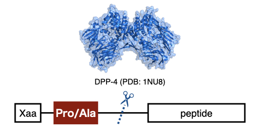
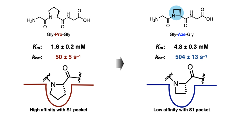
Figure 2. Enzymatic reaction parameters (Km, kcat) of Gly-Pro-Gly and Gly-Aze-Gly
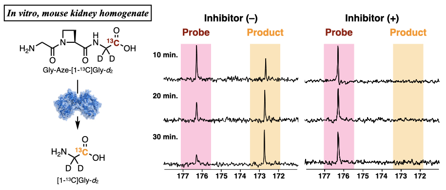
Figure 3. In vitro experiments of conversion of Gly-Aze-[1-13C]Gly-d2 in mouse kidney homogenate. To 680 µL of Gly-Aze-[1-13C]Gly-d2 solution (100 mM PB, pH = 7.4, with or without anagliptin) preincubated at 37 ºC for 10 min, 120 µL of mouse kidney homogenate preincubated at 37 ºC for 10 min was added. To the sampled solution (10, 20, 30 min.), 50 µL of 50 mM NaOH aq, 50 µL of 50 mM HCl aq. and 150 µL of D2O were added and the resulting solution was subjected to 13C NMR measurements. The NMR spectra were acquired using a 9.4 T NMR (256 scan).
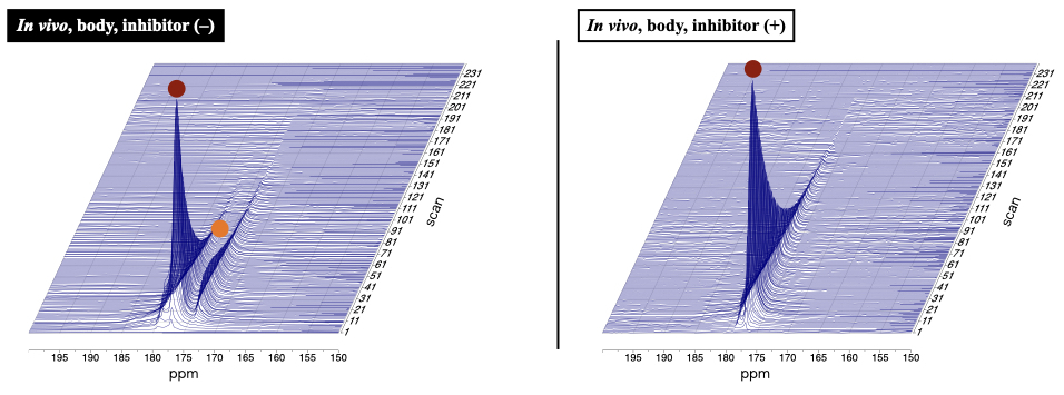
Figure 4. Hyperpolarized experiments for in vivo detection of DPP-4 activity with Gly-Aze-[1-13C]Gly-d2. 50 μL of probe solution (H2O, ca. 1.6 M) containing 30 mM OX063 was hyperpolarized using SpinAligner and dissolved by 3.2 mL DPBS containing 0.68 mM EDTA. The spectra were obtained using a 3 T MRI scanner (TR 1s, flip angle 10º), following intravenous administration of 400 μL of the hyperpolarized solution into the tail vein. In inhibitor experiments, DPP-4 inhibitor, anagliptin (10 mg/kg), was injected intravenously 1 h before injection of the hyperpolarized solution.