3179
Early Detection Of Heart Failure By Visualizing Redox Metabolism Using In Vivo Dynamic Nuclear Polarization MRI1Tumor pathology, Gifu university, 1-1 Yanagido, Gifu, Japan, 2Center for One Medicine Innovative Translational Research (COMIT), Institute for Advanced Study, Gifu University, 1-1 Yanagido, Gifu, Japan, 3Radiology, Frontier Science for Imaging, Gifu University, 1-1 Yanagido, Gifu, Japan, 4Radiology, Gifu University, 1-1 Yanagido, Gifu, Japan
Synopsis
Keywords: Molecular Imaging, Molecular Imaging
Motivation: The diagnosis of heart failure is based on clinical symptoms because noninvasive methods for early diagnosis are still insufficient.
Goal(s): Considering reactive oxygen species contribute to heart failure, we hypothesized much earlier signs of heart failure could be captured by focusing on redox metabolism.
Approach: We used a doxorubicin-induced mouse model of heart failure and analyzed at an early stage. To visualize redox metabolism, we used in vivo dynamic nuclear polarization MRI. We also evaluated the cardiomyocytes microscopically.
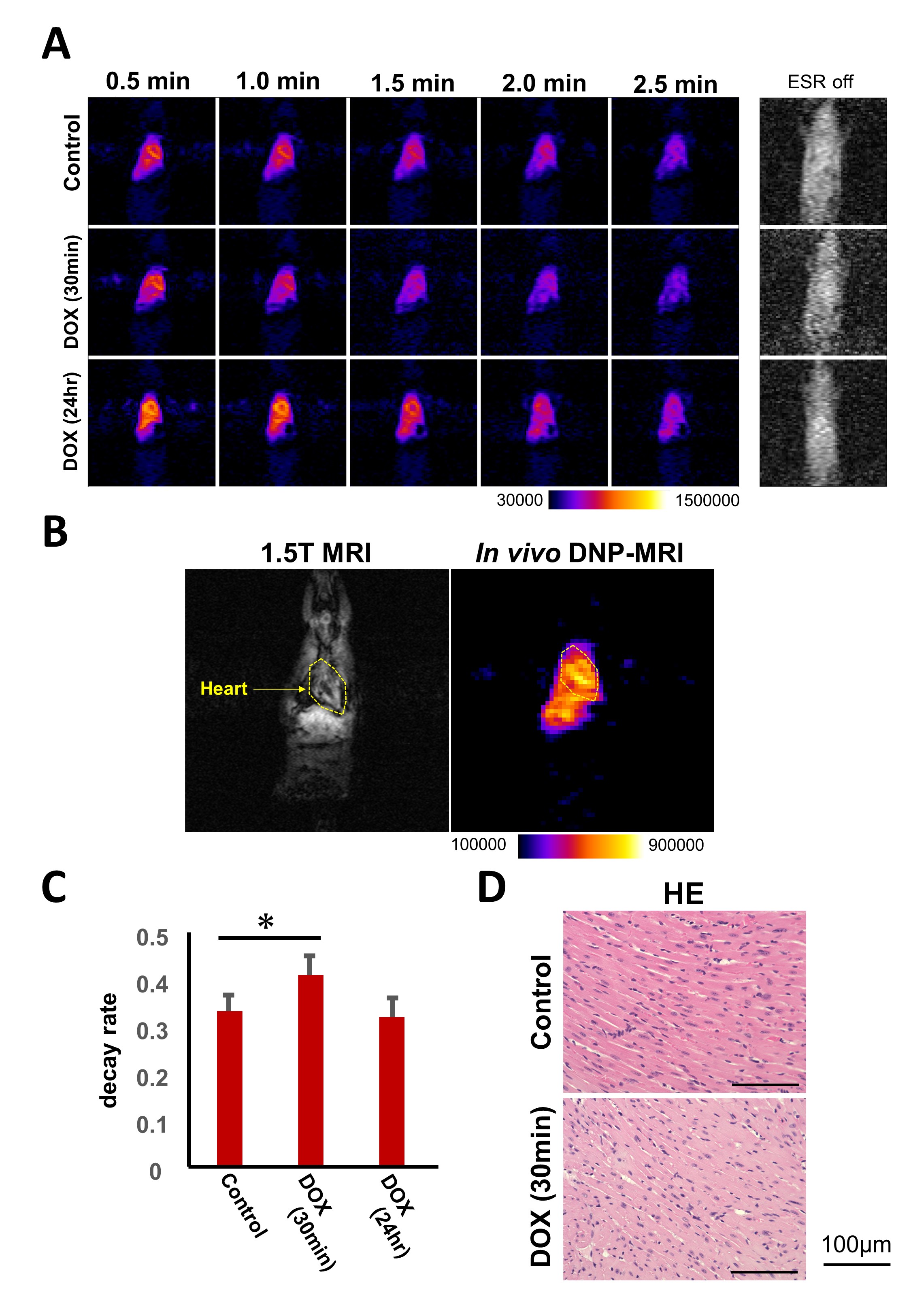
Results: The intracardiac redox metabolism was enhanced in 30 min after doxorubicin administration compared to the control group although no significant cardiomyocyte changes were observed.
Impact: In the early stages of heart failure, redox metabolism is altered even before the morphological changes are observed microscopically. These data provide a technique for detecting heart failure earlier and less invasively than conventional testing methods.
Introduction
The prevalence of heart failure is increasing with the aging of the population with more than 64 million people affected worldwide in 20171. The technology that detects heart failure at an earlier stage is of great value, as the diagnosis is made even after a decline in ejection fraction, or clinical symptoms2. The biochemical mechanisms of heart failure include reactive oxygen species and mitochondrial dysfunction3,4 and it has been reported that redox metabolism in cardiomyocytes is suppressed at late phase5. However, noninvasive methods and biomarkers for early diagnosis of heart failure are still insufficient. We attempted to detect earlier signs of heart failure that could be captured by focusing on redox metabolism.Methods
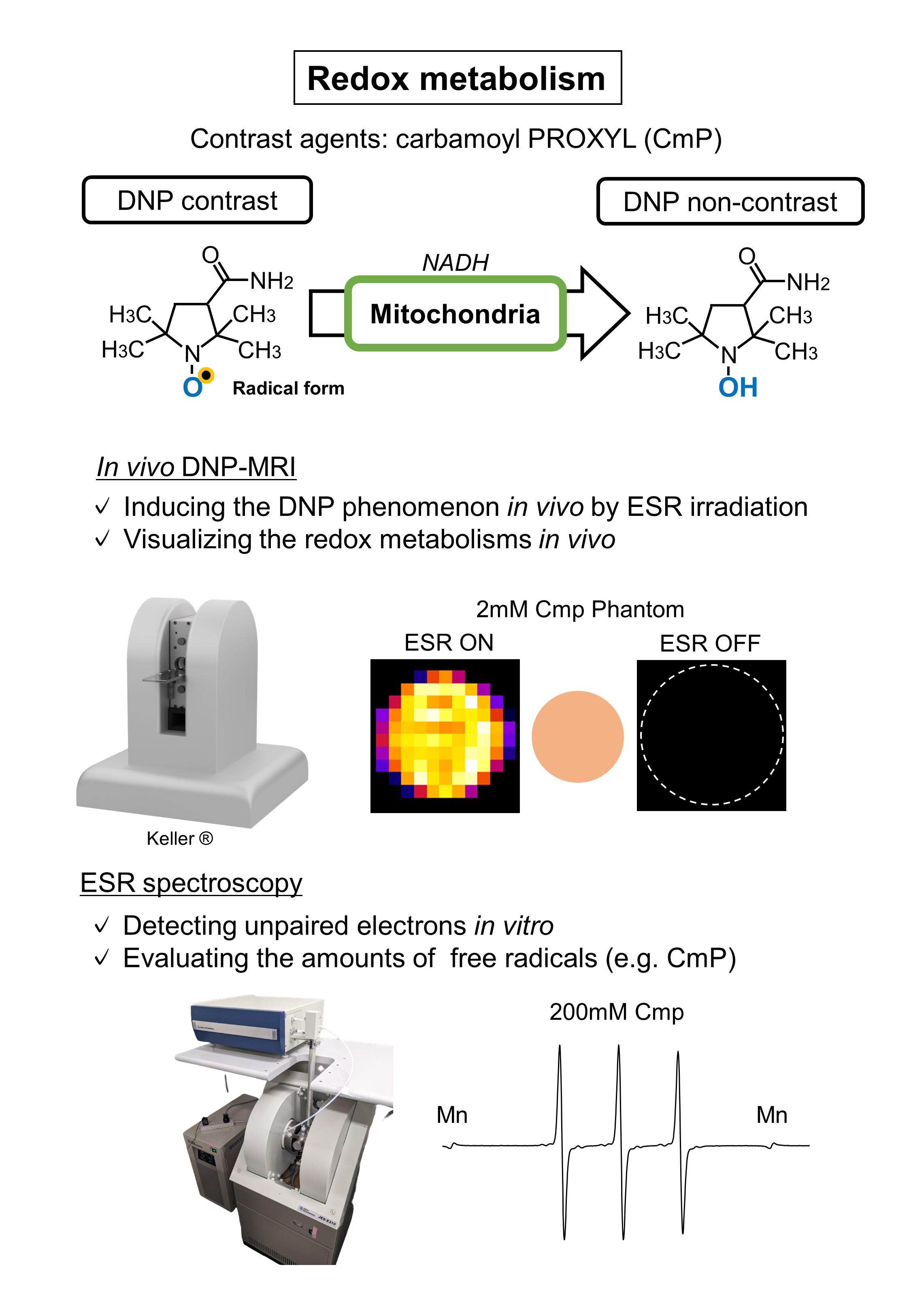
We used doxorubicin-induced heart failure model mice and analyzed at half an hour and one day after intraperitoneal administration (10mg/kg). In the same experimental systems epirubicin, which belongs to the anthracyclines but has weaker cardiotoxicity than doxorubicin, was also investigated. To visualize redox metabolism non-invasively, we used in vivo dynamic nuclear polarization (DNP) MRI and carbamoyl-PROXYL (Cmp) as a probe6,7 (Figure.1). The region of interest (ROI) of the heart was selected on the MRI images and decay rates were analyzed (Figure.2B). Then, we analyzed whether the drugs accumulated in the heart using fluorescence microscopy, because doxorubicin and epirubicin are fluorescent compounds8. We prepared heart homogenate solution taken at 30 minutes after intraperitoneal administration and, as an in vitro study, heart homogenates were mixed with doxorubicin and the CmP signal was monitored by EPR spectroscopy9. Next, we hypothesized that anaerobic metabolism was enhanced due to mitochondrial dysfunction, as in red blood cells without mitochondria10 and investigated the chemical shift from pyruvate to lactate in the early phase heat failure using DNP polarizer (Hypersense), 13C MRS and 13C NMR. The 13C MRS was performed on the hearts of living mice, as in vivo and 13C NMR was performed on the heart homogenates, as ex vivo. We also microscopically evaluated the heart tissue at the same time point. Because it has been reported that cardiomyocytes undergo microscopic degenerative changes also occurred in the late phase of heart failure5, we investigated whether these changes are also occurred in early phase cardiomyocytes.Results
The redox metabolic rate in the heart increased at 30 minutes after doxorubicin administration compared to the control group and returned to the same level within one day (Figure.2A and C). On the other hand, the metabolic rate at 30 minutes after epirubicin administration is the same level or slightly slower than that in the control group. We observed cardiac tissues by fluorescence microscopy and then doxorubicin and epirubicin accumulated in the heart at 30 minutes after intraperitoneal administration. By EPR spectroscopy, ex vivo, free radicals were metabolized faster in the doxorubicin-treated group than in the control group. The redox metabolism was suppressed by potassium cyanide (KCN). In vitro, free radical metabolism was much faster and similarly suppressed by KCN. On the other hand, the 13C-pyruvate/13C-lactate ratio did not change one day after doxorubicin administration compared to the control group as measured by 13C MRS as in vivo and 13C NMR spectroscopy as ex vivo. Histologically, no significant cardiomyocyte changes were observed 30 minutes after doxorubicin administration, although some cardiomyocytes were degenerated within one day. Degenerating cardiomyocytes showed abnormal alignment and vacuolation.Discussion
It is known that the doxorubicin acts on mitochondria and generates ROS through the complexes in the electron transport chain11. On the other hand, CmP is reduced in mitochondria but also has antioxidant properties12,13. Specifically, it reduces ROS (especially •NO2 and ROO•) and yields oxoammonium cations and reduced to hydroxylamine form, which does not induce DNP. Based on these facts, we hypothesized that the ROS produced by doxorubicin in the early phase would be rapidly reacted by CmP. Because KCN inhibits the functions of complex IV, this is consistent with the ex vivo and in vitro results that redox metabolism is suppressed by KCN. We also suggested that redox metabolism was altered earlier than histological changes or in vivo LDH activity.Conclusions
Visualization of redox metabolism in the heart is useful for early detection of heart failure, because oxidative stress or mitochondrial dysfunction contributes to the early stage of heart failure in general, not only by doxorubicin14,15.Acknowledgements
No acknowledgement found.References
1. Gianluigi S, Peter M, Lars H, et al. Global burden of heart failure: a comprehensive and updated review of epidemiology. Cardiovasc Res. 2023 Jan 18;118(17):3272-3287.
2. Theresa A, Marco M, Marianna A, et al. 2021 ESC Guidelines for the diagnosis and treatment of acute and chronic heart failure. Eur Heart J. 2021 Sep 21;42(36):3599-3726.
3. Bo Z, Rong T, et al. Mitochondrial dysfunction in pathophysiology of heart failure. J Clin Invest. 2018 Aug 31;128(9):3716-3726.
4. Yating T, Wenlong X, Yu L, et al. Autophagy protects mitochondrial health in heart failure. Heart Fail Rev. 2023 Oct 12.
5. Yukie M, Tomohiko A, Hinako Eto, et al. Noninvasive Diagnosis of the Mitochondrial Function of Doxorubicin-Induced Cardiomyopathy Using In Vivo Dynamic Nuclear Polarization-Magnetic Resonance Imaging. Antioxidants (Basel). 2022 Jul 26;11(8):1454.
6. Norikazu K, Fuminori H, Ryota I, et al. Spatiotemporal imaging of redox status using in vivo dynamic nuclear polarization magnetic resonance imaging system for early monitoring of response to radiation treatment of tumor. Free Radic Biol Med. 2022 Feb 1:179:170-180.
7. Hideo U, Fuminori H. Free Radical Imaging Using In Vivo Dynamic Nuclear Polarization-MRI. Methods Enzymol. 2015:564:553-71.
8. In Y, Wooshik C, Seung J, et al. TIMP-1-expressing breast tumor spheroids for the evaluation of drug penetration and efficacy. Bioeng Transl Med. 2021 Dec 31;7(2):e10286.
9. Ken-Ichiro M, James B, Murali C Krishna. Comparative studies with EPR and MRI on the in vivo tissue redox status estimation using redox-sensitive nitroxyl probes: influence of the choice of the region of interest. Free Radic Res. 2018 Feb;52(2):248-255.
10. Erica A, Mark P. Biochemistry, Anaerobic Glycolysis. StatPearls Publishing; 2023 Jan. 2023 Jul 31.
11. Bin B, Kam T, et al. Mitochondrial-Targeted Therapy for Doxorubicin-Induced Cardiotoxicity. Int J Mol Sci. 2022 Feb; 23(3): 1912.
12. Amram S, Sara G. Hydroxylamines inhibit tyrosine oxidation and nitration: The role of their respective nitroxide radicals. Free Radic Biol Med. 2020 Nov 20:160:837-844.
13. Sara G, Amram S. Kinetics and mechanism of peroxyl radical reactions with nitroxides. J Phys Chem A. 2007 Feb 15;111(6):1066-72.
14. Yi C, Xiaoyun G, et al. Oxidative stress induces mitochondrial iron overload and ferroptotic cell death. Sci Rep. 2023 Sep 19;13(1):15515.
15. Alessia M, Alessandro M, et al. Mitochondrial epigenetics in aging and cardiovascular diseases. Front Cardiovasc Med. 2023 Jul 13:10:1204483.
Figures