3175
Manganese-based nanoparticles for 1H/19F MRI and immune-activated co-therapy for tumor microenvironment response1Innovation Academy for Precision Measurement Science and Technology, Chinese Academy of Sciences, Wuhan, China
Synopsis
Keywords: Molecular Imaging, Molecular Imaging
Motivation: Simultaneous enhancing MRI signals and effectively generating and activating immune cells is a significant challenge when using one contrast agent.
Goal(s): This work aims to develop nanoparticles of guiding tumor synergistic therapy using 1H/19F MRI while activating the STING pathway.
Approach: FMBI nanoparticles were designed for enhancement of 1H/19F MRI and activation of the immune system.
Results: FMBI nanoparticles responsively release Mn ions in the tumor microenvironment to enhance the signals of 1H/19F MRI and improve immune pathway activity and anti-tumor efficacy.
Impact: Our synthesized FMBI nanoparticles are a STING pathway-activatable contrast agent that can be used for 1H/19F MRI-guided tumor therapy with high efficacy, which shows promise for effective tumor immunotherapy.
Introduction
Most clinical contrast agents are molecular-based gadolinium chelates lacking target specificity with a short in vivo circulation time and a relatively low longitudinal relaxation rate. In contrast, MnOx is rapidly reduced to O2 and Mn2+ in the tumor microenvironment [1]. The released Mn ions show powerful MR imaging effects and can act as a switch for 19F MRI signals by the paramagnetic relaxation enhancement (PRE) effect [2]. The emerging Mn ion immunotherapy offers an alternative way of utilizing the immunomodulatory function of metal ions in tumor treatment. Mn2+ is a promising cGAS activator, which can activate the cGAS-STING pathway and heighten cGAS sensitivity to dsDNA [3]. The utilization of manganese -based nanoparticles can potentially aid in the diagnosis of tumors and activate the immune system synergistically therapy.Methods
FS@ICG nanoparticles were synthesized using the CTAB, PFOB and ICG. FMBI nanoparticles were further decorated by a MnOx shell layer, and BSA was adsorbed onto the outlayer to improve the biocompatibility. The characterization including TEM, EDS and XPS shows the successful synthesis of the nanoparticles. The 1H/19F relaxation rates of FMBI NPs were measured in vitro and in vivo. FMBI nanoparticles were evaluated for their immune activation abilities in B16F10 cells using Western blot and immunofluorescence.Results and Discussion
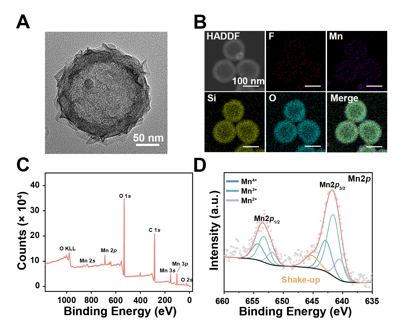
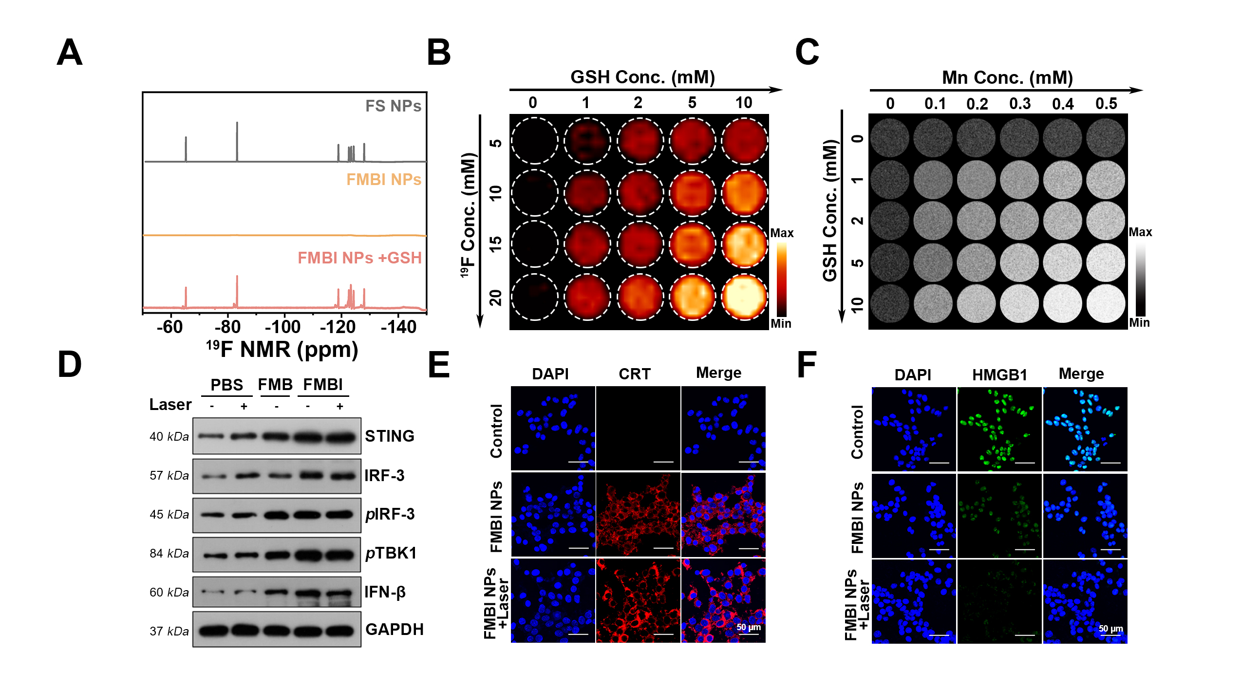
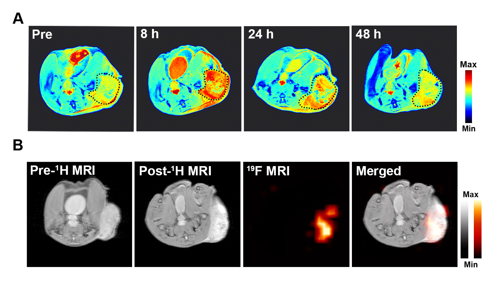
The FMBI NPs exhibit a core-shell-shell like structure (Fig. 1A) with Mn and F uniformly dispersed within the nanoparticles (Fig. 1B). The X-ray photoelectron spectroscopy results showed that Mn and O elements are present in the FMBI NPs (Fig. 1C). The Mn2p spectrum indicates that the predominant forms are Mn3+ and Mn4+ with relative intensities of 48.47% and 26.98% respectively (Fig. 1D). The 19F NMR spectrum of FS nanoparticles exhibited the same signal as PFOB, demonstrating the successful encapsulation of PFOB within the FS nanoparticles. Following MnOx modification, the 19F NMR signal vanished due to the PRE effect. Subsequently, manganese ions were released upon the addition of glutathione, which turn on the corresponding 19F NMR signal (Fig. 2A). The phantom experiments demonstrated that the signal intensity of both T1-weighted and 19F MRI were dependent on GSH concentration (Fig. 2B and 2C). To evaluate the impact on the cGAS-STING pathway, western blot was employed to detect related protein expression in B16F10 cells (Fig. 2D), which showed that FMBI NPs promoted the upregulation of these proteins. Overall, these findings suggested that FMBI NPs showed potential as a therapeutic approach for cancer treatment. In addition, confocal images demonstrated an increased expression of CRT for immunogenic cell death as well as a decreased expression of HMGB1 (Fig. 2E and 2F). T1-weighted in vivo MRI signals in the tumor region was enhanced, and the most significant signal enhancement appeared at 8 hours after i.v. injection of FMBI NPs (Fig. 3A). Moreover, 19F MRI imaging of mice showed a vivid 19F signal at the tumor site after in situ injection of FMBI NPs (Fig. 3B).Conclusion
The FMBI NPs were developed that can activate the cGAS-STING pathway for tumor diagnose and therapy. The FMBI NPs can release Mn2+ in the tumor microenvironment and enhance the signals of 1H/19F MRI in tumor areas, with the cGAS-STING pathway being activated. Combining photosensitizer-induced photothermal and photodynamic therapy with TME activation can convert immunosuppressed TME to immune-activated TME to enhance the anti-tumor efficacy. In summary, utilizing TME-activated manganese-based nanoparticles for MRI can potentially serve as an effective immunostimulatory method for anti-tumor treatment.Acknowledgements
This work is supported by National Key R&D Program of China (2018YFA0704000), and National Natural Science Foundation of China (81625011, 91859206, 81730048, 81971705).
References
[1] Sun Z, Wang Z, Wang T, et al. Biodegradable MnO-Based Nanoparticles with Engineering Surface for Tumor Therapy: Simultaneous Fenton-Like Ion Delivery and Immune Activation. ACS Nano. 2022;16(8):11862-11875.
[2] Zhang Y, Ma Q, Yan Y, et al. Intratumoral Glutathione Activatable Nanoprobes for Fluorescence and 19F Magnetic Resonance Turn-On Imaging. Anal Chem. 2020;92(23):15679-15684.
[3] Zhang K, Qi C, Cai K. Manganese-Based Tumor Immunotherapy. Adv Mater. 2023;35(19):e2205409.
Figures