3172
Acute changes of cerebral hemodynamics, metabolism and blood-brain barrier permeability in response to aerobic exercise1Department of Biomedical Engineering, Zhejiang University, Hangzhou, China, 2Department of Radiology, Johns Hopkins University, Baltimore, MD, United States
Synopsis
Keywords: Oxygenation, Oxygenation
Motivation: How does single bout of exercise affect brain oxygen metabolism and BBB permeability in addition to perfusion remains unclear.
Goal(s): This study aims to dynamically monitor the acute changes in cerebral physiology subsequent to a singular aerobic exercise training session
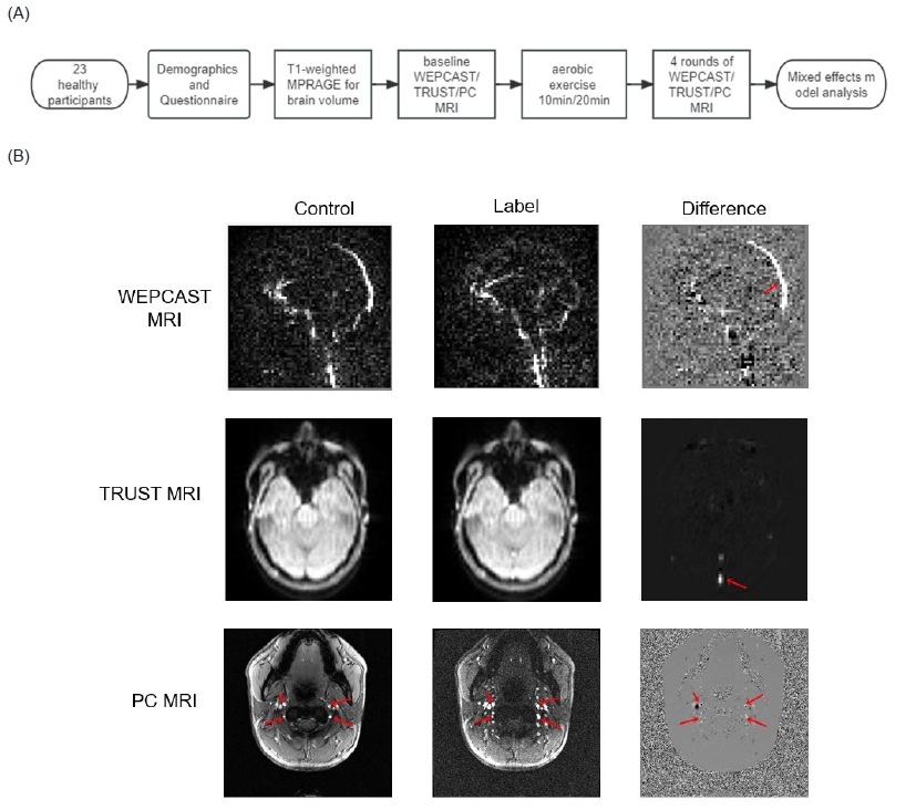
Approach: Multiple indices were quantified, including CBF as gauged by PC MRI, Yv and CMRO2 as assessed by TRUST MRI, BBB E and PS as determined via WEPCAST MRI.
Results: We found a significant increase in participants' CBF and CMRO2 post-exercise, post-exercise stability in E and a significant increase in PS were also observed.
Impact: Our findings suggest that a singular bout of moderate-intensity aerobic exercise can induce acute alterations in cerebral hemodynamics, metabolic processes, and blood-brain barrier permeability.These findings may shed light on the initial stages of the clinical implications of aerobic exercise.
Introduction
Previous studies have demonstrated the longitudinal benefit of aerobic exercise on cerebrovascular health1, thereby playing a pivotal role in thwarting the onset of a multitude of neurological disorders, including Alzheimer's disease. Nevertheless, the literature remains deficient in comprehensive understanding of the alterations engendered by short-term aerobic exercise interventions . Existing studies, employing alternative techniques, primarily focused on cerebral blood flow(CBF), neglecting other cerebral physiological factors such as oxygen metabolism and blood-brain barrier(BBB) permeability due to limited methodologies. Recent advances in MRI techniques allows a completely non-contrast assessment of these physiological parameters within ten minutes. Thus, in this study, we investigated the dynamic alterations in a multitude of physiological parameters in healthy individuals, following a single regimen of moderate-intensity aerobic exercise.Methods
The study engaged 23 healthy participants (10 females, 13 males), within an age range of 18-35 years.The participants were stratified into two cohorts. The first cohort underwent a regimen of aerobic exercise training for a duration of 10 minutes, while the other was subjected to a training session lasting 20 minutes The experiment was conducted in a sequence of steps as shown in Figure 1.A.MRI acquisitions and data processing:
Similar to previous studies2, PC MRI was performed at four major feeding arteries (left/right internal carotid arteries and left/right vertebral arteries) to quantify global CBF. The following parameters were used: encoding velocity (Venc) = 40 cm/s. Total flux of these four arteries was normalized by individual brain volume obtained from a 3D T1-weighted MPRAGE scan with the following parameters: shot interval = 2100 ms, inversion time (TI)= 1100 ms.
Additionally, venous oxygenation (Yv) was measured by TRUST MRI 3 : four effective TEs (eTE = 0, 40, 80, and 160 ms) with a 𝜏CPMG of 10 ms, labeling thickness = 100 mm. CMRO2 was then calculated by Fick equation4 as follows.
$$CMRO_{2} = CBF \times (Y_{a}-Y_{v})\times Ch$$To assess BBB permeability, WEPCAST MRI 5 was conducted in mid-sagittal plane with a labeling duration (𝜏) of 4000 ms and a post-labeling delay (PLD) of 3000 ms. Other imaging parameters were as follows: single-shot gradient EPI readout, Venc = 20 cm/s. E and PS was then quantified from the labeled spin signals at superior sagittal sinus.According to the Renkin-Crone Model6, PS can be calculated as$$PS=-\ln(1-E)\times f$$
Statistical analysis:
The time dependence of the physiological parameters was studied with a mixed-effect model, in which the age, gender, and exercise duration of the subjects were incorporated as fixed effects, while individual differences among the subjects were accounted for as random effects.
Results and Discussion
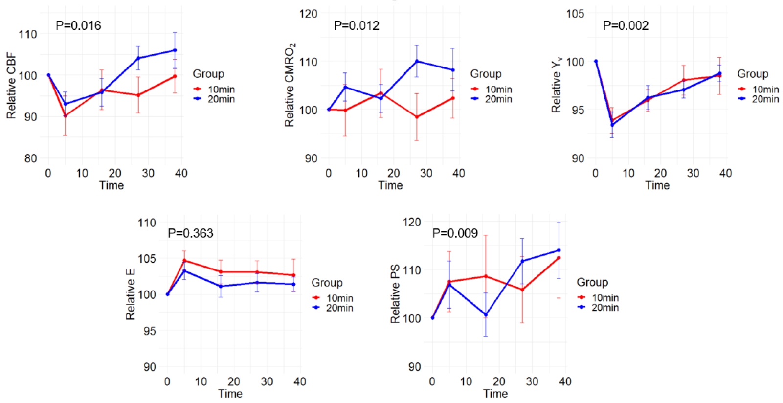
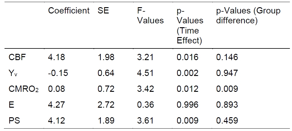
Figure 2 delineates the experimental outcomes, while Table 3 elucidates the analytical findings derived from the linear mixed-effects model. As depicted in Figure 2, there was first drop followed by a significant augmentation in the participants' CBF (p=0.016), while Yv showed a significant decrease (p=0.002), i.e. an increased oxygen extraction, and returned to baseline level at later time points. CMRO2 gradually increased (p=0.012), which could be attributable to an escalation in metabolic activity within the body and a higher demand for oxygen. The intergroup difference is only significant between the CMRO2 of the two groups, indicating a complex interaction between oxygen metabolism and exercise duration.In addition, our data showed that E remains relatively stable post-exercise (p=0.36), while PS significantly increased (p=0.009). It could due to the elevation of blood pressure after exercise, leading to a stretching of endothelial and vascular smooth muscles.
Conclusion
The findings of our investigation suggest that a singular bout of moderate-intensity aerobic exercise can induce acute alterations in cerebral hemodynamics, metabolic processes, and blood-brain barrier permeability. These findings may shed light on the initial stages of the clinical implications of aerobic exercise.Acknowledgements
Acknowledgments / Funding Information: This work is supported by College of Biomedical Engineering & Instrument Science, Zhejiang university.References
1. B. P. Thomas et al., "Brain Perfusion Change in Patients with Mild Cognitive Impairment After 12 Months of Aerobic Exercise Training," J Alzheimers Dis, vol. 75, no. 2, pp. 617-631, 2020, doi: 10.3233/JAD-190977.
2. S. L. Peng et al., "Age-related increase of resting metabolic rate in the human brain," Neuroimage, vol. 98, pp. 176-83, Sep 2014, doi: 10.1016/j.neuroimage.2014.04.078.
3. H. Lu and Y. Ge, "Quantitative evaluation of oxygenation in venous vessels using T2-Relaxation-Under-Spin-Tagging MRI," Magnetic Resonance in Medicine, vol. 60, no. 2, pp. 357-363, 2008, doi: https://doi.org/10.1002/mrm.21627.
4. F. Mielck, H. Stephan, W. Buhre, A. Weyland, and H. Sonntag, "Effects of 1 MAC desflurane on cerebral metabolism, blood flow and carbon dioxide reactivity in humans," Br J Anaesth, vol. 81, no. 2, pp. 155-60, Aug 1998, doi: 10.1093/bja/81.2.155.
5. Z. Lin et al., "Non-contrast MR imaging of blood-brain barrier permeability to water," Magnetic Resonance in Medicine, vol. 80, no. 4, pp. 1507-1520, 2018, doi:https://doi.org/10.1002/mrm.27141.
6. Renkin, E. M. (1959). Transport of potassium-42 from blood to tissue in isolated mammalian skeletal muscles. American Journal of Physiology-Legacy Content, 197(6), 1205-1210.
Figures