3170
Resting-state fMRI localization of tongue motor areas with a patient-based probabilistic functional atlas1Department of Imaging Physics, University of Texas MD Anderson Cancer Center, Houston, TX, United States, 2The University of Texas MD Anderson Cancer Center UTHealth Graduate School of Biomedical Sciences, Houston, TX, United States, 3NordicNeuroLab Inc, Milwaukee, WI, United States, 4Department of Neuro-Oncology, University of Texas MD Anderson Cancer Center, Houston, TX, United States, 5Department of Neurosurgery, University of Texas MD Anderson Cancer Center, Houston, TX, United States, 6Department of Neuroradiology, University of Texas MD Anderson Cancer Center, Houston, TX, United States
Synopsis
Keywords: fMRI Analysis, fMRI, Motor function
Motivation: To benefit patients who needs presurgical mapping of tongue motor areas but have limited tb-fMRI.
Goal(s): To create a probabilistic tongue motor atlas and evaluate its use for guiding rs-fMRI seed-based correlation (SBC) analysis.
Approach: Presurgical tb-fMRI of 30 brain tumor patients were used to create the atlas. This atlas was transferred to a different set of patients’ individual space for guiding rs-fMRI analysis. Locations of functional connectivity detected in the ipsilateral hemisphere were compared with tb-fMRI results.
Results: A tongue motor atlas was developed, which can effectively guide the rs-fMRI analysis with results similar to when seeding based on the tb-fMRI activation.
Impact: The probabilistic functional atlas enables automated seed-based analysis for rs-fMRI localization of tongue motor areas in patients undergoing brain tumor resection. It also provides a functional localizer which can be used for various quantitative image analyses of tongue motor areas.
Introduction
Task-based (tb) fMRI is a common procedure for mapping eloquent brain areas, including the tongue motor area, for pre-operative evaluation of brain tumor resections. When tb-fMRI is limited, resting-state (rs) fMRI can serve as an alternative for the presurgical mapping. Feasibility of rs-fMRI localization of ventral somatomotor network, including tongue, mouth, and face areas, has been demonstrated by using seed-based correlation (SBC) analysis1, 2, independent component analysis2-4, and deep learning methods5. While SBC is an intuitive approach, it requires a priori knowledge of the seed location which has relied on either anatomical knowledge1,2 from subdural-electrode recording6, or tb-fMRI activations2. This study aimed to develop a tongue motor atlas and test its use for guiding the SBC analysis.Methods
Tongue motor presurgical fMRI of 30 brain tumor patients (3T, GRE-EPI, TR/TE=3000/25 ms, voxel size = 1.7 x 1.7 x 4 mm3) were utilized to build the atlas. The paradigm started with a 15-s control, followed by six cycles of 15-s task and control blocks. fMRI data were processed using SPM12, including motion correction, co-registration with 3D T1, normalization to MNI space, and 6-mm FWHM spatial smoothing. General linear model was applied to generate activation t maps which were thresholded at t=5.36 (p<0.05, FWE-corrected). The probabilistic tongue motor atlas was obtained by overlaying the binary mask of activation maps for each patient.A separate dataset, including tb- and rs-fMRI, of 8 patients were used for testing the rs-fMRI analysis. For the tb-fMRI, a 50% AMPLE threshold7 was also applied. The rs-fMRI was performed using GRE-EPI (TR/TE=2000/25 ms, voxel size = 3.4 x 3.4 x 4 mm3, 180 dynamics) with eyes open and fixated. Image preprocessing included slice timing, motion correction, aligning to 3D T1, de-spiking, detrending, nuisance regression, band-pass filtering (0.01–0.08 Hz), and spatial smoothing. The atlas was spatially transferred to individual’s image space using ANTs software. Three seeds in contralateral hemisphere were selected: (1) a sphere (r=6 mm) centered at peak tb-fMRI activation, (2) a sphere (r=6 mm) centered at the peak probability in atlas, and (3) a binary mask generated from the atlas (>30%), and used to calculate functional connectivity (FC). The FC map thresholded with z=0.8-1.2 to optimize the detection of ipsilateral tongue motor area.
Results
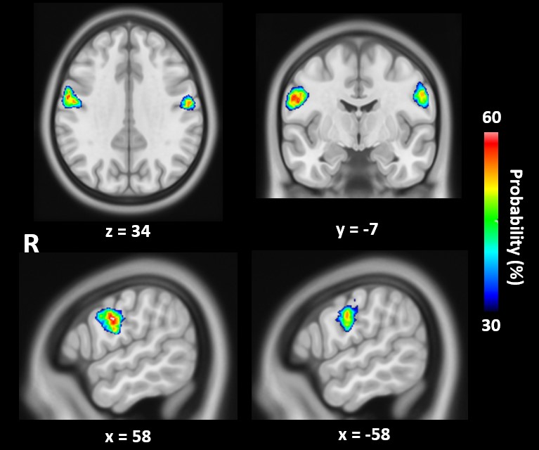
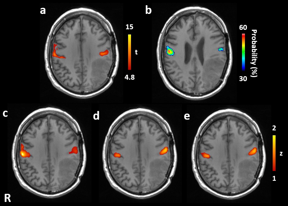
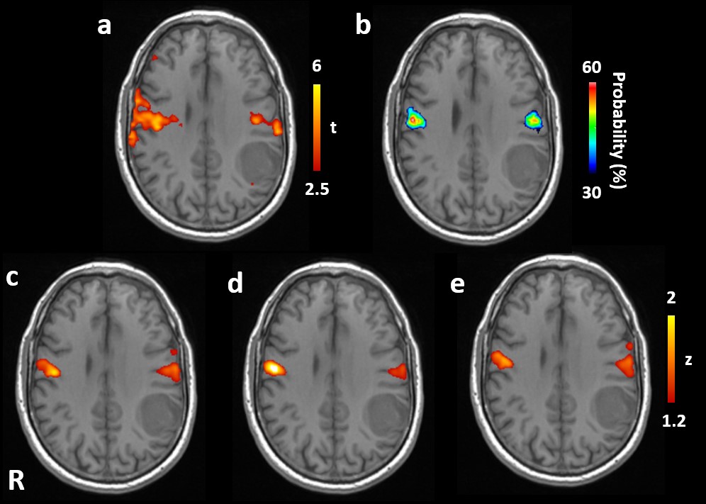
The atlas demonstrated tongue motor areas with peak probability (MNI coordinate) of 53% (-58, -8, 36) in the left and 63% (58, -6, 32) in the right hemisphere (Fig. 1). Distances between the peaks and Sylvian fissure (SF) were 23 and 26 mm in left and right hemispheres, respectively.Tb-fMRI of testing patients had significant activations (t>5.36) in bilateral tongue motor areas, except for 2 patients who only had highest t values between 4-5 in the ipsilateral hemisphere. Rs-fMRI with the three seeds were all able to localize ipsilateral tongue motor areas which overlapped with tb-fMRI activations with AMPLE threshold. Figure 2 shows a patient who had significant tb-fMRI activations in bilateral tongue motor areas (a), and all three FC maps (c-e) agreed well with tb-fMRI. Figure 3 demonstrates results of another patient whose tb-fMRI activation was not detected in the ipsilateral hemisphere with the standard threshold. An AMPLE t-threshold=2.5 yielded noisy tb-fMRI results (a), but agreed with the locations detected by rs-fMRI (c-e).
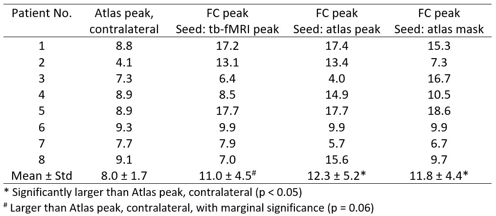
The distance between atlas and tb-fMRI peaks in the contralateral hemisphere (i.e. distance between seeds of the first two methods) was 8.0 ± 1.7 mm (Table). Similar FC-to-tb activation distances were obtained among the three methods (11.0 ± 4.5, 12.3 ± 5.2, and 11.8 ± 4.4, respectively) without statistically significant differences (p>0.37).
Discussion
The tongue motor areas in our atlas are generally consistent with locations from the subdural-electrode study (2.05 cm without lesion, 2.74 cm with fronto-parietal lesions)6. Interestingly, higher probability values were obtained in right hemisphere than left in the atlas, which will be studied with a larger patient cohort and lesion analysis. Seed selections based on the atlas resulted in similar FC-to-tb activation distances comparing to seeding based on the tb-fMRI, which suggests that tb-fMRI may not be needed to aid the SBC analysis. This result needs to be validated in more patients with wider distributions of lesion characteristics. Furthermore, the atlas can provide functional localizers that may help quantitative and automated fMRI analysis.Conclusion
A probabilistic functional atlas of tongue motor areas was developed from presurgical fMRI of brain tumor patients. This atlas can effectively guide the seed selection for rs-fMRI localization of tongue motor areas with results similar to when seeding based on the tb-fMRI activation in the contralateral hemisphere.Acknowledgements
This study was supported by NIH/NCI under award number R01 CA258788.References
- Liu H, Buckner RL, Talukdar T, Tanaka N, Madsen JR, Stufflebeam SM. Task-free presurgical mapping using functional magnetic resonance imaging intrinsic activity. J Neurosurg 2009;111:746-754.2.
- Rosazza C, Aquino D, D'Incerti L, Cordella R, Andronache A, Zacà D, Bruzzone MG, Tringali G, Minati L. Preoperative mapping of the sensorimotor cortex: comparative assessment of task-based and resting-state FMRI. PLoS One 2014 10;9(6):e98860.3.
- Schneider FC, Pailler M, Faillenot I, Vassal F, Guyotat J, Barral FG, Boutet C. Presurgical Assessment of the Sensorimotor Cortex Using Resting-State fMRI. AJNR Am J Neuroradiol 2016;37:101-107.4.
- Yahyavi-Firouz-Abadi N, Pillai JJ, Lindquist MA, Calhoun VD, Agarwal S, Airan RD, Caffo B, Gujar SK, Sair HI. Presurgical Brain Mapping of the Ventral Somatomotor Network in Patients with Brain Tumors Using Resting-State fMRI. AJNR Am J Neuroradiol 2017;38:1006-1012.5.
- Nandakumar N, Manzoor K, Agarwal S, Pillai JJ, Gujar SK, Sair HI, Venkataraman A. Automated eloquent cortex localization in brain tumor patients using multi-task graph neural networks. Med Image Anal 2021;74:102203.6.
- Urasaki E, Uematsu S, Gordon B, Lesser RP. Cortical tongue area studied by chronically implanted subdural electrodes--with special reference to parietal motor and frontal sensory responses. Brain 1994;117 (Pt 1):117-132.7.
- Voyvodic JT, Petrella JR, Friedman AH. fMRI activation mapping as a percentage of local excitation: consistent presurgical motor maps without threshold adjustment. J Magn Reson Imaging 2009;29:751–759.
Figures