3162
Altered functional connectivity of emotional circuits and default model network in postpartum women: A Resting-State fMRI Study1School of Psychology, Shandong Normal University, JiNan, China, 2Philips Healthcare, Beijing, China, 3Department of Psychology,Shanghai University of Sport, Shanghai, China
Synopsis
Keywords: Functional Connectivity, fMRI (resting state), Functional connectivity;Postpartum women
Motivation: Resting-state functional connectivity can elucidate the brain intrinsic functional connectivity patterns of postpartum women, providing new evidence for functional plasticity in postpartum women.
Goal(s): Our goal is to explore functional connectivity patterns of postpartum women.
Approach: Postpartum women were imaged using rs-fMRI and compared with nonmothers by functional connectivity analysis.
Results: Brain functional remodeling does occur in the postpartum women, as evidenced by changes in intrinsic functional connectivity patterns.
Impact: We used rs-FC to demonstrate that brain functional placity in postpartum women during raising infants, providing new ideas and directions for studying the neural mechanisms of maternal behavior.
Introduction
Women undergo enormous changes in their bodies and brains during the postpartum period, mainly including behavioral, cognition and emotion. Previous neuroimaging studies have shown that the maternal brain exhibits structural and functional plasticity using task-based functional MRI and VBM1,2. Resting-state fMRI (rs-fMRI) has mostly focused on postpartum depression using regional homogeneity (ReHo) and amplitude of low-frequency fluctuation (ALFF)3,4. However, the studies on the intrinsic functional connectivity pattern of healthy postpartum women are relatively lacking. On the basis of previous studies, we selected the brain regions associated with emotional networks and cognitive processing circuits as the regions of interest for rs-FC analysis5–7. In the current study, we intend to explore brain functional connectivity pattern relevant to maternal behaviors in mothers during the postpartum period using resting-state functional connectivity (rs-FC).Methods
Thirty-eight new mothers (age: 30.59 ± 2.95 years) and twenty-five nonmothers (age: 26.60 ± 1.90 years) participated in the current study. All subjects underwent resting-state functional magnetic resonance imaging (fMRI) examinations and completed the Interpersonal Reactivity Index (IRI), a questionnaire that assesses empathy. Using resting-state functional connectivity (rs-FC) and correlation analysis to evaluate the brain functional framework of postpartum women and neural mechanisms of maternal behavior.Results
Compared to nonmothers, mothers exhibited increased functional connectivity in regions associated with emotional processes (insula, hippocampus, amygdala, posterior cingulate gyrus, and medial frontal gyrus), while functional connectivity in the default mode network (inferior parietal lobule and precuneus) decreased significantly (see Figure 1 and Table 1).Discussion
The rs-FC results showed changed functional connectivity were primarily identified in the limbic system, amygdala, insula, medial frontal cortex, inferior parietal lobule and precuneus in postpartum women. Maternal behavior are associated with the theory of mind (ToM) networks and default mode network, which are related to emotional and cognitive processing circuits8–10. Rs-FC value between the left medial orbitofrontal cortex and left postcentral gyrus as well as between the left superior frontal gyrus and right precuneus showed a significant correlation with PD subscale scores. These findings provide further evidence to support the plasticity of brain function in postpartum women, which is beneficial for understanding the neural mechanisms of maternal behavior.Conclusion
In conclusion, our study shows that there are significant differences in resting-state functional connectivity between mothers and nonmothers, which were mainly located in the regions corresponding to the emotional circuit and DMN. These results demonstrate that functional connectivity in mothers shows plastic changes during the process of raising infants. These alterations in mothers improve the response to their infants, cultivate mother-infant attachment and enhance the quality of parenting.Acknowledgements
This work was fnancially supported by the National Natural Science Foundation of China (Grant Nos. 32200915), and the Shandong Provincial Natural ScienceFoundation Project (No. ZR2022QC251).
References
1. Kim, P., Strathearn, L. & Swain, J. E. The maternal brain and its plasticity in humans. Hormones and Behavior 77, 113–123 (2016).
2. Pereira, M. Structural and Functional Plasticity in the Maternal Brain Circuitry. New Directions For Child and Adolescent Development 153, 23-46 (2016).
3. Zheng, J.-X. et al. Disrupted Spontaneous Neural Activity Related to Cognitive Impairment in Postpartum Women. Front Psychol 9, 624 (2018).
4. Zhang, S. et al. Aberrant resting-state interhemispheric functional connectivity in patients with postpartum depression. Behavioural Brain Research 382, 483 (2020).
5. Rigo, P. et al. Specific maternal brain responses to their own child’s face: An fMRI meta-analysis. Developmental Review 51, 58–69 (2019).
6. Zhang, K. et al. Brain Responses to Emotional Infant Faces in New Mothers and Nulliparous Women. Sci Rep 10, 9560 (2020).
7. Hoekzema, E. et al. Mapping the effects of pregnancy on resting state brain activity, white matter microstructure, neural metabolite concentrations and grey matter architecture. Nat Commun 13, 6931 (2022).
8. Schaafsma, S. M., Pfaff, D. W., Spunt, R. P. & Adolphs, R. Deconstructing and reconstructing theory of mind. Trends Cogn Sci 19, 65–72 (2015).
9. Schurz, M. et al. Toward a hierarchical model of social cognition: A neuroimaging meta-analysis and integrative review of empathy and theory of mind. Psychol Bull 147, 293–327 (2021).
10. Mars, R. B. et al. On the relationship between the “default mode network” and the “social brain”. Front. Hum. Neurosci. 6, (2012).
Figures
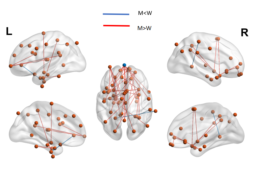
Figure 1. Areas of altered FC between new mothers and nonmothers. L: left hemisphere, R:right hemisphere, cluster-level FWE correction at p<0.05. M = new mothers. W =nonmother women.
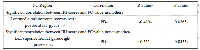
Table 1.Significant correlations between IRI scores and rs-FC in mothers and nonmothers. PD: personal distress scale. Comparisons were calculated using paired t tests and reported at P < 0.05 with Bonferroni correction. “*” indicates that the difference was < 0.05.
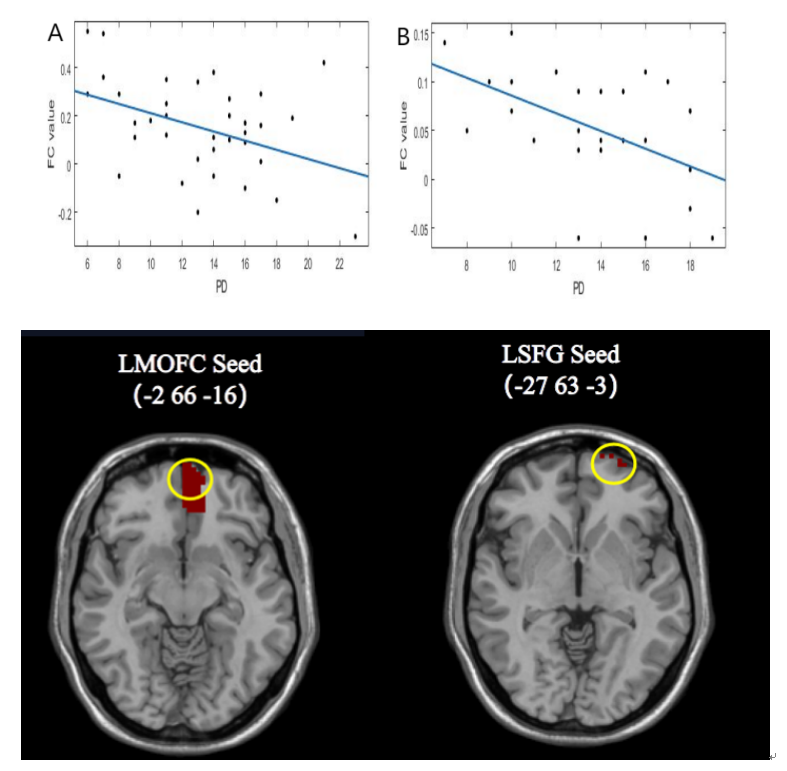
Figure 2. Correlation between FC values and IRI scores in mothers and nonmothers. (A): Correlation between FC values and PD scores in new mothers when the left medial orbitofrontal cortex (LMOFC) was used as the seed. (B): Correlation between FC values and PD scores in nonmothers when the left superior frontal gyrus (LSFG) was used as the seed.