3161
Cortical Sulcus Depth Alterations in Patients with Tinnitus Before and After Sound Therapy: A Surface-Based Morphometry Study1Radiology, Beijing Friendship Hospital, Capital Medical University, Beijing, China
Synopsis
Keywords: fMRI Analysis, Head & Neck/ENT
Motivation: This study aimed to explore alterations in brain surface-based morphometry sulcal depth in patients with idiopathic tinnitus before and after 24 weeks of sound therapy.
Goal(s): This study aimed to explore alterations in brain surface-based morphometry sulcal depth in patients with idiopathic tinnitus before and after 24 weeks of sound therapy.
Approach: Thirty-three tinnitus patients underwent magnetic resonance imaging scans at baseline and after 24 weeks of sound therapy.
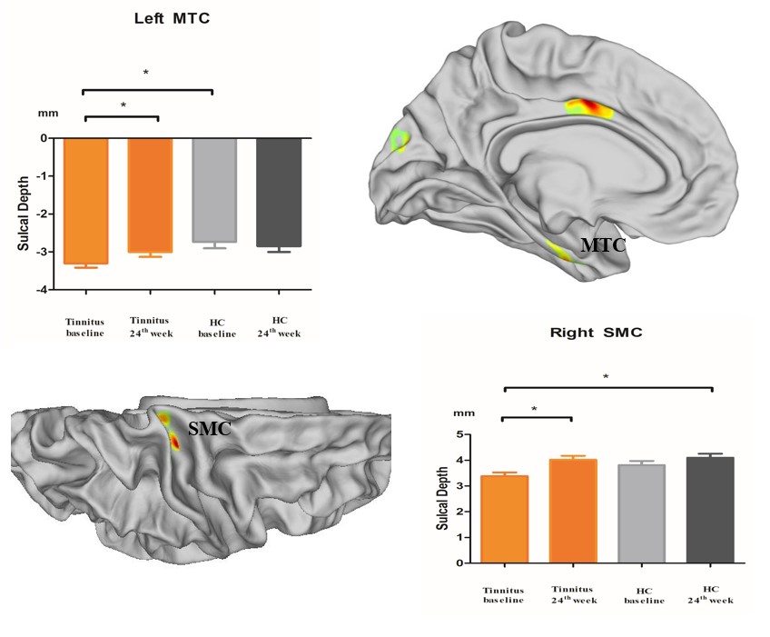
Results: Compared to HCs, those in the tinnitus group at baseline had significantly lower sulcal depth in the left medial temporal cortex (MTC) and right somatosensory and motor cortex (SMC).
Impact: The remodeling of sulcal depth after sound therapy is an indicator for effective sound therapy.
Purpose
As one of the quantitative measures of the cerebral cortex, sulcal depth has been widely used in brain morphology research. This study aimed to explore alterations in brain surface-based morphometry sulcal depth in patients with idiopathic tinnitus before and after 24 weeks of sound therapy.Methods
Thirty-three tinnitus patients underwent magnetic resonance imaging scans at baseline and after 24 weeks of sound therapy. Twenty-six age- and sex-matched healthy control (HC) individuals also underwent two scans over a 24-week interval. For all participants, 3.0T magnetic resonance imaging (MRI) and high-resolution 3D structural images were acquired with a 3D-BRAVO pulse sequence. Structural image data preprocessing was performed using the DPABISurf toolbox. Tinnitus Handicap Inventory (THI) scores were used to assess the severity of tinnitus before and after treatment. Two-way mixed model analysis of variance (ANOVA) and Pearson’s correlation analysis were used in the statistical analysis. Student-Newman-Keuls (SNK) tests were used in the post hoc analysis.Results
Compared to HCs, those in the tinnitus group at baseline had significantly lower sulcal depth in the left medial temporal cortex (MTC) and right somatosensory and motor cortex (SMC). After 24 weeks of sound therapy, the tinnitus patients demonstrated significantly increased sulcal depth in the left MTC and right SMC. There were no significant differences in sulcal depth between the tinnitus patients after treatment and HCs. (Fig1-Fig.3)Conclusions
The remodeling of sulcal depth after sound therapy is an indicator for effective sound therapy. These brain regions may serve as potential neuroimaging biomarkers for the evaluation of tinnitus treatment effects.Acknowledgements
We especially thank Beijing Friendship Hospital, Capital Medical University, for their support with our research.References
1. Henry, J. A., Schechter, M. A., Nagler, S. M., & Fausti, S. A. (2002). Comparison of tinnitus masking and tinnitus retraining therapy. J Am Acad Audiol, 13(10), 559-581.
2. Hickok, G. (2012). The cortical organization of speech processing: feedback control and predictive coding the context of a dual-stream model. J Commun Disord, 45(6), 393-402. doi:10.1016/j.jcomdis.2012.06.004
3. Husain, F. T., Medina, R. E., Davis, C. W., Szymko-Bennett, Y., Simonyan, K., Pajor, N. M., & Horwitz, B. (2011). Neuroanatomical changes due to hearing loss and chronic tinnitus: a combined VBM and DTI study. Brain Res, 1369, 74-88. doi:10.1016/j.brainres.2010.10.095
4. Im, K., Lee, J. M., Seo, S. W., Hyung Kim, S., Kim, S. I., & Na, D. L. (2008). Sulcal morphology changes and their relationship with cortical thickness and gyral white matter volume in mild cognitive impairment and Alzheimer's disease. Neuroimage, 43(1), 103-113. doi:10.1016/j.neuroimage.2008.07.016
5. James, C., Lepore, F., Collignon, O., Gajawelli, N., Lepore, N., & Coulon, O. (2017). Central sulcus depth and sulcal profile differences between congenitally blind and sighted individuals. Annu Int Conf IEEE Eng Med Biol Soc, 2017, 3008-3011. doi:10.1109/EMBC.2017.8037490
6. Kippenhan, J. S., Olsen, R. K., Mervis, C. B., Morris, C. A., Kohn, P., Meyer-Lindenberg, A., & Berman, K. F. (2005). Genetic contributions to human gyrification: sulcal morphometry in Williams syndrome. J Neurosci, 25(34), 7840-7846. doi:10.1523/JNEUROSCI.1722-05.2005
7. Kragel, P. A., Kano, M., Van Oudenhove, L., Ly, H. G., Dupont, P., Rubio, A., . . . Wager, T. D. (2018). Generalizable representations of pain, cognitive control, and negative emotion in medial frontal cortex. Nat Neurosci, 21(2), 283-289. doi:10.1038/s41593-017-0051-7
8. Krick, C. M., Grapp, M., Daneshvar-Talebi, J., Reith, W., Plinkert, P. K., & Bolay, H. V. (2015). Cortical reorganization in recent-onset tinnitus patients by the Heidelberg Model of Music Therapy. Front Neurosci, 9, 49. doi:10.3389/fnins.2015.00049
10. Langguth, B., Kreuzer, P. M., Kleinjung, T., & De Ridder, D. (2013). Tinnitus: causes and clinical management. Lancet Neurol, 12(9), 920-930. doi:10.1016/S1474-4422(13)70160-1
Figures