3160
An fMRI single-subject study with severe reading impairment – a new paradigm towards personalised medicine1Neuroimaging Unit, Scientific Institute, IRCCS Eugenio Medea, Bosisio Parini (LC), Italy, 2Department of Informatics, Systems and Communication, University of Milan-Bicocca, Milan, Italy, 3Unit of rehabilitation of rare diseases of the central and peripheral nervous system, Scientific Institute, IRCCS Eugenio Medea, Bosisio Parini (LC), Italy, 4Department of Information Engineering, University of Padova, Padova, Italy, 5Department of Brain and Behavioral Sciences, University of Pavia, Pavia, Italy, 6Child Psychopathology Unit, Scientific Institute, IRCCS Eugenio Medea, Bosisio Parini (LC), Italy, 7Diagnostic Imaging and Neuroradiology Unit, Scientific Institute, IRCCS Eugenio Medea, Bosisio Parini (LC), Italy
Synopsis
Keywords: Task/Intervention Based fMRI, Brain, Neuroimaging, Pediatrics, Personalised Medicine
Motivation: Typically, fMRI studies predominantly rely on group comparisons, often lacking direct applications at an individual subject level, whether for research or clinical purposes.
Goal(s): We aimed to unveil the power of fMRI studies using an fMRI experiment to highlight the brain’s area of abnormalities in a 9 years old child experiencing severe reading impairment.
Approach: Three fMRI tasks were used to characterize different reading network sections that were compared to an age matched control group via Crawford-Howell t-test.
Results: The study subject showed a different activation pattern in the right insula, an area associated with reading comprehension.
Impact: fMRI experiments can compare and emphasize differences in brain function at an individual subject basis in comparison to a control population. This evidence suggests the potential use of fMRI as an informative tool within the precision medicine framework.
Introduction
Functional magnetic resonance imaging (fMRI) has become one of the most common and powerful tools for studying in vivo the cognitive processes in the human brain. Neurosciences and psychiatry/psychology represent the fields that benefits most from this tool, with several studies relying on group analysis [1]. While there are a few instances where fMRI is applied successfully to individual subjects, such in the pre-operative planning phase to safeguard primary functional regions, there is a general lack of fMRI studies tailored to single patients for diagnostic use and for developing suitable therapeutic strategies. In this study, we present a single-case scenario involving a 9 year old child facing severe reading difficulties without additional cognitive impairments. Our aim is to utilize an fMRI experiment to identify brain areas exhibiting functional abnormalities in comparison to a group of similarly aged children.Methods
Study participants: The considered subject is a 9 years old male child, who underwent cognitive tests showing a normal cognitive profile but strong reading difficulties and deficit procedural memory. The control group consisted of 9 typical reader (TR) children (age = 9.4 ± 0.7 years; 6/3 males/females) who are age-matched and IQ-matched (according to the Wechsler Intelligence Scale for Children – IV edition) with the study subject. All subjects were Italian native speakers.MRI acquisition protocol: MRI data were collected on a 3T Philips Achieva d-Stream with a 32-channel head coil [2]. The MRI acquisition protocol is reported in figure 1.
fMRI tasks: Three tasks were acquired, each of them meant to detect aspects related to the reading process (figure 2). (a) The Picture Identification (PID) task tests lexical processing showing an object’s image (cue) with a matching or mismatching visual or auditory stimulus. (b) The Posner cueing task targets the visuo-spatial attention network [3]. (c) The Coherent Dots Motion (CDM) detection task elicits the dorsal stream[2].
Data analysis: fMRI preprocessing involved the use of FMRIB Software Library (FSL) and the Advanced Normalization Tool (ANTs), following a standard pipeline that encompassed slice-timing correction, distortion and motion correction, registration to subject’s own structural image, spatial smoothing and intensity normalization. Outlier volumes were identified using the Artifact Detection Tools (ART) and subsequently discarded from the subsequent analyses. The first level analysis was conducted using SPM12, with a General Linear Model (GLM) using task specific conditions as predictors and motion parameters and outlier volumes as nuisance regressors. According to the specific task, contrast maps were obtained and mapped into MNI template space for comparisons. Second level analysis was performed starting from TRs to derive the population level activation maps. Activation clusters were identified for each contrast (p < 0.001, p-cluster < 0.05, FDR-corrected, cluster size > 240 mm3) and used as mask for the subsequent voxel-wise comparison between the patient and the control group using the Crawford-Howell test [4] with a significant threshold set to p <= 0.001 (uncorrected).
Results
An expert neuroradiologist read the diagnostic images that revealed no clear abnormalities (figure 3). The tasks analysis showed a significant difference in the right Insula (cluster size = 456 mm3; peak T value = 3.6; peak p-value = 2.3x10⁻⁶) for the PID task when considering the contrast map “Visuo correct > Visuo uncorrect” (figure 4), namely when the subject reads correctly versus reading mistake, thus highlighting an area involved in the correct reading process [5]. In particular, the patient showed a higher activation with respect to the control population. On the other hand, the other tasks and contrasts showed no differences between the patient and the control population, suggesting that the visuo-dorsal and the attentive networks in the patient are not compromised.Discussion
This study allowed the comparison of a single subject with a control population on an fMRI experiment. According to the results provided by the different tasks we were able to isolate a portion of the cortex involved in the reading network that shows an abnormal activation profile when performing a reading task. Specifically, the insular cortex, recognized for its role in visual integration tasks crucial for proficient reading, exhibited this abnormal activity. The localization was within an area known to be involved in the reading comprehension task, but located contralateral to the normative reading circuit, suggesting a potential language translocation. These subject’s specific findings suggest the possibility towards a precision medicine approach using fMRI and allow, for example, the tailoring and monitoring of personalised cognitive therapies.Acknowledgements
This study was partially supported by “Ricerca corrente 2023” and “5 per mille” funds provided by the Italian Ministry of Health.
References
[1] Frankford, S. A., Nieto-Castañón, A., Tourville, J. A., Guenther, F. H. (2021). Reliability of single-subject neural activation patterns in speech production task. Brain and Language, 212:104881. doi: 10.1016/j.bandl.2020.104881
[2] Mascheretti, S., Peruzzo, D., Andreola, C., Villa, M., Ciceri, T., Trezzi, V., ... & Arrigoni, F. (2021). Selecting the most relevant brain regions to classify children with developmental dyslexia and typical readers by using complex magnocellular stimuli and multiple kernel learning. Brain Sciences, 11(6), 722.
[3] Facoetti, A., Trussardi, A. N., Lorusso, M. L., Cattaneo, C., Galli, R., Molteni, M., Zorzi, M. (2010). Multisensory spatial attention deficits are predictive of phonological decoding skills in developmental dyslexia. Journal of Cognitive Neuroscience, 22(5):1011-25. doi: 10.1162/jocn.2009.21232
[4] Fehr, T., Weber, J., Willmes, K., Herrmann, M. (2010). Neural correlates in exceptional mental arithmetic – About the neural architecture of prodigious skills. Neuropsychologia, 48, 1407-1416.
[5] Landi, N., Frost, S. J., Menc, W. E., Sandak, R., Pugh, K. R. (2013). Neurobiological bases of reading comprehension: Insights from neuroimaging studies of word level and text level processing in skilled and impaired readers. Reading & Writing Quarterly, 29(2): 145-167. DOI: 10.1080/10573569.2013.758566
Figures
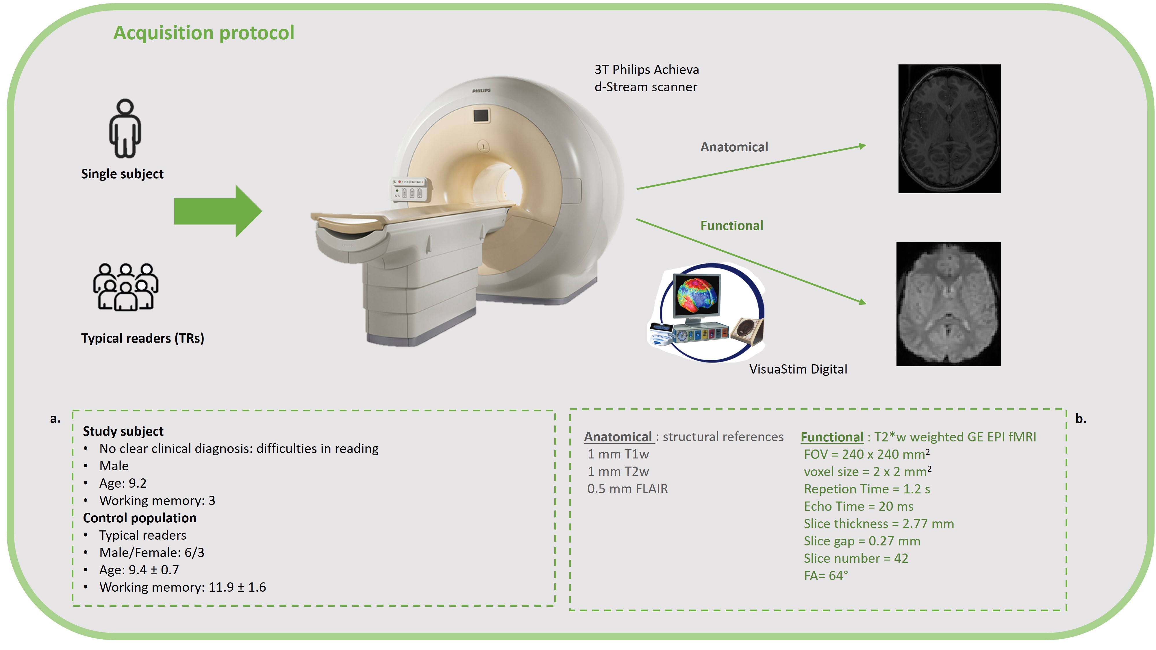
Figure 1. Technical details about study participants and MRI acquisition protocol. Subjects being age-matched with similar cognitive profiles (a) underwent an fMRI experiment. The acquisition protocol (b) included a structural acquisition session for anatomical reference and a functional acquisition session using VisuaStim Digital system with MRI-compatible pads for recording subject’s response.
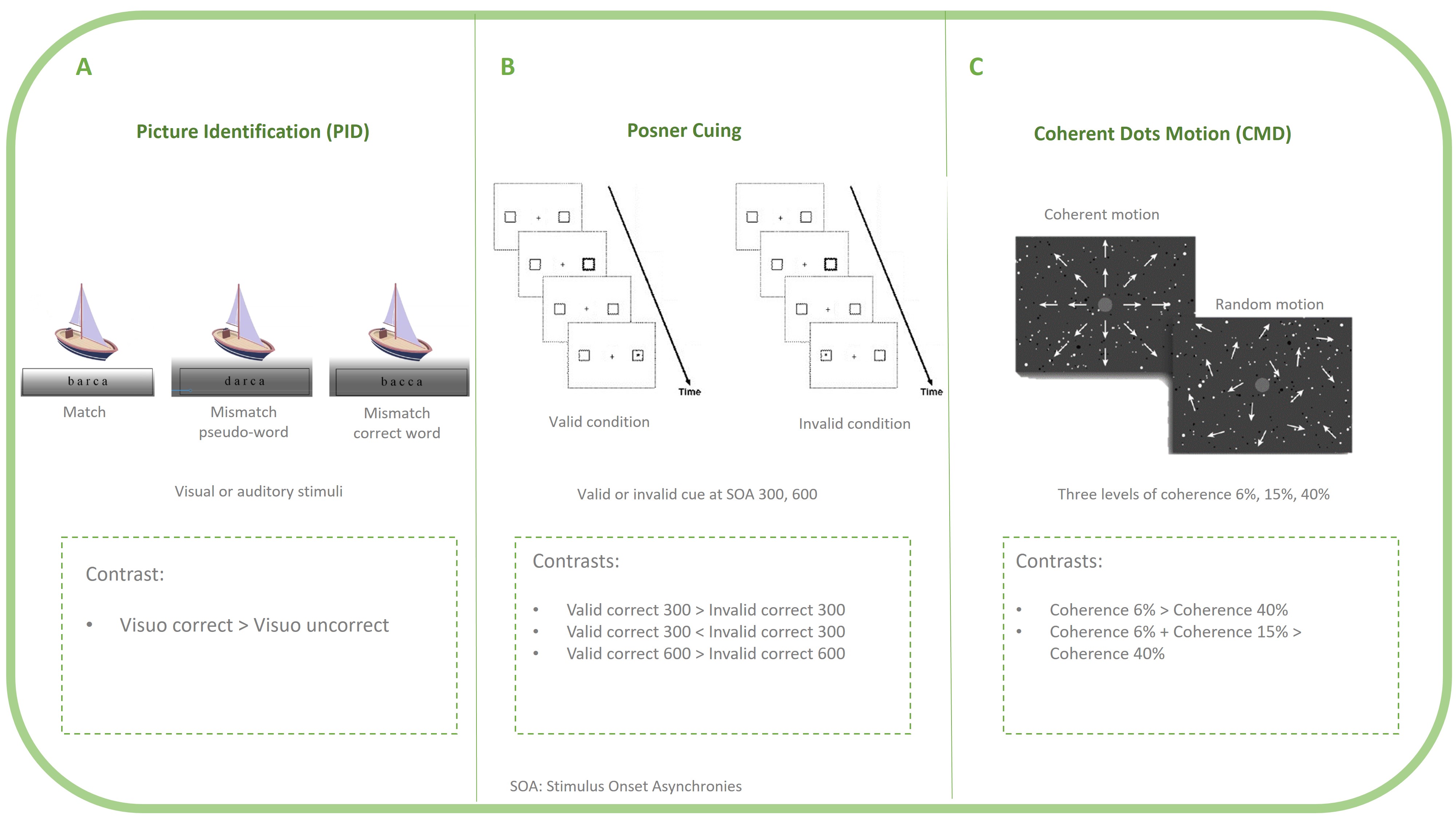
Figure 2. fMRI tasks representation and corresponding contrasts to highlight subjects’ responses. Each subject underwent three task eliciting different brain circuits. A) PID tests lexical processing. B) Posner cuing measures visuo-spatial attention. C) CDM elicits the dorsal stream, measuring the ability of motion perception.
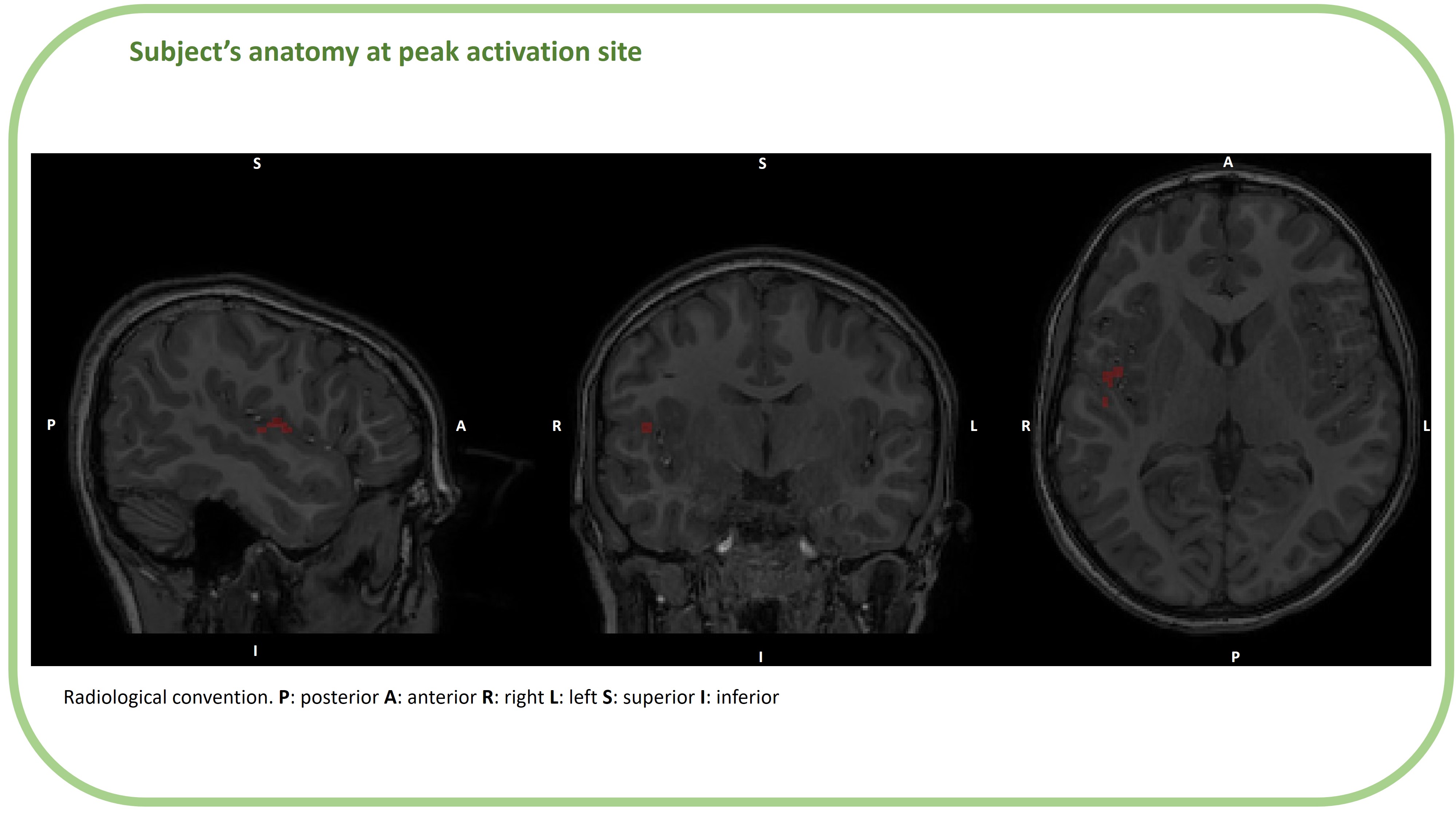
Figure 3. Study subject’s anatomical acquisition (T1w) at height of peak of activation during PID task in the three orthogonal planes: sagittal, coronal and axial, respectively from left to right. Red area represents the activation cluster area. As referred by an expert neuroradiologist, the anatomy shows no structural anomaly, leading to conduct the fMRI experiment.
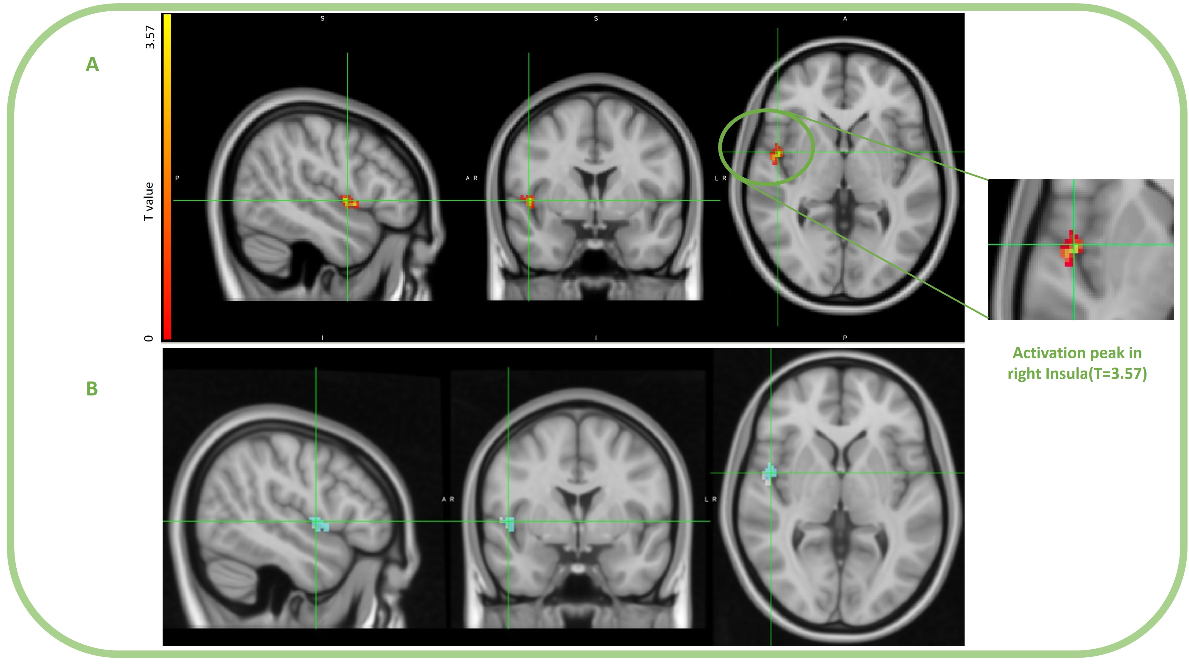
Figure 4. Results representation. Study subject shows a cluster of significant difference during PID task. A) T values in the activation cluster visualised at the peak level. The peak is located in the right Insula according to AAL atlas (T = 3.57). The cluster covers following areas in the right hemisphere: Heschl (52.6%), Insula (39.47%) and Rolandic Operculum (2.63%). B) Area of the cluster at peak level where Crawford-Howell test output is a p_value <= 0.001 (light-blue region).