3158
Neural Correlates of Transcutaneous Vagal Nerve Stimulation: Functional MRI Effects of Anatomical Site and Waveform Parameters1Radiology and Nuclear Medicine, Maastricht University Medical Center, Maastricht, Netherlands, 2Internal Medicine, Maastricht University Medical Center, Maastricht, Netherlands, 3Department of Psychiatry and Neuropsychology, Alzheimer Center Limburg, Maastricht University Medical Center, Maastricht, Netherlands, 4Centre for Neuroscience and Trauma, Queen Mary University of London, London, United Kingdom, 5Spinoza Centre for Neuroimaging, Amsterdam, Netherlands
Synopsis
Keywords: Task/Intervention Based fMRI, Brain Connectivity, Transcutaneous Vagal Nerve Stimulation
Motivation: There is marked heterogeneity in transcutaneous vagal nerve stimulation (tVNS) based on the anatomical site (cervical/auricular), and the waveform parameters (fixed/variable frequency) of the stimulation.
Goal(s): To explore the functional brain differences between cervical vagal nerve stimulation and auricular vagal nerve stimulation.
Approach: Ultra-high field functional neuroimaging is performed of the neural correlates of tVNS with disparate anatomical sites and electrical waveform characteristics during rest in healthy participants
Results: Auricular stimulation with a variable frequency was shown to result in a significant decrease of the BOLD response in the amygdala and insular cortex compared to sham stimulation.
Impact: There exist multiple knowledge gaps in the use of tVNS, which hinders the advancement of tVNS applications. This explorative study has shown the differences in neuronal response of auricular and cervical stimulation with fixed and variable frequencies.
Introduction
Transcutaneous vagal nerve stimulation (tVNS) has been applied to a number of disease areas including visceral pain, depression, migraine and epilepsy1. However, there is marked heterogeneity in these studies pertaining to the anatomical site of stimulation (eg. cervical or auricular), and the waveform parameters of the stimulating impulse (eg. fixed or variable frequency).This exploratory cross-sectional study will address these knowledge gaps by comparative functional neuroimaging of the neuronal response to tVNS with disparate anatomical sites and electrical waveform characteristics during rest in healthy participants. Subsequently, functional connectivity changes in the central autonomic network (CAN) (ie. insular cortex, anterior cingulate cortex, thalamus, amygdala, and the nucleus tractus solitarii (NTS) of the brain stem)2 will be quantified during the different stimuli.
Methods
MRI AcquisitionTo study the effect of anatomical stimulation site and waveform characteristic of the tVNS, fifteen healthy subjects (all females, mean age 24) were scanned with auricular stimulation and cervical stimulation using a fixed wave stimulation (25Hz with pulse width of 200µs) and a variable wave stimulation (oscillate between 10 to 50Hz with pulse width between 50 and 250µs)3,4. Cervical and auricular stimulation was performed in separate scan sessions with a washout period of 2 weeks. Additionally, a scan with a sham (stimulating the hand) stimulation design was added. For each stimulation, a block design was used with five alternating periods of rest and stimuli lasting 60 seconds each.
For each stimulation, BOLD fMRI data were collected on a Siemens Magnetom 7T scanner (Siemens Healthineers, Erlangen, Germany) using a 32-channel Head Coil. Functional MRI data were acquired with a gradient echo EPI sequence using a multi-band factor of 2. This sequence used the following parameters: 1.25mm pixel size, 1.25mm slice thickness, repetition time (TR) = 2s, echo time (TE) = 19ms, flip angle = 80°, and 300 dynamics. Slices were tilted to also fully incorporate the brainstem5. Five additional volumes with opposite phase encoding were acquired for susceptibility distortion correction.
Additionally, for anatomical reference and segmentation, magnetization-prepared 2 rapid-acquisition gradient-echoes (MP2RAGE) MRI (TR = 5000ms, TE = 2.47ms, TI1/T12 = 900/2750ms, 0.7mm cubic voxel size).
Analysis
The fMRI data was first corrected for susceptibility-induced distortion (FSL’s topup6). Next, they were slice-time and motion corrected using FSL, and normalized to MNI space. Subsequently, the dataset was denoised using the anatomical component based correction method (aCompCor)7. Using aCompCor, the BOLD signal is cleaned using nuisance regressors related to motion and non-neuronal BOLD signals as described previously5. Finally, a band-pass filter of 5-100 mHz, and a 6 mm full-width at half maximum (FWHM) Guassian smoothing filter were applied to all datasets.
To visualize the effect of stimulation site and waveform characteristic, the BOLD response was correlated with the block design of the stimulation (convolved with the hemodynamic response function). The resulting Pearson's correlation coefficients were transformed to z-scores using Fisher's r-to-z transformation. Subsequently, a dependent sample student t-test was used to determine whether the BOLD response of the stimuli was significantly different from the sham stimulation, on a voxel-wise basis.
Next to visualization of the BOLD response, a quantitative region-wise analysis was performed. From the AAL-atlas9, the insular cortex, anterior cingulate cortex, thalamus and amygdala regions were extracted. Furthermore, the NTS of the brainstem was manually drawn in the MNI-space based on a prior study10. Mean BOLD response of the stimuli were calculated for these regions, and compared to the sham stimulation using dependent sample t-tests.
Results
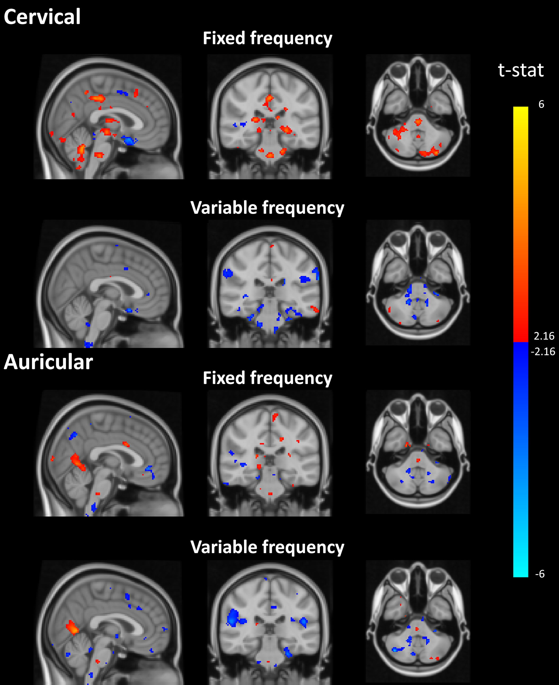
The BOLD response of the stimuli with respect to the sham stimulation is shown in Figure 1.Region-wise analysis revealed that auricular stimulation with a variable frequency resulted in a reduced BOLD connectivity in the amygdala (-.042 vs .031, p<0.01) and insular cortex (-.053 vs .008, p=0.02). Auricular stimulation with a fixed frequency, as well as cervical stimulation did not reveal significant differences on a region-wise basis.
Discussion & Conclusion
In this explorative study, the effects of stimulation site and waveform characteristics of tVNS are investigated. Interestingly, the fixed frequency tends to result in an increased BOLD response, while variable frequency stimulation showed mostly decreases in BOLD activity with respect to sham stimulation.Moreover, auricular stimulation with a variable frequency was shown to result in a significant decrease of the BOLD response in the amygdala and insular cortex compared to sham stimulation. These preliminary results already provide valuable insights into the neuronal effects of tVNS stimulation site and waveform characteristics. However, further work should also focus on the role of the CAN regions in the cerebral functional network, for example using nodal importance metrics such as the betweenness centrality.
Acknowledgements
No acknowledgement found.References
1. Yap JYY, Keatch C, Lambert E, Woods W, Stoddart PR, Kameneva T: Critical Review of Transcutaneous Vagus Nerve Stimulation: Challenges for Translation to Clinical Practice. Front Neurosci 2020; 14(April).
2. Benarroch EE. The Central Autonomic Network: Functional Organization, Dysfunction, and Perspective. Mayo Clin Proc 1993; 68:988–1001.
3. Farmer AD, Albu-Soda A, Aziz Q: Vagus nerve stimulation in clinical practice. Br J Hosp Med 2016; 77:645–651.
4. Gooneratne IK, Green AL, Dugan P, et al.: Comparing neurostimulation technologies in refractory focal-onset epilepsy. J Neurol Neurosurg Psychiatry 2016; 87:1174–1182.
5. Beckers AB, Drenthen GS, Jansen JFA, Backes WH, Poser BA, Keszthelyi D: Comparing the efficacy of data-driven denoising methods for a multi-echo fMRI acquisition at 7T. Neuroimage 2023; 280(August):120361.
6. Andersson JLR, Skare S, Ashburner J: How to correct susceptibility distortions in spin-echo echo-planar images: Application to diffusion tensor imaging. Neuroimage 2003; 20:870–888.
7. Behzadi Y, Restom K, Liau J, Liu TT: A component based noise correction method (CompCor) for BOLD and perfusion based fMRI. Neuroimage 2007; 37:90–101.
8. Puonti O, Iglesias JE, Van Leemput K: Fast and sequence-adaptive whole-brain segmentation using parametric Bayesian modeling. Neuroimage 2016; 143:235–249.
9. Rolls ET, Huang CC, Lin CP, Feng J, Joliot M: Automated anatomical labelling atlas 3. Neuroimage 2020; 206(August 2019):116189.
10. Garcia RG, Lin RL, Lee J, et al.: Modulation of brainstem activity and connectivity by respiratory-gated auricular vagal afferent nerve stimulation in migraine patients. Pain 2017; 158.
Figures