3144
Alterations of functional segregation and integration in children with autism spectrum disorder: A resting-state MRI study.1Department of Radiology, Shanghai Tenth People’s Hospital, Tongji University School of Medicine, Shanghai, China, 2Philips Healthcare, Shanghai, China
Synopsis
Keywords: fMRI Analysis, fMRI (resting state), Autism
Motivation: Neuroimaging analysis of brain functional changes can provide a new imaging perspective for understanding the neural mechanism and providing biomarkers for the clinical diagnosis of ASD.
Goal(s): This study aimed to explore the brain functional changes in children with autism spectrum disorder (ASD).
Approach: All subjects underwent brain resting state functional MRI (rs-fMRI) scans.The data were analyzed using various methods including functional segregation and functional integration.
Results: The results showed the significant higher and lower activities in many brain regions, hypo-connectivity between different brain networks and only hyper-connectivity between DAN and DMN.
Impact: The functional changes of the brain may be the pathogenesis for children with ASD accompanied, which could become the biomarkers in future clinical diagnosis.
Introduction
Autism spectrum disorder (ASD) has been widely recognized as a complex neurodevelopmental disorder characterized by impaired social interaction and repetitive behaviors. Several children with ASD have while few studies have focused on the situation. This study aimed to explore the brain functional alterations in children with autism spectrum disorder (ASD).Methods
This prospective study enrolled 185 children with ASD (155 boys and 30 girls) and 73 healthy controls (HCs) (41 boys and 32 girls). Children with ASD were diagnosed according to the autistic disorder criteria of the Diagnostic and Statistical Manual-V (DSM-V). All subjects underwent cognitive assessments and brain resting state functional MRI (rs-fMRI) scans.The preprocessing of resting-state fMRI data was performed in MATLAB R2013b (Mathworks, Natick, MA) with RESTplus software. The first ten volumes of fMRI data were removed. Because of using multiband scanning, slice timing was not performed1. After head motion correction and realignment, the images were normalized by the DARTEL toolbox using T1 image new segment. Resultant images were smoothed with a Gaussian kernel full width at a half-maximum of 6 mm (for ReHo and DC, this step was performed during calculation). The Friston 24 motion parameters, white matter, and cerebrospinal fluid signals were regressed as covariates. Finally, band-pass filtering (0.01–0.08 Hz) was performed to remove the effects of high-frequency noise (except for ALFF). ICA was analysed by the GIFT toolbox and the number of independent components (IC, N = 32) was estimated automatically by the software. Then, we used the GIFT to calculate the spatial correlation coefficients between the specific resting-state network templates and independent components, and selected the independent components of the largest spatial correlation coefficient2. This selection procedure resulted in 7 functional networks out of the 32 independent components obtained: default mode networks (DMN), left frontoparietal networks (LFPN), right frontoparietal networks (RFPN), auditory network (AN), dorsal attention networks (DAN), visual networks (VN) and salience network (SN).
Results
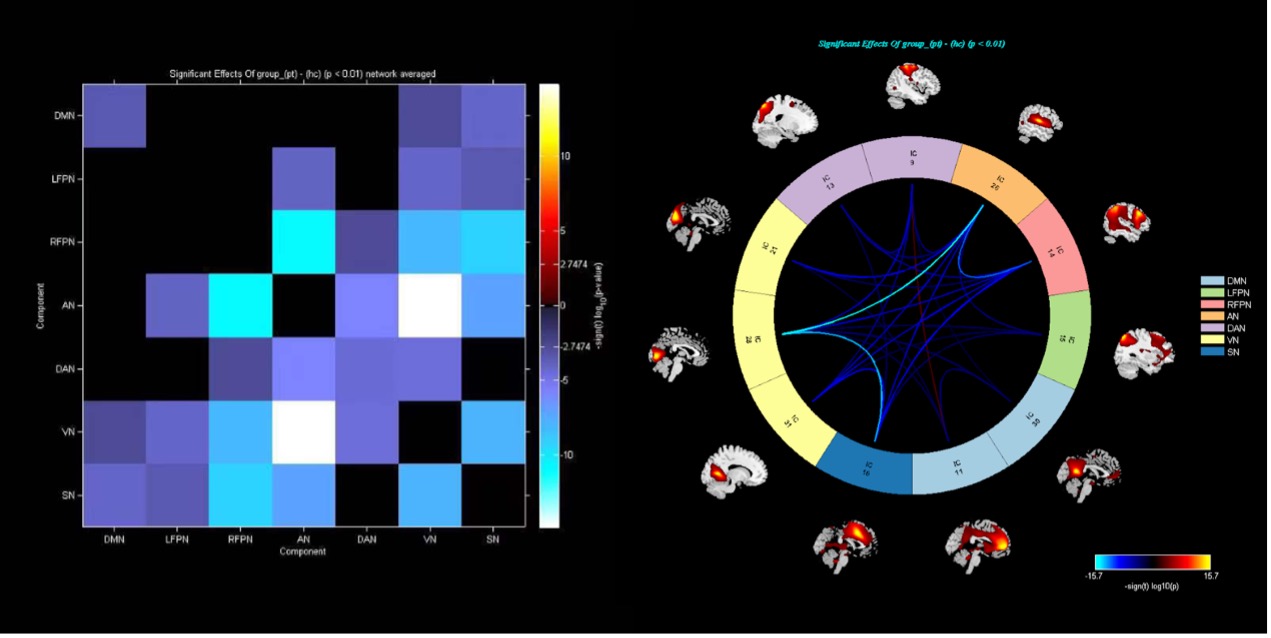
ASD showed lower ALFF in the bilateral cerebellum crus1 and anterior cingulate, and higher ALFF in the bilateral precentral gyrus and calcarine fissure. Compared with the HCs, ASD exhibited decreased ReHo in the bilateral cerebellum crus1, left anterior cingulate gyrus, and right insula and increased ReHo in the bilateral calcarine and postcentral gyrus. The DC values decreased in the bilateral cerebellum crus1, bilateral precentral gyrus, and right insula, while increased in the left middle occipital gyrus and right calcarine fissure. (voxel-wise p < 0.001, FDR-corrected cluster-wise p < 0.05) (Figures 1). There were 32 independent components extracted by ICA, 7 of which were believed to correspond to the DMN (IC11, IC30), LFPN (IC15), RFPN (IC14), AN (IC26), DAN (IC9, IC13), VN (IC21, IC28, IC31) and SN (IC16) by template matching analysis. Correlation analyses revealed significant correlations between internetwork functional connectivity (p <0.01, FDR corrected; Figure 2). Results showed negative correlations with functional connectivity between most brain functional networks. There was only a positive correlation with functional connectivity between IC9 and IC11 (t = 3.26, p = 0.0013).Discussion
This study found spontaneous higher and lower activity alterations in multiple brain regions and many brain regions included bilateral cerebral hemispheres. The ICA results showed significant hypo-connectivity between different brain networks and only hyper-connectivity between DAN (IC9) and DMN (IC11). Most previous literature results are hypo-connectivity3, which is consistent with our results. DMN is a key brain network involved in the processing of information and its dysfunction may manifest in interaction with other brain systems and be be a cause of social impairment in ASD subjects4. DMN, FPN and SN play important roles in cognition and self-monitoring processes. The SN serves as a switch between the DMN and FPN, in line with salience and cognitive demand. Dysfunctions in SN may be connected to a broad spectrum of deficits and maladaptive behavioral patterns in ASD5. The weak coherence among different network would mean less coordination and hence less than optimal output, which can explain the language, social and communication impairments in ASD.Conclusion
The alterations of functional segregation and integration in the brain may be the pathogenesis for children with ASD, which could help understand the neural mechanism and become the potential biomarkers in future diagnosis.Acknowledgements
We would like to thank Shanghai Tenth People’s Hospital and Philips Healthcare for supporting this research. We also thank all participants and their families for their cooperation in this study.
References
1. Smitha KA, Arun KM, Rajesh PG, et al. Multiband fMRI as a plausible, time-saving technique for resting-state data acquisition: Study on functional connectivity mapping using graph theoretical measures. Magn Reson Imaging, 2018, 53: 1-6.
2. Shirer WR, Ryali S, Rykhlevskaia E, et al. Decoding subject-driven cognitive states with whole-brain connectivity patterns. Cereb Cortex, 2012, 22: 158-165.
3. Wei L, Zhang Y, Zhai W, et al. Attenuated effective connectivity of large-scale brain networks in children with autism spectrum disorders. Front Neurosci, 2022, 16: 987248.
4. Washington SD, Gordon EM, Brar J, et al. Dysmaturation of the default mode network in autism. Hum Brain Mapp, 2014, 35: 1284-1296.
5. Schimmelpfennig J, Topczewski J, Zajkowski W, et al. The role of the salience network in cognitive and affective deficits. Front Hum Neurosci, 2023, 17: 1133367.
Figures
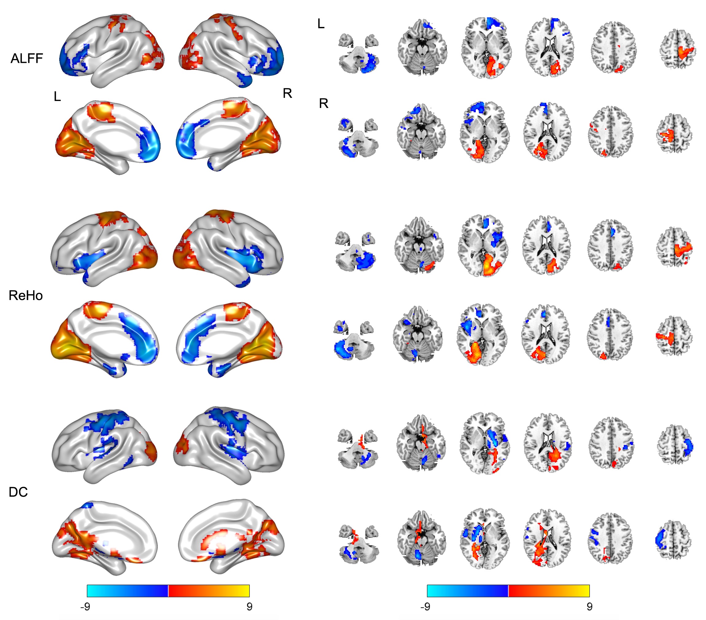
Figure 1 Significant differences in ALFF, ReHo and DC between ASD and HCs. The warm colors denote higher values, and the cool colors denote lower values in ASD (p < 0.05, FDR corrected).