3143
Aberrant Dynamic Functional Connectivity In Childhood Basic Intermittent Exotropia: an exploration in pathogenesis of visual center pathway1Beijing Tongren Hospital, Capital Medical University, Beijing, China, 2Chinese University of Hong Kong (Shenzhen) School of Science and Engineering, Shenzhen Research Institute of Big Data, People's Republic of China, Shenzhen, China, 3MR Research Collaboration, Siemens Healthineers Ltd, Beijing, China, 4Chinese University of Hong Kong (Shenzhen) School of Medicine, Shenzhen Research Institute of Big Data, People's Republic of China, Shenzhen, China, 5Chinese University of Hong Kong (Shenzhen) School of Medicine, People's Republic of China, Shenzhen, China, 6Shenzhen Research Institute of Big Data, People's Republic of China, Shenzhen, China
Synopsis
Keywords: Functional Connectivity, fMRI (resting state)
Motivation: To explore the pathogenesis and functional abnormalities in visual center pathway in children with basic intermittent exotropia and develop an accurate approach for early diagnosis.
Goal(s): This study aims to uncover the neural basis of IXT by investigating the functional network alterations.
Approach: We used dynamic functional connectivity (dFC) analysis of resting state fMRI data to detect the brain network changes in IXT.
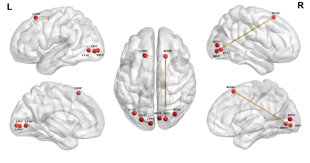
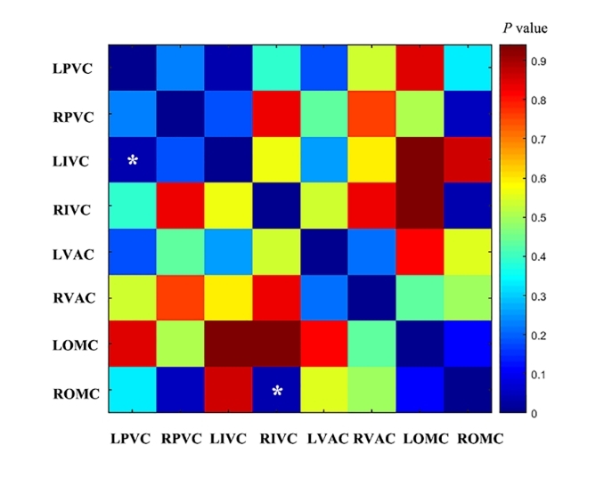
Results: Decreased dFC Variance between the left primary visual cortex and the left intermediate visual cortex, between the right ocular motor cortex and the right intermediate visual cortex were found in IXT children compared with HCs.
Impact: Our study was the first to investigate the dynamic brain network changes among primary visual cortex secondary visual cortex, and ocular motor cortex in IXT. This enhanced understanding of the neuropathological mechanisms underlying visual and oculomotor impairments in IXT children.
Introduction
Intermittent exotropia (IXT) is a common developmental disease of children involving ocular motility impairment1-2. It includes 4 sub-types and the basic type is the most common type of IXT3. The underlying pathological mechanisms of IXT remain unclear. Accumulating evidences indicate that the presence of IXT is associated with the abnormal changes of the visual center, and furthermore, it was suggested that IXT is first caused by defective binocular fusion in the primary visual cortex, which makes the secondary visual cortex unable to form normal stereoscopic vision and ultimately leads to eyes deviation controlled by ocular motor cortex. All these studies suggest the importance of studying the links between different visual centers. But ordinary functional connection analysis can only reflect the average value of the correlation between fluctuation signals cannot reflect the dynamics and effectiveness of information interaction in the brain at different time periods4. Dynamic functional connection analysis is to dynamically examine the dynamic functional connections at different time points or time periods within the scanning time5. Many studies have confirmed that dynamic functional connection analysis can provide more valuable information for pathological exploration and auxiliary diagnosis of diseases. Thus, in this study, using dynamic functional connectivity (dFC) analysis, we explore the morphological changes of the primary visual cortex, secondary visual cortex, eye movement cortex pathway in children with IXT and their correlation with the severity of the disease based on the high temporal and high spatial resolution SMS-EPI sequence.Methods
Data were collected using a 3T MRI scanner (Siemens Healthcare, Erlangen, Germany) with a 64-channels head/neck coil. A total of 31 children and adolescent patients with IXT and 37 age-, sex-, handedness-, and education-matched healthy controls (HCs) were recruited in this study. Resting-state fMRI was obtained using an simultaneous multi-slice echo planar imaging (SMS-EPI) sequence with the following parameters: repetition time (TR)=1500ms, echo time (TE)= 30 ms, flip angle = 70°, FOV = 220 mm × 220 mm, matrix = 110×110, and 340 time points. slice thickness = 2 mm. Three-dimensional T1-weighted anatomical images were acquired with 3D magnetization-prepared rapid acquisition gradient echo (MPRAGE) sequence with the following parameters: TR = 2000ms, TE = 2.25 ms, TI= 900ms, matrix = 256 × 256, FOV = 256mm×256mm, slice thickness = 1.0 mm, flip angle = 8°, 192 slices.Resting-state fMRI data was preprocessed using the Data Processing Assistant for Resting State fMRI (http://www. restfmri.net/forum/DPARSF) that works with SPM12 on the Matlab 2021b platform. The first 10 volumes were discarded, followed by temporal correction, realignment, realignment, Gaussian smoothing with a full-width half-maximum of 6x6x6 mm3, and the removal of non-brain tissues. Both sides of BA17, BA18, BA19 and BA8 were chosen as region of interests (ROIs). MATLAB 2021b and Dynamic BC 2.2 software were used for analysis using the sliding time window technique with a window length of 50 TR and a step size of 1TR. 181 sliding Windows were obtained for each subject. FC variation was calculated for each of the 181 Windows for each subject, resulting in a final 8*8 dFC variance matrix for each subject, which was used for statistical analysis. The variance index of dFC between the two groups was used to do a two-sample T test. Correction for multiple comparisons was performed with a threshold of 0.05 after NBS correction.Results
Compared with HCs, IXT children had decreased dFC values between the left primary visual cotex and the left intermediate visual cortex, between the right ocular motor cortex and the right intermediate visual cortex.Discussion & Conclusion
Our finding of decreased dFC values between the left primary visual cotex and the left intermediate visual cortex was related to the abnormality of binocular fusion. As well all know, the primary visual cortex receiving inputs from both eyes is the first step of vision processing, then enters the intermediate visual center for visual fusion6. Abnormal dFC Variance between the right intermediate visual cortex and right ocular motor cortex was associated with eye movement. If the input signals from binocular vision cannot be converted into appropriate eye movement signals, it can lead to dysfunctional visual perceptual eye movement regulation, which may explain the continuous progression of the disease. IXT children exhibited decreased dFC between the primary visual cortex, secondary visual cortex and ocular motor cortex, which are associated with the impaired binocular fusion and deviation of eye position.Acknowledgements
No acknowledgement found.References
[1] Ouyang, J., Yang, L., Huang, X., Zhong, Y. L., Hu, P. H., Zhang, Y., et al. (2017). The atrophy of white and gray matter volume in patients with comitant strabismus: evidence from a voxel-based morphometry study. Mol. Med. Rep.16, 3276–3282.
[2] Li, D., Li, S., and Zeng, X. (2018). Analysis of alterations in white matter integrity of adult patients with comitant exotropia. J. Int. Med. Res. 46, 1963–1972.
[3] Pan CW, Zhu H, Yu JJ, Ding H, Bai J, Chen J, et al. Epidemiology of Intermittent Exotropia in Preschool Children in China. Optometry and vision science: official publication of the American Academy of Optometry 2016; 93:57–62.
[4] Preti MG, Bolton TA, Van De Ville D. The dynamic functional connectome: State-of-the-art and perspectives. Neuroimage. 2017;160:41-54.
[5] Chen JE, Rubinov M, Chang C. Methods and considerations for dynamic analysis of functional MR imaging data. Neuroimaging Clin N Am. 2017;27(4): 547-560.
[6] Scholl B, Burge J, Priebe NJ. Binocular integration and disparity selectivityin mouse primary visual cortex. J Neurophysiol. (2013) 109:3013–24.doi: 10.1152/jn.01021.2012.
Figures