3141
Study on the changes in low-frequency amplitude and regional homogeneity of the cerebral cortex in children with congenital deafness.1Department of Radiology, Affiliated Hospital of Guizhou Medical University, Guiyang, China, 2Guizhou Medical University, Guiyang, China, 3Philips Healthcare, Shanghai, China
Synopsis
Keywords: Functional Connectivity, fMRI (resting state), congenital deafness, functional magnetic resonance imaging, cochlear implantation
Motivation: To investigate the changes in low frequency fluctuation amplitude (mALFF) and regional homogeneity (ReHo) in the brains of children with congenital Sensorineural hearing loss (SNHL) by resting state fMRI.
Goal(s): The primary objective of this research was to improve understanding of the changes in neural functional activity within the cortical regions of SNHL patients during a resting state.
Approach: The study's goal is to establish a neuroimaging pathological foundation, which could potentially provide significant neuroimaging biomarkers.
Results: These biomarkers could aid in the clinical selection of appropriate candidates for cochlear implantation (CI), thereby enhancing the effectiveness of this therapeutic intervention.
Impact: The findings offer a fresh insight into the neuropathological mechanisms that drive functional reorganization in the cerebral cortex of children with congenital deafness after auditory deprivation. Moreover, these results provide valuable neuroimaging evidence for assessing the prognosis of post-CI patients.
Introduction
Congenital deafness (CD) leads to varying degrees of hearing loss in individuals after birth, with about 90% of cases being severe1. Cochlear implantation is the primary treatment for children with severe hearing loss. Delayed intervention can affect a child's cortical function development and post-surgery auditory rehabilitation2. Therefore, assessing preoperative brain function in hearing loss patients is critical. Resting-state fMRI3 is a reliable and repeatable method that detects neural activity based on changes in cerebral blood oxygen levels. It can identify changes in brain function under different conditions, offering valuable biomarkers for preoperative evaluation in cochlear implant patients.Materials and Methods
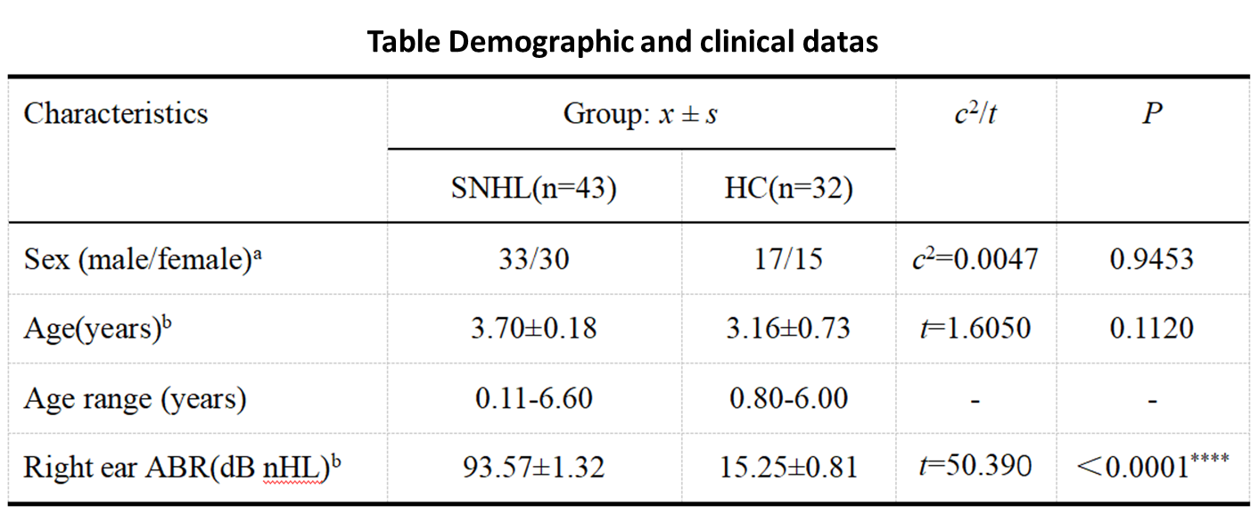
We collected resting-state functional magnetic resonance imaging (rs-fMRI) data from 43 individuals diagnosed with congenital sensorineural hearing loss (SNHL) and 32 healthy control (HC) subjects of similar age between May 2017 and June 2020. After cochlear implantation (CI), a 6-month follow-up was conducted to assess auditory behavioral grading scale scores (Clinical Assessment of Prelingual Auditory Performance, CAP). The rs-fMRI data was preprocessed using DPABI and SPM12 software. Whole-brain functional parameters, such as the amplitude of mALFF and ReHo, were calculated. Subsequently, two-sample t-tests were performed on mALFF and ReHo values, with gender, age, and head motion parameters as covariates. Cluster-level family-wise error (FEW) or false discovery rate (FDR) correction was applied. For brain regions that exhibited statistically significant differences in mALFF and ReHo, Spearman correlation analysis was conducted to examine the relationship between their values and the clinical CAP scores. Results with P < 0.05 were considered statistically significant.Results
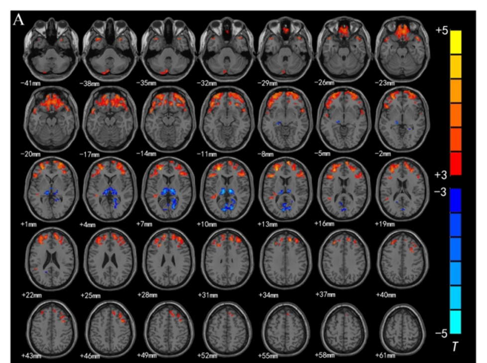
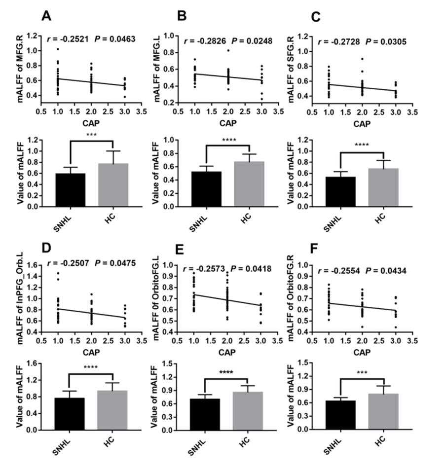
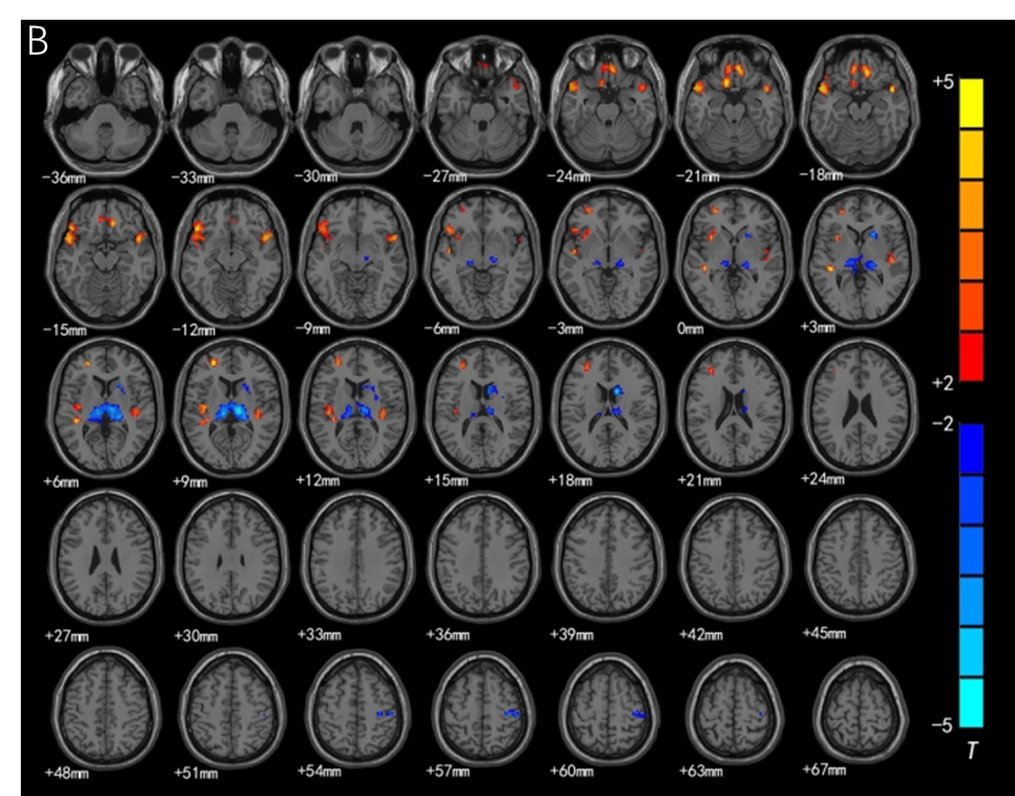
Compared to controls, the SNHL group showed decreased mALFF in regions including the bilateral superior frontal gyrus (BA9), middle frontal gyrus (BA10), and superior temporal gyrus (BA38) among others, but increased mALFF in the bilateral thalamus and fusiform gyrus (BA17). (Figure 1). The ReHo analysis showed that compared to the healthy control (HC) group, the SNHL group exhibited decreased synchronized ReHo in the bilateral superior temporal gyrus (BA38, 41), bilateral BA11, BA10, BA47, and left insula (BA13). However, there was an increased ReHo in the bilateral thalamus, right caudate nucleus, and right precentral gyrus (BA3) (Figure 3). Correlation analysis revealed the following: The mALFF values of the left/right middle frontal gyrus (r = -0.2826, P = 0.0248; r = -0.2521, P = 0.0463), right superior frontal gyrus (r = -0.2728, P = 0.0305), left inferior frontal gyrus (r = -0.2507, P = 0.0475), right orbitofrontal gyrus (r = -0.2554, P = 0.0434), and left orbitofrontal gyrus (r = -0.2573, P = 0.0418) were negatively correlated with CAP scores (Figure 2). However, there was no correlation between ReHo values in different brain regions and clinical CAP scores.Dicussion
Functional MRI (fMRI) is a non-invasive technique used to study changes in brain function in various diseases. It provides valuable information for understanding the development of these diseases and guiding clinical practice4. fMRI, which is based on Blood oxygen level dependent (BOLD) signals, has been shown to reflect underlying neuronal activity. In this study, we examined the neural activity in the auditory, visual, and somatosensory cortices of individuals with congenital deafness. The results revealed significant changes in neuronal activity in these regions. Specifically, there was a significant decrease in the mean amplitude of mALFF in the auditory cortex, while the mALFF activity in the visual and somatosensory regions showed a significant increase5-7. Furthermore, we observed abnormal desynchronization between the auditory cortex and other cortical regions, characterized by a decrease in regional homogeneity8 in the auditory cortex. After the loss of auditory function in individuals with congenital deafness, the prefrontal cortex (PFC) underwent functional alterations9. Correlation analysis demonstrated a negative correlation between the mALFF values of the bilateral middle frontal gyrus (MFG), right superior frontal gyrus (SFG), left inferior prefrontal gyrus (InPFG_Orb), bilateral orbitofrontal gyrus (OrbitoFG), and clinical scores on the CAP evaluation (A-F).Conclusion
This study used resting-state fMRI to observe brain function changes in children with congenital deafness. Findings include: (1) Abnormal functional changes in motor-related cortical regions were noted following auditory loss in pre-school deaf children, possibly due to cross-modal reorganization; (2) Decreased activity was observed in the prefrontal cortex, suggesting that auditory loss may affect cognitive functions; (3) Correlation analysis showed a decrease in cognitive functions mediated by the prefrontal cortex after auditory loss, and compensatory changes in other sensory-motor cortices may not benefit auditory-language function recovery post cochlear implantation.Funding
This study is supported by the Guizhou Provincial Science and Technology Plan Project (Contract No.: Qiankehe Foundation-ZK[2023] General 355) and the Guizhou Medical University National Natural Science Foundation Cultivation Project (Project No.: 22NSFCP53).Acknowledgements
We are grateful to all the participants for their cooperation and patience.References
1. Chadha S, Cieza A. World Health Organization and Its Initiative for Ear and Hearing Care. Otolaryngologic clinics of North America 2018;51:535.
2. Benchetrit L, Ronner EA, Anne S, Cohen MS. Cochlear Implantation in Children With Single-Sided Deafness: A Systematic Review and Meta-analysis. JAMA Otolaryngol Head Neck Surg 2021;147:58.
3. Raimondo L, Oliveira Ĺ AF, Heij J, et al. Advances in resting state fMRI acquisitions for functional connectomics. NeuroImage 2021;243:118503.
4. Huang R, Zhou X, Chen G, et al. Advances of functional nanomaterials for magnetic resonance imaging and biomedical engineering applications. Wiley Interdiscip Rev Nanomed Nanobiotechnol 2022;14:e1800.
5. Putkinen V, Nazari-Farsani S, Seppälä K, et al. Decoding Music-Evoked Emotions in the Auditory and Motor Cortex. Cerebral cortex (New York, NY : 1991) 2021;31:2549-60.
6. Li YT, Chen JW, Yan LF, et al. Dynamic alterations of functional connectivity and amplitude of low-frequency fluctuations in patients with unilateral sudden sensorineural hearing loss. Neurosci Lett 2022;772:136470.
7. Chen J, Hu B, Qin P, et al. Altered Brain Activity and Functional Connectivity in Unilateral Sudden Sensorineural Hearing Loss. Neural plasticity 2020;2020:9460364.
8. Liu L, Fan J, Zhan H, et al. Abnormal regional signal in the left cerebellum as a potential neuroimaging biomarker of sudden sensorineural hearing loss. Frontiers in psychiatry 2022;13:967391.
9. Luan Y, Wang C, Jiao Y, Tang T, Zhang J, Teng GJ. Prefrontal-Temporal Pathway Mediates the Cross-Modal and Cognitive Reorganization in Sensorineural Hearing Loss With or Without Tinnitus: A Multimodal MRI Study. Frontiers in neuroscience 2019;13:222.
Figures