3139
Human Layer-specific VASO fMRI at 5.0T Whole-body MRI Scanner: A Preliminary Study1Shenzhen Institute of Advanced Technology, Chinese Academy of Sciences, Shenzhen, China
Synopsis
Keywords: Task/Intervention Based fMRI, High-Field MRI
Motivation: This study seeks to address the challenge of human layer-specific fMRI at 5.0T whole-body MRI scanner.
Goal(s): Develop Layer-specific VASO fMRI based on the slice-selective slab-inversion vascular space occupancy (SS-SI VASO) pulse sequence at 5.0T scanner.
Approach: The SS-SI VASO pulse sequence with GOIA RF pulse at 5.0T scanner has been developed with the five processing steps, including noise reduction with distribution corrected (NORDIC), motion correction, BOLD correction and contrast combination, quality measures, and functional activity.
Results: The preliminary results have shown that the in vivo experiment of finger tapping for VASO fMRI at 5.0T scanner is feasible as similar as BOLD fMRI.
Impact: The preliminary experiments provided by layer-specific VASO fMRI at 5.0T scanner can help scientists and doctors in accurately identifying and diagnosing primary motor cortex or other brain applications.
Introduction
The study of human brain function at mesoscale between micro and macro is the frontier of brain science. Brain functional imaging should not be derived from large venous vessels, but from small blood vessels in the local tissue of the corresponding brain activation region[1]. At present, layer-specific function magnetic resonance imaging (fMRI) at ultra-high fields brings a new light to the study of mesoscopic human brain function. In this study, we proposed a method of layer-specific fMRI at 5.0T whole-body scanner, which is based on the slice-selective slab-inversion vascular space occupancy (SS-SI VASO) pulse sequence[2,3]. The in vivo experiments on human brains at 5.0T scanner showed that VASO fMRI could work well as same as BOLD fMRI at 5.0T Whole-body Scanner.Methods
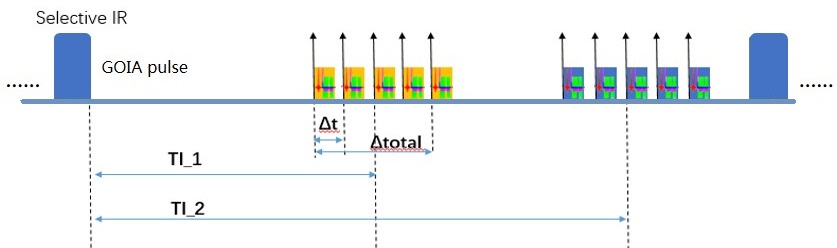
Here, we designed a slice-selective slab-inversion vascular space occupancy (SS-SI VASO) pulse sequence [2,3] at 5.0T whole-body scanner, which utilized the gradient-modulated offset-independent adiabaticity (GOIA) RF pulses, as seen as Fig.1. The SS-SI VASO pulse sequence widens the longitudinal relaxation difference between tissue and blood at the zero point of blood and increases the signal-to-noise ratio by changing the inversion pulse of the non-selected layer to the inversion pulse of the selected layer and adding the saturation pulse of the selected layer before inversion. The hybrid image of VASO and BOLD and the image of BOLD were acquired twice before and after acquisition, and the BOLD Correction was performed with post-processing to remove the pollution of the BOLD signal. The VASO data have been processed with the five steps, including noise reduction with distribution corrected (NORDIC), Motion correction, BOLD correction and contrast combination, Quality measures, and Functional activity. NORDIC was as same as the method [4]. A manual mask is defined to avoid faulty motion estimation due to variable distortions outside the FOV. The non-steady-state images (the first four time points) are overwritten with steady-state images. The SS-SI VASO sequence acquires images with and without blood nulling interleaved. Thus, the motion correction is applied separately for BOLD and VASO. Ideally, the motion parameters should be very similar for BOLD and VASO. This needs to be checked manually every time. The blood-nulled image was separated from the not-nulled BOLD image. Since the blood-nulled and not-nulled image are acquired interleaved, the time series are temporally upsampled (in afni) and shifted with respect to each other, such that the respective contrasts refer to the same point in time. The division happens in the LAYNII program LN_BOCO. First average trials and then do the BOLD correction. One way to estimate the signal change is to just subtract the signal during activity with the signal during rest. Quality measures include MEAN, tSNR, Skew and kurtosis, Auto-correlation as same as [5]. Another way is to use a GLM implemented in all the mayor software packages. Results of depth-dependent GLMs can be hard to interpret for the following reasons: 1) signal amplitude, quality and stability are heterogeneous across cortical depths; 2) the hemodynamic response function varies across cortical depths. Layering includes Upscaling, Manual delineation of GM, Calculation of cortical depths in ROI, Extracting functional data based on calculated cortical depths.Experiments
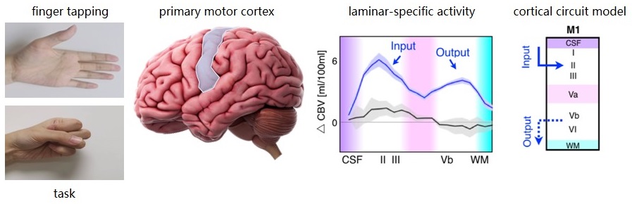
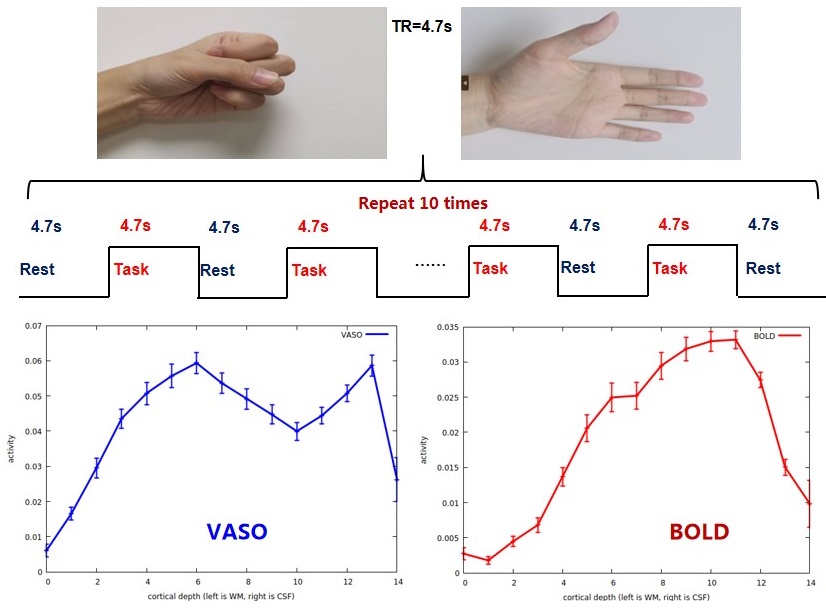
The in vivo head experiments from SS-SI VASO were acquired on a 5.0T MRI system (uMR Jupiter, United Imaging Healthcare, Shanghai, China) and the protocols were approved by the Institutional Reviews Board (IRB). A 48-channel head coil (24-channel Open-transmit and flexible receiver head coil) was used on a healthy volunteer, and the whole-brain scanning sequence parameters were set as: in-plane resolution = 0.80× 0.80 mm2; slice thickness = 1.5 mm; TRVASO = 2350 ms; TRVASO+BOLD = 4700 ms. The classical experiments of finger tapping (as seen as Figure 2) were done to study the feedforward and feedback effects in S1 and the input and output in M1 in human brain. For 0.7-0.8 mm resolution datasets, an upscaling factor of 4-5 was recommended.Results
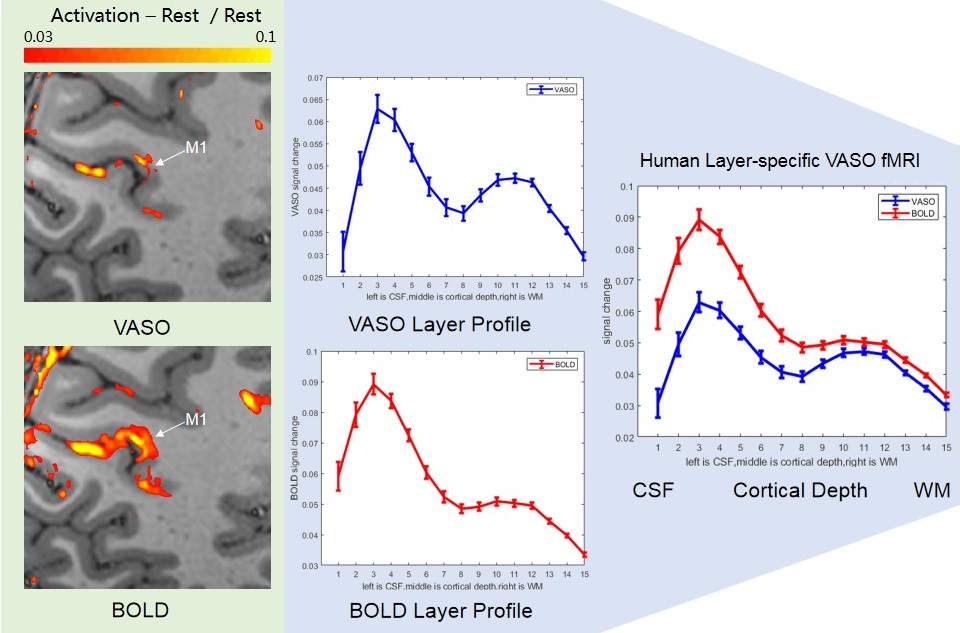
Figure 1 showed the timing diagram of the SS-SI VASO pulse sequence at 5.0T scanner. Figure 2 demonstrated the proposed experiment of finger tapping based on laminar-spedific activity of primary motor cortex. Figure 3 was the human in vivo experiment of finger tapping at 5.0T scanner, compared between VASO and BOLD. Figure 4 was the human layer-specific VASO and BOLD study at 5.0T scanner with the open-face birdcage transmit coil and the 24-channel fMRI head receiver coil.Discussion and Conclusion
In this study, the layer-specific fMRI of the SS-SI VASO sequence has been implemented and tested at 5.0T scanner. All of the results have shown the VASO and BOLD experiments are feasible at 5.0T whole-body scanner.Acknowledgements
This work was partially supported by the National Natural Science Foundation of China (62271474), the National Key R&D Program of China (2023YFB3811400), the High-level Talent Program in Pearl River Talent Plan of Guangdong Province (2019QN01Y986) and the Shenzhen Science and Technology Program (KQTD20180413181834876 and JCYJ20210324115810030).References
[1] Huber L, et al. Techniques for blood volume fMRI with VASO: From low-resolution mapping towards sub-millimeter layer-dependent applications. Neuroimage. 2018 Jan 1:164:131-143.
[2] Huber L, et al. High-Resolution CBV-fMRI Allows Mapping of Laminar Activity and Connectivity of Cortical Input and Output in Human M1. Neuron. 2017 Dec 20;96(6):1253-1263.e7.
[3] Huber L, et al. Slab-selective, BOLD-corrected VASO at 7 Tesla provides measures of cerebral blood volume reactivity with high signal-to-noise ratio. Magn Reson Med. 2014 Jul;72(1):137-48.
[4] Moeller S, et al. NOise reduction with DIstribution Corrected (NORDIC) PCA in dMRI with complex-valued parameter-free locally low-rank processing. Neuroimage. 2021 Feb 1:226:117539.
[5] Huber LR, et al. LayNii: A software suite for layer-fMRI. Neuroimage. 2021 Aug 15;237:118091.
Figures