3138
Acquisition and characterisation of sub-millimetre resolution GE-BOLD laminar fMRI data at 3T and 7 T1Krembil Brain Institute, University Health Network, Toronto, ON, Canada, 2Center for Neuroscience Imaging Research, Institute for Basic Science, Sungkyunkwan University, Suwon, Korea, Republic of, 3Department of Biomedical Engineering, Sungkyunkwan University, Suwon, Korea, Republic of, 4Department of Medical Biophysics, University of Toronto, Toronto, ON, Canada
Synopsis
Keywords: fMRI Acquisition, fMRI, Laminar fMRI
Motivation: To explore the potential of layer-specific imaging of the brain at standard clinical field (3T) and ultra-high field strengths (7T)
Goal(s): To assess the feasibility of sub-millimetre BOLD fMRI at 3T compared to 7T and characterise activation maps and laminar profiles
Approach: 12 healthy participants scanned using product 2D-EPI at four resolutions on both 3T and 7T scanners. Data were meticulously processed using ANTs and AFNI following NORDIC-PCA denoising.
Results: We demonstrate that sub-millimetre GE-BOLD fMRI at 3T is feasible using a standard fMRI sequence, has sufficient tSNR and reliable BOLD activation maps.
Impact: Our work broadens accessibility to high-resolution fMRI at 3T. Thus, encouraging new directions in neuroimaging and enabling a deeper understanding of human brain function in health and disease, impacting not only neuroscientists but also the broader scientific and medical communities.
Introduction
The study aims to, first, investigate the feasibility of sub-millimetre functional magnetic resonance imaging (fMRI) in human subjects using gradient-echo BOLD (GE-BOLD) contrast at standard clinical field strengths of 3T, and, second, to characterise the patterns of activation and temporal signal-to-noise ratio (tSNR) values in the same subjects scanned on both 3 and 7T field strengths. The feasibility of sub-millimetre resolution GE-BOLD fMRI enables the investigation of layer-specific differences in wider cohorts than is currently possible at ultra-high field strengths due to the ubiquity of 3T MRI scanners as well as the GE-BOLD fMRI sequence available on all vendor platforms.Methods
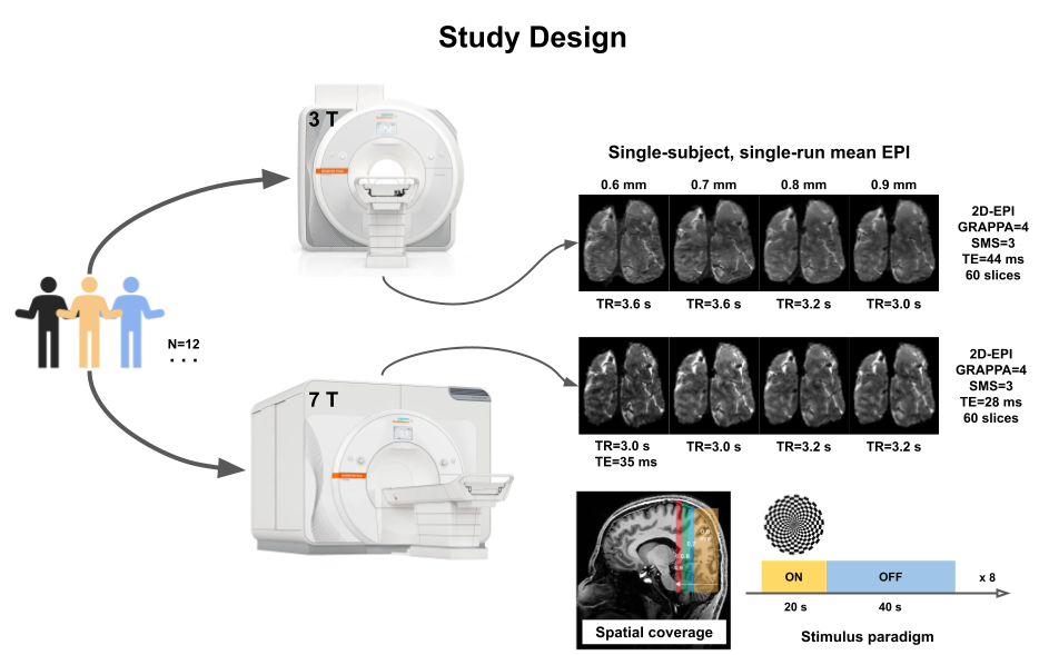
12 healthy participants were scanned on a Siemens 3T Prisma and 7T Terra using a 2D-EPI sequence at nominal resolutions of 0.6, 0.7, 0.8 and 0.9 mm isotropic, respectively. Data were acquired oblique coronal covering at least V1 and V2 (60 slices). Other sequence parameters were adapted to enable scanning within SAR limits with the TE/TR being 44/3000-3600 ms and 28-35/2500-3000 ms at 3 and 7T, respectively. A 0.6 mm isotropic MP2RAGE acquired at 7T served as the anatomical reference. All participants were shown eight blocks of 20 (on) - 40 (off) flickering checkerboard per run to elicit visual activation (Figure 1). Data from both scanners were corrected for gradient nonlinearities using the HCP’s grad_unwarp tool and denoised using NORDIC1. A high-resolution slab-optimised pipeline using ANTs2 was used for data pre-processing and AFNI3 for statistical analyses.Results
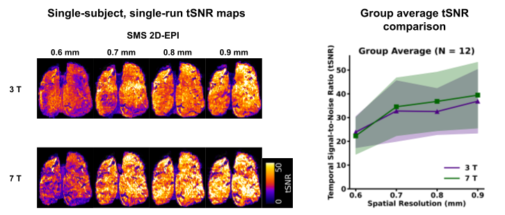
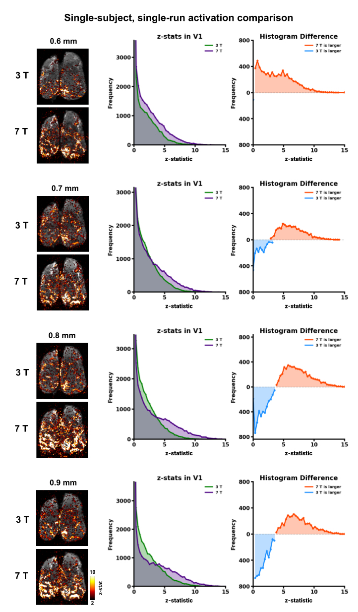
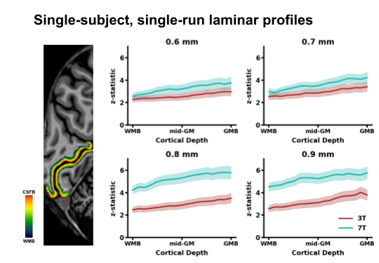
Figure 2 illustrates on a single subject that tSNR maps for resolutions 0.6-0.9 mm isotropic are highly comparable between 3 and 7T and is indicative of the robust performance of product 2D-EPI sequence. Figure 3 shows that robust activation maps can be obtained in a single run in less than 10 min, even at 0.6 mm at 3T. Consistent with expectations, 7T yields increased contrast-to-noise (CNR) of the BOLD signal (z-statistics) compared to 3T as is evident from the histogram difference plots. Laminar profiles in Figure 4 show the characteristic GE-BOLD signal increase to the pial surface for all data and consistent with activation maps in Figure 3, 7T has a higher dynamic range across depths than at 3T.Discussion
Numerous fMRI studies have shown that spatial patterns of neuronal activation can be resolved at a mesoscale (< 1 mm) using the GE-BOLD signal. However, mesoscale fMRI at 3T (and 7T) has been fraught with challenges particularly due to low SNR at sub-millimetre resolutions. While there exists previous work on attempting laminar fMRI at 3T, they often require bespoke coils, offline reconstruction, have limited spatial coverage and/or temporal resolutions4-10. We demonstrate the feasibility of acquisition of highest spatial resolution fMRI at 3T (0.6 mm isotropic) using the vendor sequence. We show that novel denoising methods such as NORDIC-PCA enables robust high-resolution fMRI at 3T for resolutions 0.6-0.9 mm isotropic with activation maps that are comparable to those acquired at 7T.Summary and Outlook
The present work successfully pushes the boundary of what has been thus far considered infeasible or difficult to achieve at standard clinical field strengths, showcasing a systematic characterisation of sub-millimetre resolution fMRI data. Even though 7T MRI is superior for layer fMRI, this demonstration of robust, reliable activation achievable at 3T will pave the way for future mesoscale studies of human brain function in health and disease.Acknowledgements
We would like to thank Dr. Prathamesh Chopade for help with participant recruitment and data acquisition.References
[1] Vizioli, L., et al. Nat Commun 12, 5181 (2021) [2] Avants, B. B., et al. Insight j 2.365: 1-35 (2009) [3] Cox, R.W., Comput Biomed Res. 29(3):162-73 (1996) [4] Ress, D. et al. Neuroimage 34.1 74-84 (2007) [5] Koopmans, P.J., et al. Hum. Brain Mapp., 31: 1297-1304 (2010) [6] Markuerkiaga, I., et al. Sci. Rep. 11.1: 1862 (2021) [7] Knudsen, L., et al. NeuroImage, 271, 120011 (2023) [8] Huber, L., et al, Aperture Neuro 3 1:17 (2023) [9] Huber, L. et al., #1149 Proc. of ISMRM (2023) [10] Chang, W., et al., bioRxiv (2023)Figures