3137
Functional line-scanning of cortical layers at 3T and 7T1CiNet, NICT, Osaka, Japan, 2Graduate School of Frontier Biosciences, Osaka University, Osaka, Japan, 3Tohoku Fukushi University, Sendai, Japan
Synopsis
Keywords: fMRI Acquisition, fMRI, Highe resolution fMRI
Motivation: Recently, line-scanning fMRI has been used to map laminar BOLD response, because it can enhance spatial resolution to sub-millimetre with sub-second TR. Some animal studies at high field scanner have been reported.
Goal(s): Line-scan scheme that can keep thin line profiles and provide multi T2* relaxed conditions for BOLD signal measuring is requested for layer fMRI.
Approach: We developed a multi-shot spin echo EPI based line-scan scheme with a readout train after spin echo time.
Results: Our 1D line-scanning finger tapping/grabbing fMRI results show the capability of this sequence to study differences in cortical layers in the human cortex.
Impact: The proposed line-scanning sequence can detect different BOLD response at different layers at M1/S1 in high resolution fMRI studies at 3T and 7T
Introduction
The detectability of variations in task-elicited functional activity is low when using normal EPI owing to the reduced temporal SNR with high temporal and high spatial resolution. We have introduced a multi shot EPI technique called block-interleaved segmented EPI (BISEPI) [1,2] to enable the detection of evoked BOLD signals in 0.4 mm resolution. But for some research targets, not isotropic but only high resolution in one direction is requested severely, like cortical layer studies. 1D line-scanning fMRI provides the capability to detect the BOLD signals with very high resolution in one specified direction [3,4]. In this work, we introduce a multi-shot spin echo EPI based line-scan method to measure BOLD signal at different T2* relaxed situation to improve BOLD sensitivity with good line selection performance. Two different orientations of the alpha and pi gradients were used to excite and refocus perpendicular planes. A sinc-shaped refocusing pulse and gradient crushers were used to remove the signal from spurious echoes and keep thin line profiles and executable at 3T and 7T. Our 1D line-scanning fMRI results show the capability of this sequence to study differences in cortical layers in the human cortex.Materials & Methods
The fMRI data were acquired using a Siemens MAGNETOM Prisma 3T and a Terra 7T MRI scanner (Siemens, Erlangen, Germany). The line-scanning sequence was based on a multi-shot spin echo EPI excitation and readout with the refocus gradient was moved from the slice-selection direction to the phase-encoding direction. For 1D line-scanning, spin echo line was set to the first line in readout train. Readout direction was oriented to the M1/S1 cortical layers based on turbo spin echo anatomical images and normal EPI fMRI experimental results. After a 2D line-scanning for conforming the location, A 1D line-scanning was performed with the phase-encoding gradient turned off. The imaging parameters for 3T and 7T are: 0.6 x 4 x 4/0.5 x 4 x 4 mm3 voxel size, TR of 500/1000 ms, echo time of 15/23.3 ms, FOV readout of 154/128 mm, flip angle of 130°/90°, bandwidth of 130/260 Hz/pixel, and 16 readout lines after one excitation (the first readout line is the refocused spin echo). The raw data were transformed by 1D FFT but reconstructed to 2D image liked (read out direction and readout line direction) complex time-series. Each coil channel of this image time-series was denoised separately by NORDIC Raw. Finally, the denoised data was combined by sum-of-squire (SOS) to one image series for next step analyzing. Using BrainVoyager, the data were then temporally filtered using a GLM Fourier basis set of four sine/cosines and temporal Gaussian smoothing with an FWHM kernel with three datapoints. Two subjects were scanned under 16s/16s ON/OFF finger tapping/grabbing task of total 8 minutes at 3T and 7T scanner.Results and Discussion
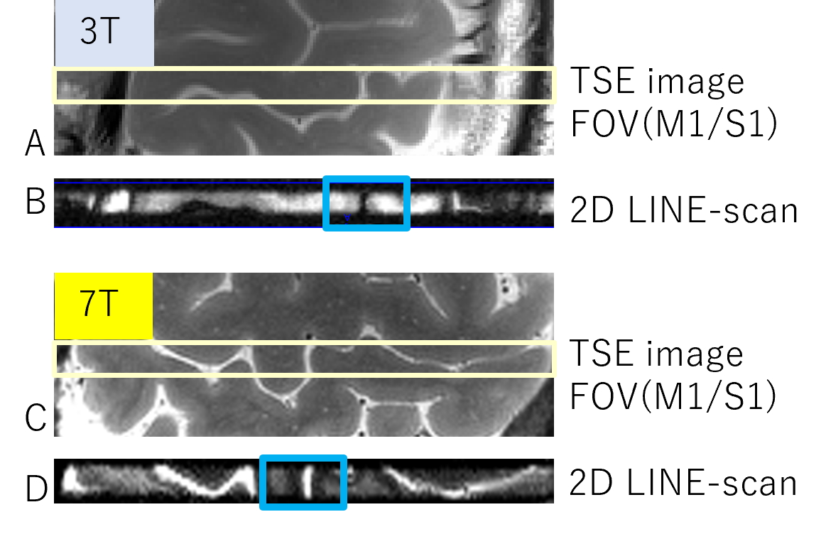
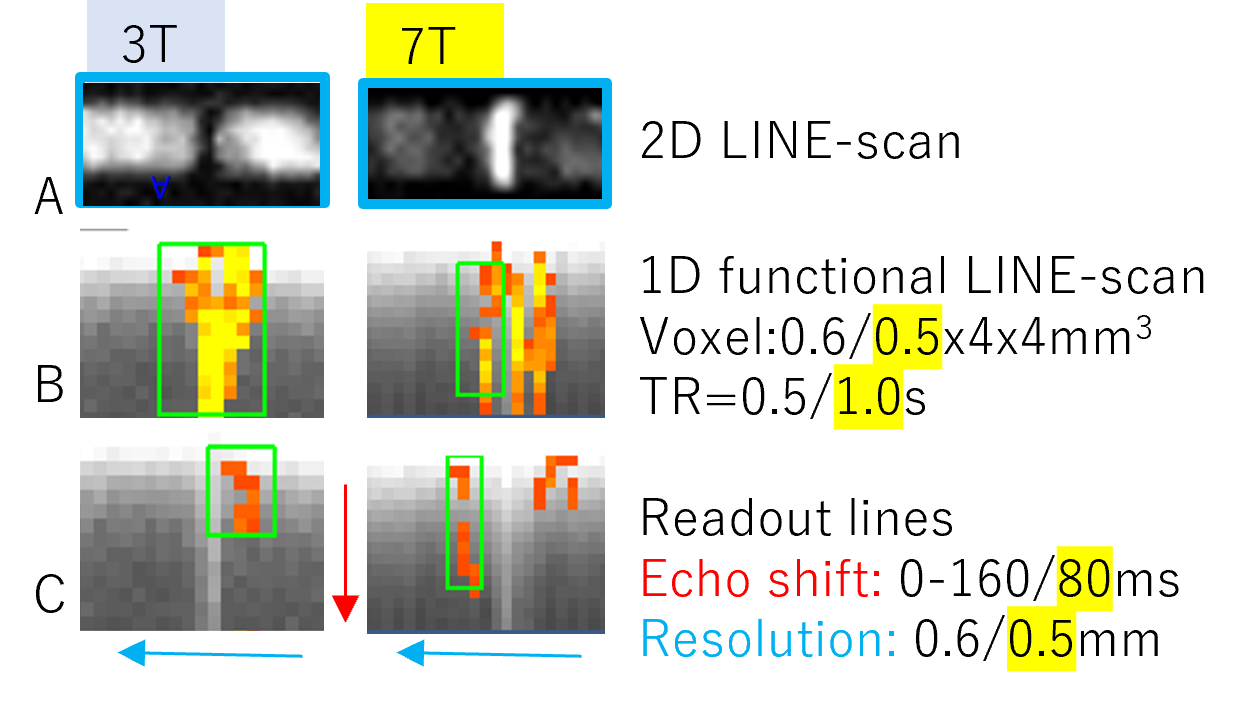
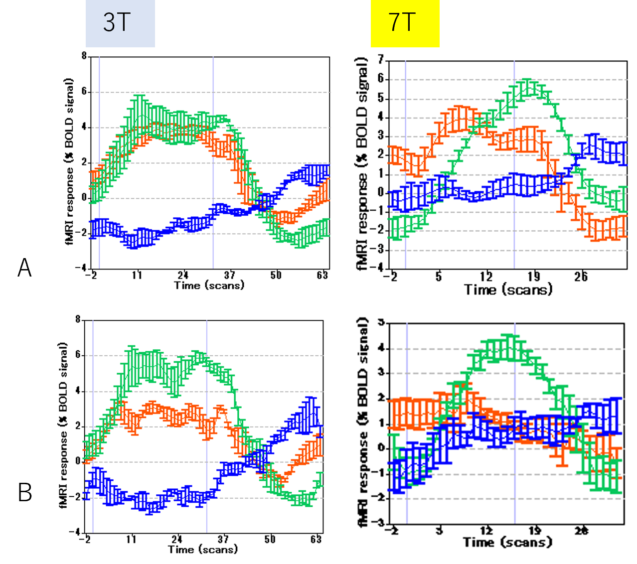
The performance of our 2D excitation can be confirmed by comparing the images acquired by turbo spin echo sequence and our 2D line-scan sequence. Figure 1 shows that only the sharp, targeted line was excited and acquired. The blue region in Figure 1B and 1D were enlarged and shown in Figure 2A, which includes M1 and S1 as ROI for analyzing. The reconstructed 1D line-scanning data of this ROI was shown at Figure 2B and 2C with a red arrow indicates the readout line direction (in normal phase-encode direction). Two GLM tests were performed, the targets were: 1, contralateral finger tapping or grabbing (Figure 2B), 2, difference between contralateral finger tapping and grabbing (Figure 2C). From Figure 2B and 3A, we can see, BOLD signal can be detected at the pial surface and deep layers of both M1 and S1. Figure 2C and 3B show that, at deep layer of S1 (and M1 at 7T data), finger tapping task elicited more BOLD response than grabbing task significantly. Ipsilateral finger tapping elicited negative or zero BOLD responses. Figure 2B (7T) presents high level BOLD responses at pial surface layer of M1, surface and deep layers of S1 than at middle S1 layer [6,7]. Our fMRI experimental results show that activation caused signal changes in the fMRI time course at 0.5 mm level in one direction were obviously visible with the use of multi shot spin echo EPI based 1D line-scanning.One benefit of the proposed method is that MR signal at different T2* relaxed condition (time shift from spin echo time) can be acquired including actual gradient TE=0. Data with multi T2* relaxed conditions avoid MR signal lose and improve BOLD sensitivity. Using this method, pure spin echo BOLD and gradient BOLD can be compared deeply in future works.
Summary of Main Findings
Multi-shot spin echo EPI based line-scanning can detect different BOLD response at different layers at M1/S1 in high resolution fMRI studies at 3T and 7T.Acknowledgements
No acknowledgement found.References
1. Liu G, Shah A, Ueguchi T. Block-Interleaved Segmented EPI for voxel-wise high-resolution fMRI studies at 7T. Proc. Intl. Soc. Mag. Reson. Med. 26; 2018; 5450
2. Liu G, Ueguchi T, Ogawa S. BISEPI high-resolution fMRI at 7T with NORDIC. ISMRM 2023
3. Yu, X., Qian, C., Chen, D.Y., Dodd, S.J., Koretsky, A.P., 2014. Deciphering laminar-specific neural inputs with line-scanning fMRI. Nat Methods 11. https://doi.org/10.1038/nmeth.2730
4. Raimondo, L., Heij, J., Knapen, T. et al. Towards functional spin-echo BOLD line-scanning in humans at 7T. Magn Reson Mater Phy 36, 317–327 (2023). https://doi.org/10.1007/s10334-022-01059-7
5. Vizioli, L., Moeller, S., Dowdle, L. et al. Lowering the thermal noise barrier in functional brain mapping with magnetic resonance imaging. Nat Commun 12, 5181 (2021). https://doi.org/10.1038/s41467-021-25431-8
6. Huber L, Finn ES, Chai Y, et al. Layer-dependent functional connectivity methods. Prog Neurobiol. 2020:in print. Doi:j.pneurobio.2020.101835
7. Yu Y, Huber L, Yang J, Fukunaga M, Chai Y, Jangraw DC, Chen G, Handwerker DA, Molfese PJ, Ejima Y, Sadato N, Wu J, Bandettini PA. Layer-specific activation in human primary somatosensory cortex during tactile temporal prediction error processing.
Figures