3135
Investigating concurrent neuronal activities and hemodynamic signals with simultaneous fMRI and wild-field cortical optical imaging in mice1Emory University/Georgia Institute of Technology, Atlanta, GA, United States
Synopsis
Keywords: fMRI Acquisition, fMRI
Motivation: To identify the best sequence for future multimodal studies, the simultaneous fMRI and cortical optical imaging in mice were used for evaluation of consistency of neuronal activation with EPI or ZTE scans.
Goal(s): The goal of this work is to establish a general framework for simultaneous fMRI and wide-field optical imaging.
Approach: ZTE and EPI were evaluated during sensory stimulation and at rest with simultaneously wide field optical imaging.
Results: High quality images were obtained from 7 mice, 4 with calcium indicators and three with voltage indicators. The localized neuronal activations at cortex have great consistency with both EPI and ZTE scans.
Impact: The validation opens a pathway for further imaging cell-specific fluorescence activities. The integration of these two complementary neuroimaging modalities opens a broad range of future applications for systems level neuroscience and preclinical studies of pathological brain function.
INTRODUCTION
Functional MRI is most commonly based on detection of the BOLD signal using echo-planar imaging (EPI). Recently, distortion-free zero echo time sequences, such as MBSWIFT or ZTE, have been introduced and successfully applied to obtain functional contrast1,2. Both types of fMRI signal reflect some aspect of the hemodynamic response to activity, making them difficult to interpret. Optical fluorescence imaging, in contrast, can capture signals directly related to neuronal activity and can provide important insight into how different types of activity are reflected by hemodynamics. We have optimized important aspects of these multimodal studies, including the cranial window method for optical access, surface coil, rodent cradle and MRI-comparable optical system3,4. The main goal of the current stage of this work is to validate our approach and establish a general framework that allows concurrent wide-field cortical neuronal activities recorded during a whole-brain fMRI study.METHODS
Seven mice were used in the simultaneous fMRI and optical imaging setup, including 4 with neuronal calcium fluorescence (GCaMP6f) and 3 with neuronal voltage indicators (2 VSFPB and 1 JEDI). The cranial window for optical imaging was created weeks prior to imaging and the mice were recovered completely4.EPI and ZTE fMRI: All mice were imaged on the recently upgraded Bruker-BioSpin 9.4T scanner with AVANCE NEO console and Paravision360v3.4. EPI and ZTE were set to the same spatial resolution of ~ 345um isotropic voxels with whole brain coverage and temporal sampling rate of 2s per brain volume scan. The FOV saturation pulses were applied at the brain sides for minimizing non-brain signal pickup. Single-shot gradient echo EPI: TR=2000ms/TE=15ms for 18 axial slices. ZTE: TR: 0.673ms, flip angle 3.7°, bandwidth 187.5kHz, oversampling 4, matrix size 72×72×72, field-of-view 25×25×25 mm3, polar under sampling factor 5.64, and number of projections 2460. The 10min scan sessions were conducted alternating between ZTE and EPI for right forepaw electric stimulation (10 cycles of 20s on with 3.85Hz 1ms 0.5mA and 40 off), interleaved with 10min resting state scans under 1% isoflurane. The 2D-TOF angiography were conducted to image cortical vessel structure for registration with optical images. The ECG, respiration pillow signals and temperature signals were sampled every 10ms and recorded during each fMRI session. A slim and curved surface coil with compact match/tuning circuits was customized to fit the implanted window piece, allowing closely positioning to the mice head for sensitivity3.
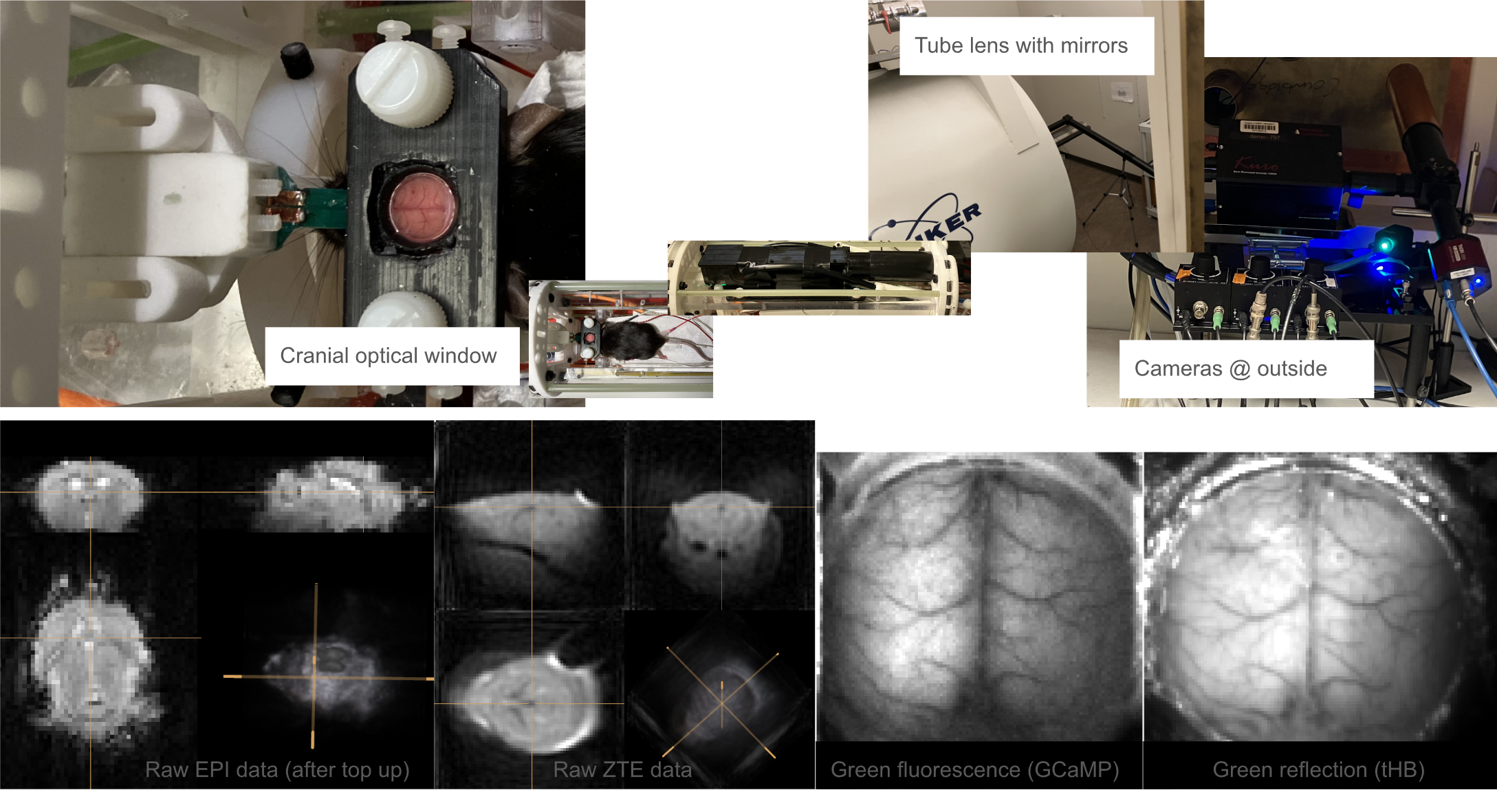
Optical imaging: The optical imaging system was designed to fit inside the magnet with a long tube lens4, shown in figure 1., slightly different from the recent pioneering work5. The wavelength was 466/40nm for fluorescence excitation, and the emitted 525/50nm were detected in a camera at 50Hz in 16bit with image matrix 110×110. Meanwhile, the green light pulses were illuminated alternatively at 50Hz and the reflection signals were detected with the same camera. In addition, NIR illuminated the cortex and detected by another camera at 50Hz over a beam split.
Data analysis: Both fMRI and optical data were preprocessed and registered to Allen atlas with a strategy of tissue boundary landmarks6. The green reflection light was used for both imaging total hemoglobin absorption of CVB signals and regression of fluorescence to minimize the influence of hemoglobin absorption and purify neuronal calcium or voltage signal detection. The block designs were convolved with the hemodynamic response function and correlated with the fMRI time courses to detect a stimulation response.
RESULTS
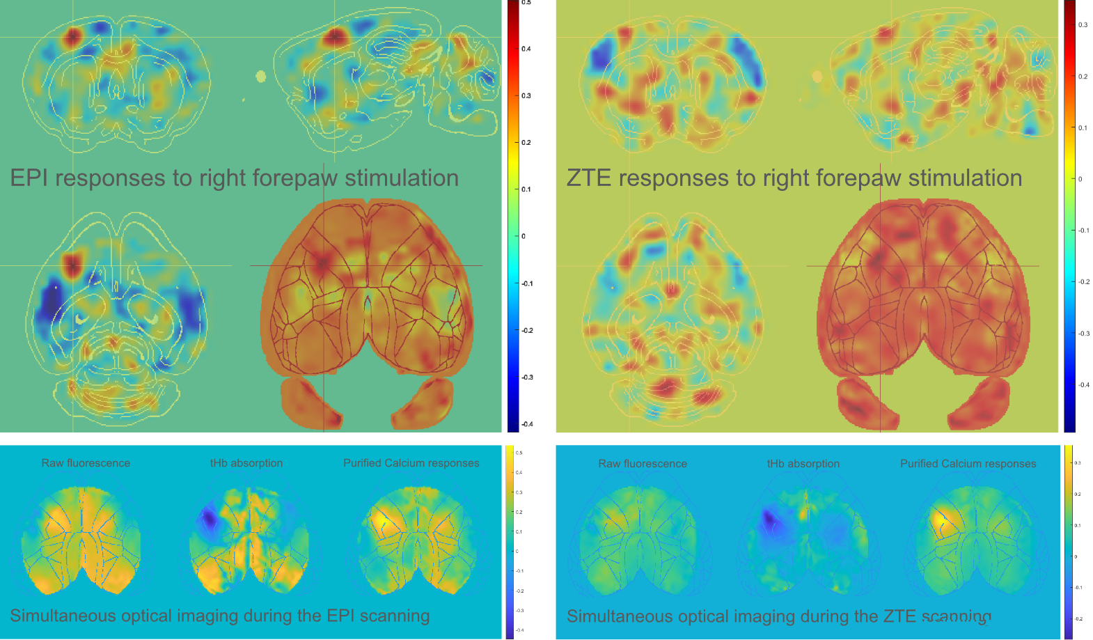
Five of the 7 animals were evaluated in this initial analysis. As a demonstrated in figure 2, the forelimb area of the contralateral hemisphere was primarily activated by forepaw stimulation in both methods of fMRI. Some negative responses in other somatosensory areas, including the mouth area, nose area and part of barrel cortex were detected by both EPI and ZTE. Further analysis are continuing to allow us to take advantage of both whole brain fMRI studies and neuronal genetic fluorescence indicators.DISCUSSION/CONCLUSION
Relative to EPI-based fMRI, ZTE-fMRI has great advantages for whole brain coverage without signal dropout/distortion. The extrinsic neuronal calcium or membrane voltage sensors in optical fluorescence may provide a great opportunity to image a specific neuronal activities. However, the green fluorescent signals may be largely modulated by hemoglobin absorption when the slow frequencies of primary interest for fMRI are examined. The setup with simultaneous same-wavelength reflection light may be helpful to regress out both slow hemodynamics and shared physiological variation, i.e. breath/cardiac pulses. The integration of these two complementary neuroimaging modalities opens a broad range of future applications for systems level neuroscience and preclinical studies of pathological brain function.Acknowledgements
This work was supported by NIH grants: r01mh111416, r01ns078095, and r01eb029857.References
1. Idiyatullin, D., Corum, C. A. & Garwood, M. Multi-Band-SWIFT. Journal of Magnetic Resonance 251, 19–25 (2015).
2. MacKinnon, M. J. et al. (ISMRM 2020) iZTE-fMRI. https://archive.ismrm.org/2020/1221.html.
3. Pan, W.-J., Zhou, L., Clavijo, G. P., Sharghi, V. K. & Keilholz, S. (ISMRM 2021) Optimized Single-loop Coil with 3D-shaped Design for Simultaneous fMRI and Optical Imaging in Rodent. https://archive.ismrm.org/2021/2511.html.
4. Pan, W.-J. et al. (ISMRM 2022) Optimization of wide-field optical imaging method towards fMRI integration in mice. https://archive.ismrm.org/2022/3331.html.
5. Lake, E. M. R. et al. Simultaneous cortex-wide fluorescence Ca2+ imaging and whole-brain fMRI. Nature Methods 17, 1262–1271 (2020).
6. Pan, W.-J., Anumba, N., Xu, N., Meyer-Baese, L. & Keilholz, S. (ISMRM2023) Manual registration and customized template for rodent fMRI data spatial normalization. https://www.ismrm.org/23/program-files/D-19.htm.
Figures