3133
Functional connectivity of medial pulvinar to limbic system in macaque monkey revealed by INS-fMRI1Key Laboratory for Biomedical Engineering of Ministry of Education, College of Biomedical Engineering and Instrument Science, Zhejiang University, Hangzhou, China, 2Department of Neurosurgery of the Second Affiliated Hospital, Interdisciplinary Institute of Neuroscience and Technology, School of Medicine, Zhejiang University, Hangzhou, China, 3MOE Frontier Science Center for Brain Science and Brain-Machine Integration, School of Brain Science and Brain Medicine, Zhejiang University, Hangzhou, China
Synopsis
Keywords: Functional Connectivity, Brain Connectivity
Motivation: To understand the circuitry underlying the pulvinar and limbic cortical areas in the brain.
Goal(s): To map at mesoscale functional connections between medial pulvinar (PM) and insular and cingulate cortices in Macaque monkey.
Approach: Infrared neural stimulation was delivered via optic fibers to sequential sites in PM in Macaques and BOLD responses at connected sites mapped in 7T MRI.
Results: We find connectivity of PM with cingulate cortex was patchy and columnar, with sequential sites in PM producing mediolateral topographic activation. A functional topology of these patches was observed in cingulate.
Impact: The specificity and mesoscale nature of functional connections from medial pulvinar to limbic systems, providing new treatment ideas for mental disorders.
Introduction
The pulvinar complex of primates has been implicated in playing important roles in visual attention, higher cognitive processing1 and emotional regulation. The medial pulvinar (PM), in particular, forms connections with the limbic system, and is also named “limbic thalamus” due to its projections to cingulate and insula2,3 . Previous anatomical tracer studies show that PM is connected primarily with posterior cingulate and posterior insula, with only sparse connections to anterior cingulate4,5. However, MRI studies6,7 have attained mesoscale resolution visualization of these connections. The precise functional connection of PM to limbic system remains to be elucidated. Here, we use a novel approach called INS-fMRI (Infrared Neural Stimulation with functional Magnetic Resonance Imaging). We have previously used this approach to study mesoscale functional topographic relationships of pulvinar with dorsal and ventral pathways of visual cortex8. Here, we examine the connectivity of those same stimulation sites in PM with cingulate and insular cortex.Method
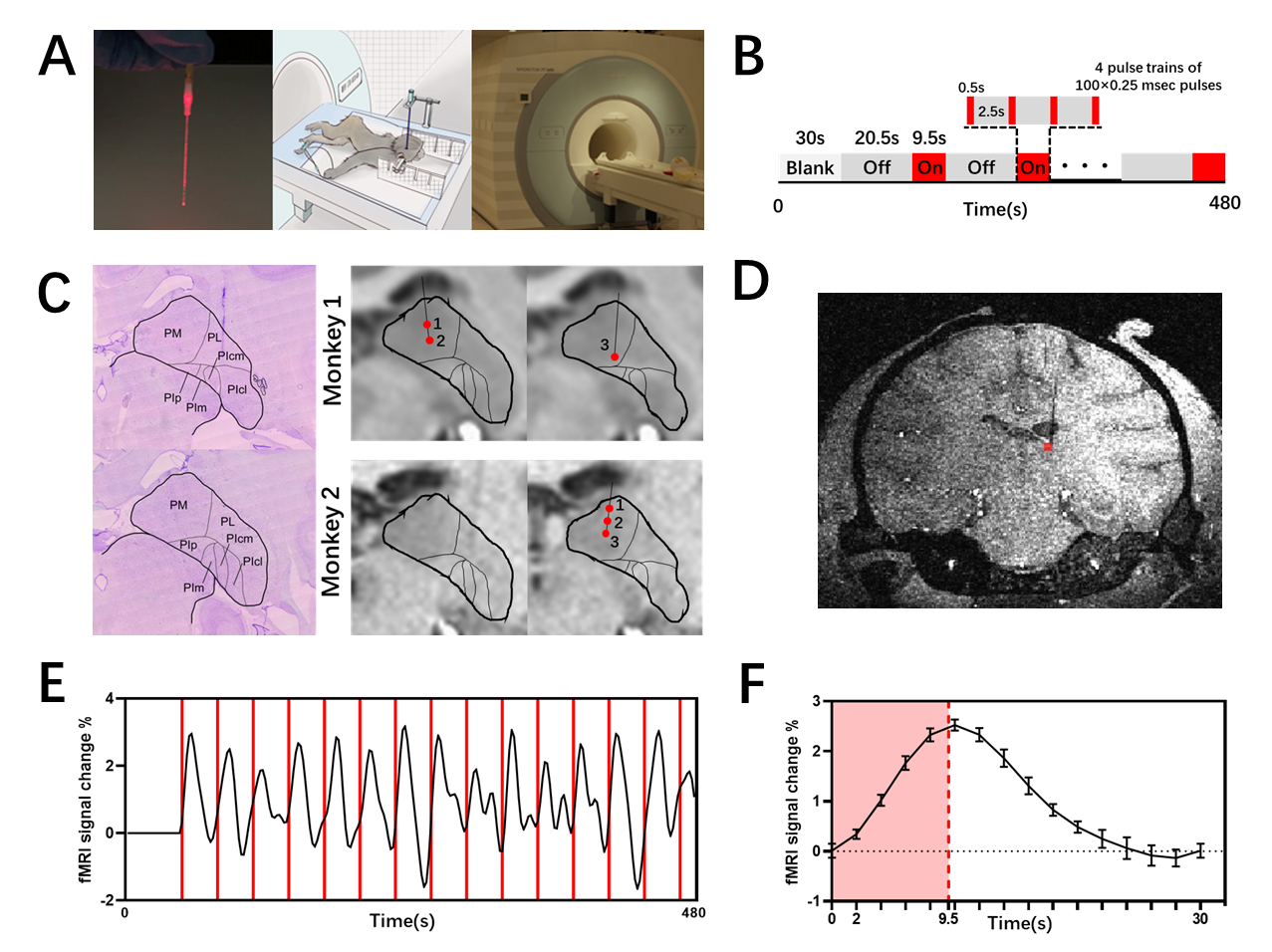
Animal preparation: Two adult female anesthetized rhesus monkeys (Macaca mulatta; Case 1: 5.9kg, Case 2: 5.5kg) were used in the study. Single optical fibers (Fig 1A) were inserted into the pulvinar and fiber tip locations confirmed by structural imaging9. Animals were anaesthetized with 1–2 µg/kg/h sufentanil and ~0.5% isoflurane.Data acquisition: MRI data were acquired on a 7T Magnetom scanner (Fig 1A), using a single loop RAPID coil. Structural images were collected at 0.3 mm isotropic resolution using a T1-weighted MPRAGE sequence. BOLD functional magnetic resonance imaging (fMRI) data were acquired at 1.5 mm isotropic resolution using an EPI (echo-planar imaging, TE = 22 ms, TR = 2000 ms).
INS paradigm: The stimulation site is determined by structural scanning prior to stimulation (Fig 1C). Data was acquired via block design (Fig 1B); each trial 30s, four INS pulse trains per trial (1875nm, 0.5sec pulse train, each pulse 0.25 msec, delivered at 200hz, intensity 0.3 J/cm2, ISI between pulse trains 2.5sec). Total 15 trials in one run, duration 480s.
Result
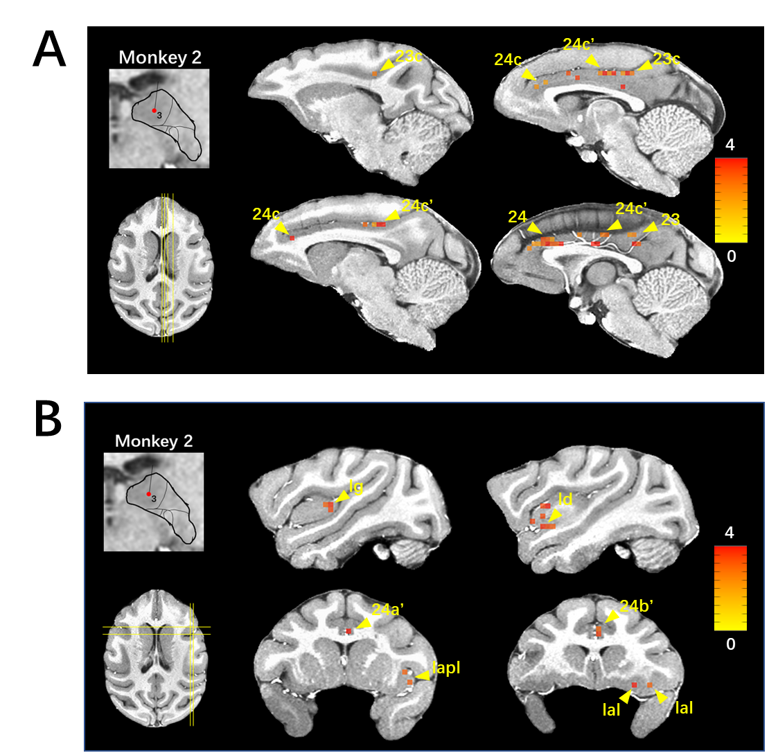
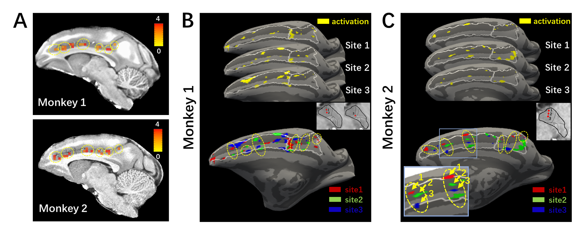
Results show that INS induces patchy activations in cingulate cortex. Both insula and cingulate revealed distinct activations in anterior vs posterior regions of each cortical area (Fig 2A and 2B).Fig 3A indicates that there are focal clusters of activation in cingulate cortex; this was consistent in two monkeys. Topographic characteristics of these activations are shown in surface view Fig 3B and 3C. Stimulation of sequential sites in PM (see inset in Fig 3C) induce 3-4 spatially segregated patches in anterior cingulate, respectively. Each patch, together with neighboring patches activated by shifting PM sites, form an activation cluster (yellow dash circle).
Discussion
We reveal focal, patchy functional connections in the anterior portions of cingulate and insula. These activations exhibit a topographic organization related to the sequential locations of stimulation sites in PM. These results provide insight into anatomical and functional connections in pulvinar-limbic network.Conclusion
As revealed by INS-fMRI, our results reveal the mesoscale organization of functional connections between PM and limbic insular and cingulate cortex. These connections have spatially shifted distribution and reveal a topographic relationship with cingulate cortex. These organizations may suggest a topography of PM-limbic circuits related to interoceptive behavior10, emotional expression, attention11.Acknowledgements
This work was supported in part by STI 2030-Major Projects (2021ZD0200401to A.W.R.), the National Natural Science Foundation of China (U20A20221, 819611280292), the Key Research and Development Program of Zhejiang Province (2020C03004), the Fundamental Research Funds for the Central Universities.References
[1] Homman-Ludiye J, Bourne JA. The medial pulvinar: function, origin and association with neurodevelopmental disorders. J Anat. 2019 Sep;235(3):507-520.
[2] Yeterian EH, Pandya DN. Corticothalamic connections of paralimbic regions in the rhesus monkey. J Comp Neurol.
[3] Froesel M, Cappe C, Ben Hamed S. A multisensory perspective onto primate pulvinar functions. Neurosci Biobehav Rev. 2021 Jun;125:231-243.
[4] Romanski LM, Giguere M, Bates JF, Goldman-Rakic PS. Topographic organization of medial pulvinar connections with the prefrontal cortex in the rhesus monkey. J Comp Neurol. 1997 Mar 17;379(3):313-32. PMID: 9067827.
[5] Thalamic connections of the insula in the rhesus monkey and comments on the paralimbic connectivity of the medial pulvinar nucleus.
[6] Barron DS, Eickhoff SB, Clos M, Fox PT. Human pulvinar functional organization and connectivity. Hum Brain Mapp. 2015 Jul;36(7):2417-31.
[7] Kagan I, Gibson L, Spanou E, Wilke M. Effective connectivity and spatial selectivity-dependent fMRI changes elicited by microstimulation of pulvinar and LIP. Neuroimage. 2021 Oct 15;240:118283.
[8] Yao S, Shi S, Zhou Q, Wang J, Du X, Takahata T, Roe AW. Functional topography of pulvinar-visual cortex networks in macaques revealed by INS-fMRI. J Comp Neurol. 2023 Apr;531(6):681-700.
[9] Saleem, K. S., & Logothetis, N. K. (2012). Atlas of the rhesus monkey brain. Academic Press.
[10] Evrard HC. The Organization of the Primate Insular Cortex. Front Neuroanat. 2019 May 8;13:43.
[11] Rolls ET. The cingulate cortex and limbic systems for emotion, action, and memory. Brain Struct Funct. 2019 Dec;224(9):3001-3018.
Figures