3132
Contrast Discrimination in Awake Macaque with 7T MRI1Key Laboratory for Biomedical Engineering of Ministry of Education, College of Biomedical Engineering and Instrument Science, Zhejiang University, Hangzhou, China, 2Department of Neurosurgery of the Second Affiliated Hospital and Interdisciplinary Institute of Neuroscience and Technology, School of Brain Science and Brain Medicine, Zhejiang University, Hangzhou, China, 3MOE Frontier Science Center for Brain Science and Brain-Machine Integration, School of Brain Science and Brain Medicine, Zhejiang University, Hangzhou, China
Synopsis
Keywords: Task/Intervention Based fMRI, fMRI (task based)
Motivation: This research aims to investigate visual perception in awake macaques using 7T MRI, enhancing our comprehension of BOLD responses during a contrast discrimination task and developing a quantifiable method for neuromodulation.
Goal(s): The specific objectives include directly observing BOLD responses at mesoscale during a contrast discrimination task.
Approach: The study utilized 7T MRI while macaques performed a 2AFC task, allowing simultaneous stimulus presentation and BOLD response measurement.
Results: The study successfully mapped BOLD responses during behavioral tasks, revealing contrast sensitivities in the visual cortex. Notably, it identified BOLD signal decreases in ambiguous contrast conditions, shedding light on perceptual uncertainty in stimulus discrimination.
Impact: These findings may advance quantification of behavioral an neural processes, helping to bridged the gap between human non-invasive and monkey invasive studies.
INTRODUCTION
The neural basis underlying visual perception remain at the forefront of neuroscience. Specifically, contrast discrimination stands as a quantitative bridge linking BOLD responses with behavioral outputs. Traditional human studies1,2 have offered valuable insights into these phenomena. However, most previous fMRI research used the 2IFC (two-interval forced choice) task, with its sequential stimulus presentation, fails to capture the concurrent comparison of neural signals that correspond to each choice in a 2AFC (two-alternative forced choice) paradigm.Addressing this gap, our research used of awake macaques conducting a contrast discrimination task in a 7T ultra-high field MRI. The superior spatial resolution of 7T MRI, unveils submilimeter functional domains such as V2 color sensitive thin stripes, which remained shrouded in lower resolutions. Crucially, our 2AFC paradigm concurrently presents two stimuli at distinct receptive fields, enabling a direct examination of the BOLD responses from each stimulus within the 2AFC framework.
Beyond their suitability for such tasks, macaques pave the way for next-phase neuromodulation applications, underscoring the broader translational relevance of this approach.
METHODS
The experiment were conducted on two rhesus monkeys in 7-Tesla MR Scanner (Siemens, Germany). We obtained functional images with a single-shot echo planar imaging (EPI) sequence (TE 28ms; TR 2000ms) with 0.8mm isotropic resolution.The monkeys were trained to perform a two-alternative forced-choice (2AFC) task to choose the stimulus with higher contrast. Each trial started with a fixation cue. Following this, two sine wave gratings of differing contrasts were displayed on either side of the fixation point. After a duration of 6 seconds, the fixation point vanished, prompting the monkey to make a choice via an eye saccade within a 2-second timeframe. Subsequently, a 10-second inter-trial interval (ITI) was introduced before the commencement of the next trial.
The time-courses of all voxels were fitted with the expected time-course given stimulus onsets and canonical hemodynamic response in generalized linear model (GLM)
RESULTS
Using 7T MRI, we have successfully obtained simultaneous BOLD and behavioral curves in macaques.During passive viewing, our analyses across functional domains revealed nuanced contrast sensitivity functions. Notably, V2 thick stripes were more sensitive to contrasts below 10%, with a notably shallower response slope at higher contrasts in comparison to V2 thin stripes. This suggests a differential, domain-specific contrast processing within the visual cortex that is now observable at submillimeter scale, and which is consistent with previous fMRI studies in humans3 and optical imaging in monkeys4.
In the behavior task phase, we employed a subtractive strategy between the BOLD signals from the right and left visual cortices. This revealed the neural processes involved in the comparison phase while subjects were fixating prior to making a choice. By assessing both hemifields, we could more accurately delineate the neural contrasts corresponding to the visual stimuli.
Further insights were gained when we focused on behaviorally correct trials. Particularly in conditions of near-equivalent contrasts—where stimuli closely approximated a 50% reference level—we identified a notable drop in BOLD signal. This may suggest that the presence of ambiguity in stimulus contrast may engage the attentional mechanisms differently5, possibly indicating a neural basis for the perceptual challenge posed by similar stimuli.
Our findings, enabled by the application of high-resolution imaging in awake macaques, pave the way for a deeper understanding of the neural dynamics underlaying contrast discrimination,aiming at relating behavioral curves, neural BOLD-based curves, and specific functional domain response.
CONCLUSION
In this study, using 7T MRI on behavioral macaques, we have studied the neural basis of contrast discrimination neural mechanisms. This will bridges human non-invasive studies with monkey invasive studies, and shed light on the functional organization in the visual cortex. Our observations of BOLD signal shifts in low contrast differential trials may offer insights into the neural processes underlying challenging perceptual judgments.Acknowledgements
This work was supported by STI 2030—Major Projects (2021ZD0200401to A.W.R.),the National Natural Science Foundation of China (U20A20221, 819611280292), the Key Research and Development Program of Zhejiang Province (2020C03004), MOE Frontier Science Center for Brain Science & Brain-Machine Integration (Zhejiang University), the Fundamental Research Funds for the Central Universities.References
1. Boynton, Geoffrey M. , et al. "Neuronal basis of contrast discrimination. " Vision Research 39.2(1999):257-269.
2. Marquardt, et al. "Cortical depth profiles of luminance contrast responses in human V1 and V2 using 7 T fMRI." Human brain mapping 39.7(2018):2812-2827.
3. Roger, B H , et al. "Columnar Segregation of Magnocellular and Parvocellular Streams in Human Extrastriate Cortex. " The Journal of neuroscience : the official journal of the Society for Neuroscience (2017).
4. Lu, Haidong D, and Anna W Roe. “Optical imaging of contrast response in Macaque monkey V1 and V2.” Cerebral cortex vol. 17,11 (2007)
5. Jiang, Yong et al. “Different roles of response covariability and its attentional modulation in the sensory cortex and posterior parietal cortex.” Proceedings of the National Academy of Sciences of the United States of America vol. 120,42 (2023)
Figures
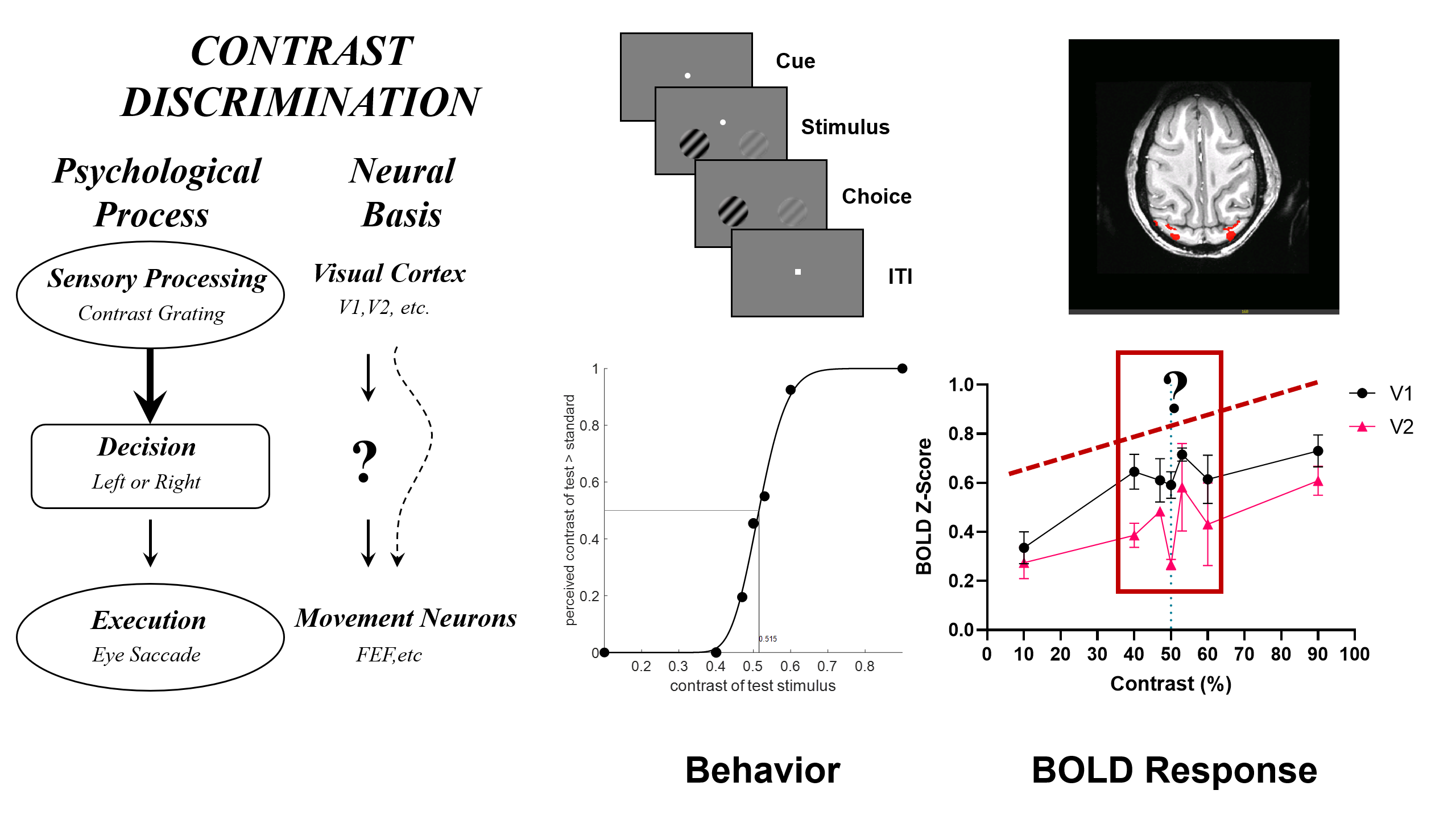
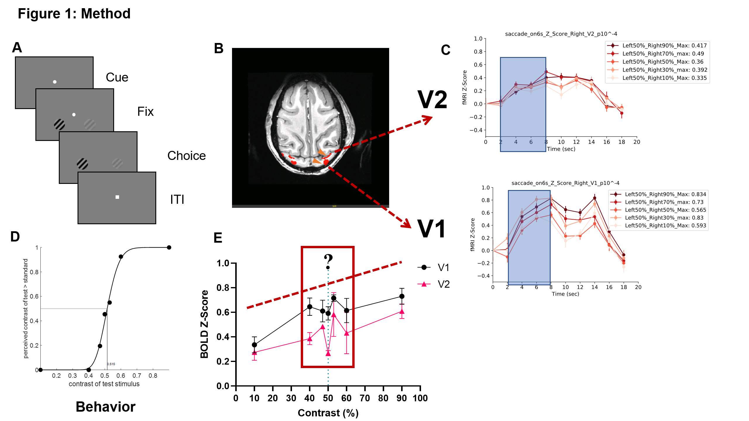
Figure 1: Methodology
(A) Illustration of the two-alternative forced-choice contrast discrimination task paradigm.(B) Activation map highlighting the regions of interest (ROI) in response to contrast stimuli.(C) Upper panel: Average BOLD time courses from Area V2. Lower panel: Average BOLD time courses from Area V1, with the stimulus period marked by a blue box.(D) Behavioral response curve of a Macaque performing the 2AFC contrast discrimination task.(E) Data points represent the average BOLD response between 2-8 seconds from the onset of the stimulus for each condition.
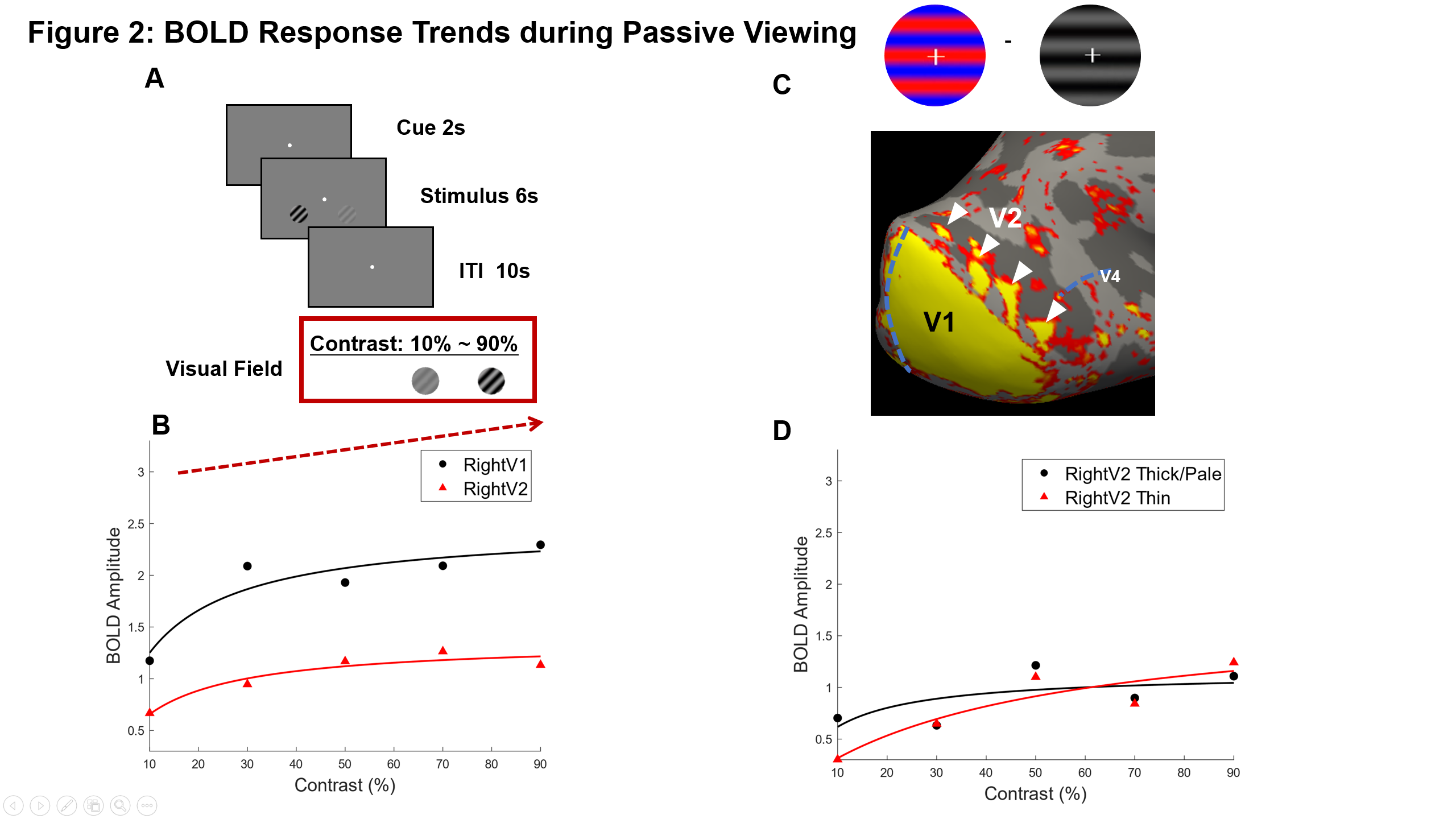
Figure 2: BOLD Response Trends during Passive Viewing
(A) The paradigm of passive viewing(B) The overall BOLD response demonstrates an ascending trend with increasing contrast of stimuli during passive viewing. In visual area V1, the fitting function's slope is steeper compared to V2, indicating a more rapid signal increase in V1. Signal saturation is observed to occur earlier in the V2 region than in V1.(C) The white triangle highlights the V2 thin stripes, a color-sensitive region. (D)The fitting function's slope of V2 thin stripes is higher than that of the V2 thick and pale stripes.