3131
Unveiling Mesoscale Finger-specific Neural Circuits through Near-infrared Stimulation and fMRI Mapping1College of Biomedical Engineering and Instrument Science, Zhejiang University, Hangzhou, China, 2Department of Neurology of the Second Affiliated Hospital, Interdisciplinary Institute of Neuroscience and Technology, Key Laboratory of Medical Neurobiology of Zhejiang Province, Zhejiang University School of Medicine, Hangzhou, China, 3MOE Frontier Science Center for Brain Science and Brain-Machine Integration, State Key Laboratory of Brain-machine Intelligence, School of Brain Science and Brain Medicine, Zhejiang University, Hangzhou, China
Synopsis
Keywords: Functional Connectivity, Neuroscience
Motivation: The dexterity of individuated finger movements is fundamental to daily hand use; yet specific brain networks serving each finger remains poorly understood.
Goal(s): Our objective is to unravel mesoscale connections associated with finger-specific neural circuits.
Approach: In 2 macaque monkeys, single digit locations were functionally mapped in cortical areas 3b via fMRI. Pulsed INS (infrared neural stimulation) targeted to these sites elicited mesoscale BOLD activation at functionally connected sites resulting in brainwide single digit networks.
Results: INS-induced BOLD activations revealed the distinct networks associated with each finger. Index finger connected with sensorimotor, multisensory and emotion circuits, supporting flexibility and stability in fine movement.
Impact: This research identifies specific brain networks underlying single digit maps in SI, and will be relevant for understanding hand dexterity, neurorehabilitation, and prosthesis development.
INTRODUCTION
The primate hand serves as an essential tool for manipulation, requiring delicate coordination and flexibility across fingers to achieve precision grasp and object manipulation1, 2, 3. Although certain neural circuits related to hand perception and control have been discovered4, little is known about how the functional networks of individual fingers in the brain. The combination of pulsed near-infrared neural stimulation with functional magnetic resonance imaging (INS-fMRI), as a novel network mapping method, provides reliable focal activation and insights into whole-brain mesoscale connections5. We hypothesize that each digit may be involved in both shared and specific connectivity networks, reflecting their distinct operational characteristics. This will shed light on the neural networks and their role in enhancing the dexterity of the primate hand.METHODS
Two female rhesus monkeys (Macaca mulatta; 5.3 kg for case 1 and 4.7 kg for case 2) were used in the study. The animals were anesthetized with 0.2-0.5% isoflurane, ketamine (10 mg/kg bolus and 4 mg/kg/hr i.v.) and vecuronium bromide (50 ug/kg/hr i.v.) and placed in a MR-compatible stereotaxic frame. MRI data were acquired using a Siemens 7T Magnetom with structural images by a T1-weighted MPRAGE sequence (TE = 2.82 ms, TR = 2590 ms, BW = 300 Hz, 0.3 mm isotropic resolution) and functional images by a single-shot EPI sequence (TE = 22 ms, TR = 2000 ms, BW = 1776 Hz, 1.5 mm isotropic resolution). A digit map was obtained by vibrotactile stimulations on thumb (D1), index finger (D2) and middle finger (D3), facilitated through an MR-compatible tactile stimulator (Figure 1A). Laser pulses (1875 nm, 0.5sec pulse train, 250 usec per pulse at 200 Hz) were transmitted through a 200-um optical fiber and intensity of laser pulses were set at 0.1 J/cm2. Each block consisted of 18-s blank, four 0.5 sec pulse trains, 2.5sec ISI; each stimulation block was repeated for 15 times per scan. Both tactile-evoked and INS-evoked BOLD signals were identified by generalized linear model with AFNI, highlighting voxels with significant p values (p < 0.005). Error bars: SEM.RESULTS & DISCUSSION
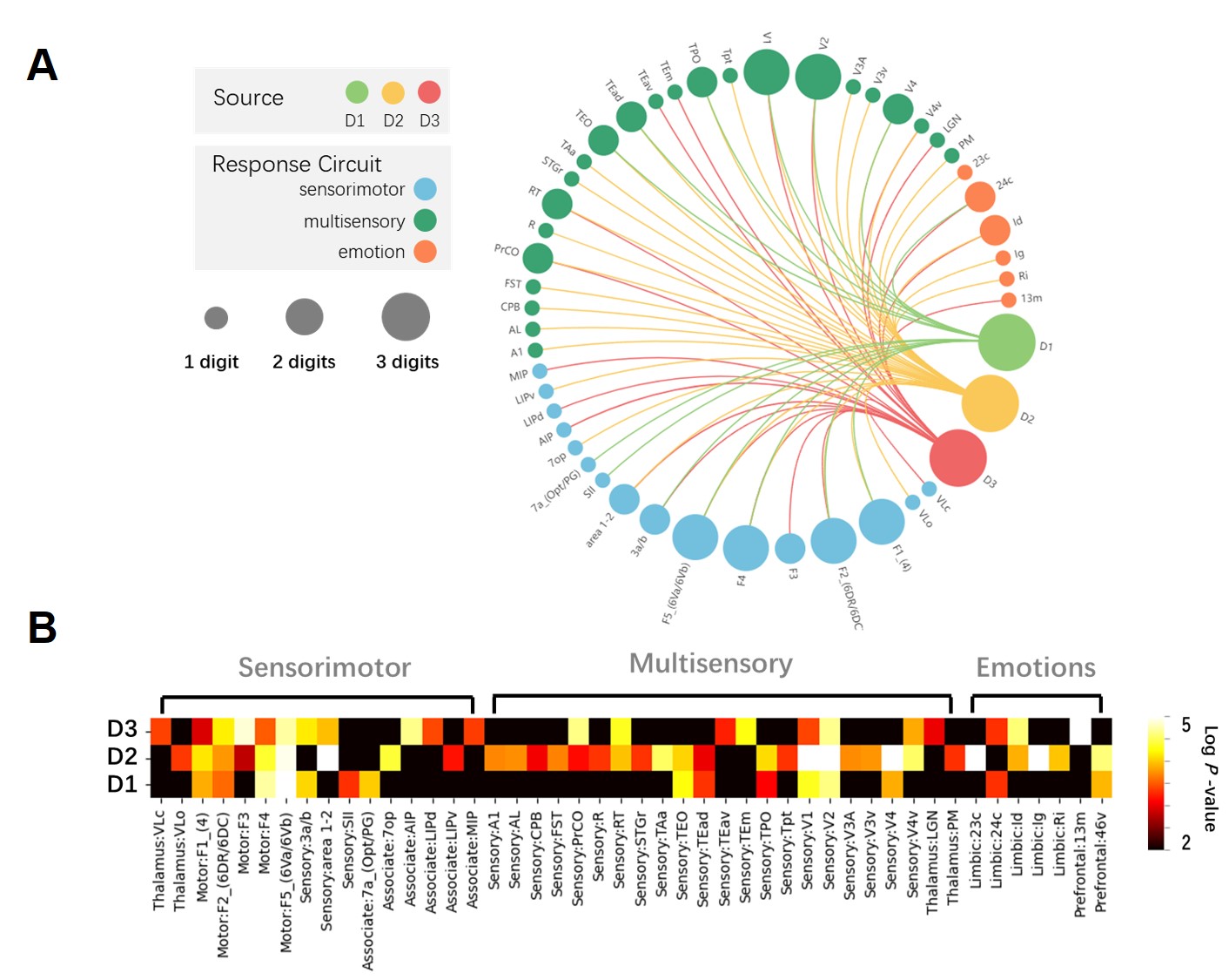
To define the implant target, we analyzed the positive BOLD responses evoked by vibrotactile stimulation at fingertips, revealing a distinct pattern from lateral to medial (Figure 1B). The results associated with D1 (yellow), D2 (green), and D3 (blue) were depicted in the expected finger-related area 3b of primary somatosensory cortex (S1), along with the averaged BOLD time courses for each digit. Figure 2A provides an example of the laser fiber in brain. Figure 2B shows INS-induced BOLD activation maps from INS at D3, revealing positive BOLD signals in sensorimotor-, multisensory- and emotion-related areas. Figure 3A shows the overall projections induced by INS at various digits (D1 in green, D2 in yellow and D3 in red) regarding sensorimotor (blue), multisensory (green), and emotion (red) circuits. Significant sharing of the sensorimotor and multisensory connections was observed among the three digits, particularly within motor and visual cortices. These results are consistent with known anatomical and functional connections6, especially regarding somatosensory projections to the motor and visual cortex. In addition, the number of activations in D2-related areas (n=34) was significantly higher than those in D1-related areas (n=15) and D3-related areas (n=22), suggesting that D2 exhibits a distinctive and complex pattern of activation (Figure 3B). Interestingly, INS at D1 showed fewer and weaker activations compared to D2 and D3; however, unique and distinct projections to higher-level somatosensory areas, including SII and 7a, were observed.The unique connections induced by various digits area in S1 provide direct evidence of the functional independence of hand use7, resulting from efficient information processing known as spatial segregation for complex inputs4. Specifically, the increased connectivity in D2-related areas would facilitate the integration of inputs from the coactivated sensory system, indicating its importance in multisensory interactions. Compared with D2-related activations, D3-related areas exhibit an overlapping but complementary pattern; the distinctions of D1 connectivity may reflect the role of thumb opposability. These specific activations begin to unveil the potential roles of each finger during manual grasp.
CONCLUSION
Use of INS-fMRI reveals precise brainwide mesoscale networks following stimulation of single digit loci. Our finding suggests that sensory inputs from different digits are processed in both cooperative and independent circuits, with specific networks reflecting their functional utility.Acknowledgements
This research was supported by National Key R&D Program of China (2021YFF0702200), STI 2030—Major Projects(2021ZD0200401), National Natural Science Foundation of China (U20A20221, 81961128029).References
1. Santello M, Flanders M, Soechting JF. Patterns of hand motion during grasping and the influence of sensory guidance. Journal of Neuroscience 22, 1426-1435 (2002).
2. Thakur PH, Bastian AJ, Hsiao SS. Multidigit movement synergies of the human hand in an unconstrained haptic exploration task. Journal of Neuroscience 28, 1271-1281 (2008).
3. Sobinov AR, Bensmaia SJ. The neural mechanisms of manual dexterity. Nature Reviews Neuroscience 22, 741-757 (2021).
4. Lazar L, Chand P, Rajan R, Mohammed H, Jain N. Somatosensory cortex of macaque monkeys is designed for opposable thumb. Cerebral Cortex 33, 195-206 (2023).
5. Shi S, et al. Infrared neural stimulation with 7T fMRI: A rapid in vivo method for mapping cortical connections of primate amygdala. NeuroImage 231, 117818 (2021).
6. Padberg J, et al. Thalamocortical connections of parietal somatosensory cortical fields in macaque monkeys are highly divergent and convergent. Cerebral cortex 19, 2038-2064 (2009).
7. Ejaz N, Hamada M, Diedrichsen J. Hand use predicts the structure of representations in sensorimotor cortex. Nature neuroscience 18, 1034-1040 (2015).
Figures
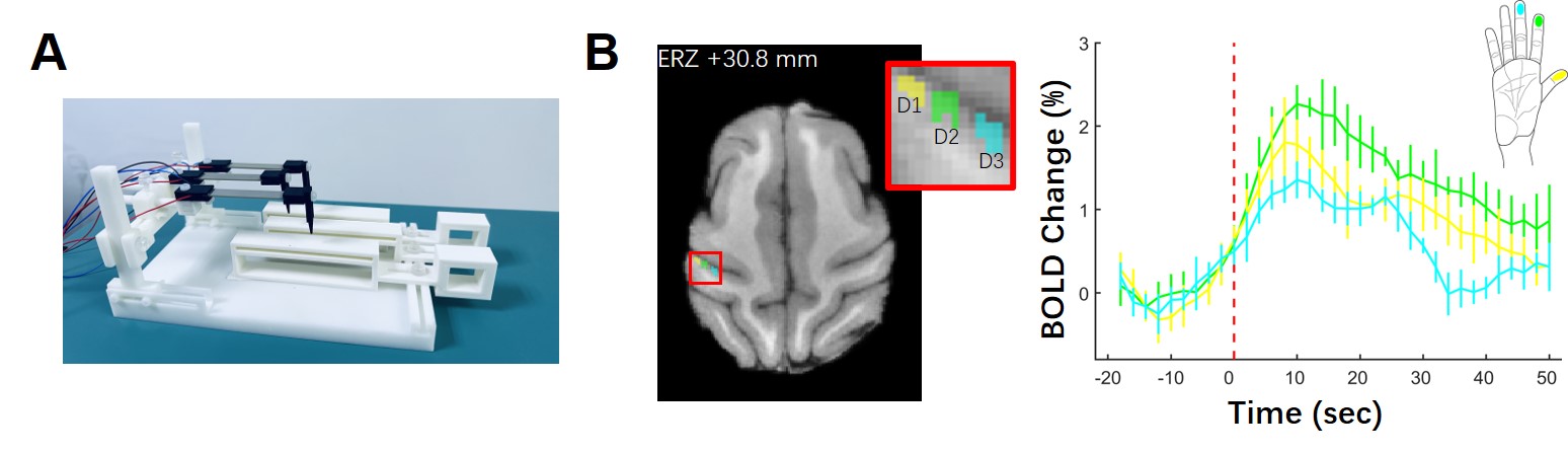
Figure 1. (A) MR-compatible stimulator. (B) Tactile-induced fMRI activation map for various digits, and their BOLD time courses. Yellow, green and blue represented the location activation for thumb (D1), index finger (D2) and middle finger (D3), receptively.
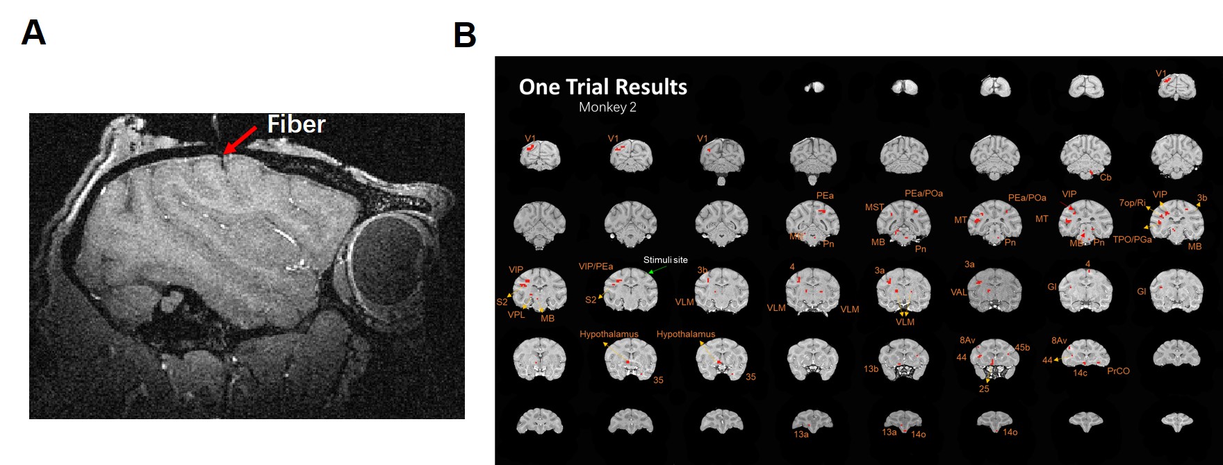
Figure 2. INS-induced activation map. (A) Location of stimulation laser fiber, indicated by red arrow, is superimposed on sagittal view of T1 image. (B) Results from a signal trial of INS-induced BOLD activations map in the middle finger of Monkey 2.