3129
Layer-fMRI in lower brain structures: why is it so hard and what can we do about it?1NIH, Bethesda, MD, United States, 2German Center for Neurodegenerative Diseases (DZNE), Bonn, Germany, 3Maastricht University, Maastricht, Netherlands, 4University College London, London, United Kingdom
Synopsis
Keywords: fMRI Acquisition, fMRI, layer-fMRI, UHF, EPI, VASO, sub-millimeter fMRI, 7 tesla
Motivation: Layer-fMRI can address questions of directional information flow, but it’s difficult in lower brain areas.
Goal(s): We want to make layer-fMRI work in low brain structures, despite commonly low tSNR, EPI distortions, and EPI phase errors.
Approach: We tested the efficacy of four advanced acquisition approaches to mitigate these challenges: pTx, dual-polarity readouts, multi-shot segmentation, and aggressive GRAPPA.
Results: We found that pTx and high GRAPPA had limited impact for improved image quality. Though, multi-shot segmentation and dual-polarity readouts allowed layer-fMRI applications in low brain areas.
Impact: This work helps to fulfill the promise of layer-fMRI beyond the top 50% of the cortex.
Purpose
High-resolution and layer-specific fMRI can unveil the intricacies of directional neural information processing within microcircuits and across the brain. While the upper regions of the brain have witnessed significant progress, as evidenced by over 240 published papers, lower brain areas remain underexplored, ultimately also hindering applications of whole brain layer-fMRI. Only 3.4% of these publications (layerfmri.com/papers) have ventured into the depths of lower brain areas (Fig 1), primarily due to substantial acquisition challenges:- Low tSNR due to distance of RF-receive elements (amplified g-factors).
- Insufficient flip angles from limited B1+.
- Geometric distortions from B0-inhomogeneities.
- EPI phase errors resulting from the necessity of large imaging matrices (long readouts).
Methods
Mitigation approaches tested here:- Parallel transmit (pTx) at 7T with the 8ch Nova coil has received CE-clearance (FDA pending) and static pTX has become more straightforwardly applicable. However, the extent to which B1-shimming enables sub-millimeter fMRI in lower brain areas remains uncertain.
- Segmentation with multi-shot approaches allows an uncoupling of TE and echo train length for EPI with large matrix sizes (250-350) that are necessary for imaging lower brain areas. Long readouts allow EPI-phase errors to accumulate over the readout increasing the artifact level in low areas (1).
- Aggressive GRAPPA, typically only employed for acceleration with a factor of 2-3 in layer-fMRI, has the potential to further minimize EPI phase errors in lower areas. Yet, the trade-off between noise amplification from high g-factors and the benefits of conventional 32ch Rx coils remains unclear.
- The application of dual-polarity GRAPPA and dual-polarity readouts can mitigate challenging B0-shimming and k-space trajectory imperfections, which lead to higher-order EPI-ghosting and shading artifacts, including low-spatial frequency "Fuzzy ripples." Dual polarity GRAPPA and dual-polarity readouts can mitigate them (2-6).
Results
We find that the strategies differ in their effectiveness of making layer-fMRI in lower brain areas more feasible. In-plane segmentation and dual-polarity imaging, although less conventional, exhibit potential for straightforward application, effectively mitigating challenges (Fig. 2). Surprisingly, more established approaches, such as pTx and high GRAPPA accelerations, were found to be less efficient (Fig. 3). We did not see VASO artifacts of inflowing fresh (non-inverted blood) across experiments.Discussion and Conclusion
Layer-fMRI in deep brain structures offers a unique opportunity to investigate fundamental hypotheses about functional neural processing across various brain regions:- Different layers in the entorhinal cortex/hippocampus/parahippocampal are responsible for memory encoding and retrieval (13).
- Different layers in FFA/PPA receive feedforward-feedback input for neural representations of faces and houses (14-17).Different sub-nuclei of the Amygdala are involved in visual perception on emotion memory vs. emotion context (18).
- Different depths of the colliculi are expected to be differently biased by vascular draining artifacts (19).
- Unique lobules of the cerebellum contain sensorimotor digit representations (20).
Acknowledgements
The research was conducted as part of the NIMH Intramural Research Program (#ZIAMH002783). We thank the HCP 7T connectome project for providing the movie stimuli used in Fig. 4 A-B.References
- Seifert and Vannesjo, 2023, 7T fMRI in the Cervical Spinal Cord Under Noxious Thermal Stimulation. ISMRM #0571.
- Hoge and Polimeni, 2016. Dual-polarity GRAPPA for simultaneous reconstruction and ghost correction of echo planar imaging data. Magnetic Resonance in Medicine 76, 32–44. https://doi.org/10.1002/mrm.25839.
- Poser et al., 2013. Single-shot echo-planar imaging with Nyquist ghost compensation: Interleaved dual echo with acceleration (IDEA) echo-planar imaging (EPI). Magn Reson Med 69, 37–47. https://doi.org/10.1002/mrm.24222.
- van der Zwaag et al., 2009. Minimization of Nyquist ghosting for echo-planar imaging at ultra-high fields based on a “negative readout gradient” strategy. J. Magn. Reson. Imaging 30, 1171–1178. https://doi.org/10.1002/jmri.21951.
- Stirnberg et al., 2022 T2*-weighted dual-polarity skipped-CAIPI 3D-EPI: 400 microns isotropic whole-brain QSM at 7 Tesla in 6 minutes. #0594, ISMRM, 2022.
- Huber et al., 2023, #1149, ISMRM, Low spatial-frequency ripple artifacts in layer-fMRI EPI: Identification, cause, and mitigation strategies with Dual-polarity readout.
- Stirnberg, R., Stöcker, T., 2021. Segmented K-Space Blipped-Controlled Aliasing in Parallel Imaging (Skipped-CAIPI) for High Spatiotemporal Resolution Echo Planar Imaging. Magnetic Resonance in Medicine 85, 1540–1551 https://doi.org/10.1101/2020.06.08.140699.
- Huber et al., 2021. Layer-dependent functional connectivity methods. Progress in Neurobiology 207. https://doi.org/10.1016/j.pneurobio.2020.101835.
- Huber et al, 2021. LayNii: A software suite for layer-fMRI. NeuroImage 237. https://doi.org/10.1016/j.neuroimage.2021.118091.
- Graedel et al., 2022. ISMRM, Why is 7T fMRI in the entorhinal cortex so difficult and what can we do about it? #1096.
- De Panfilis, C., Schwarzbauer, C., 2005. Positive or negative blips? The effect of phase encoding scheme on susceptibility-induced signal losses in EPI. NeuroImage 25, 112–121. https://doi.org/10.1016/j.neuroimage.2004.11.014.
- Weiskopf, N., Hutton, C., Josephs, O., Deichmann, R., 2006. Optimal EPI parameters for reduction of susceptibility-induced BOLD sensitivity losses: A whole-brain analysis at 3 T and 1.5 T. NeuroImage 33, 493–504. https://doi.org/10.1016/j.neuroimage.2006.07.029.
- Zhang, et al., 2023. Differential laminar activation dissociates encoding and retrieval in the human medial and lateral entorhinal cortex. J. Neurosci. JN-RM-1488-22. https://doi.org/10.1523/JNEUROSCI.1488-22.2023.
- Carricarte, et al., 2023. Laminar dissociation of feedforward and feedback signals in high-level ventral visual cortex during imagery and perception. PsyArXiv. https://doi.org/10.31234/osf.io/7zcp8.
- Persichetti et al., 2023, VSS. Investigating layer-specific responses to mental imagery and perception in ventral occipital temporal cortex.
- Dowdle, L.T., Ghose, G., Moeller, S., Ugurbil, K., Yacoub, E., Vizioli, L., 2022. Task Demands Differentiate Regional Depth-Dependent Activity Profiles Within the Ventral Visual Pathway (preprint). BioRxiv. https://doi.org/10.1101/2022.12.03.518973.
- Koiso K, Akamatsu K, Huber L, Miyawaki Y, OHBM 2023, Laminar-level object information representation in higher visual areas revealed by VASO layer fMRI, #1788.
- Geissberger, N., Tik, M., Sladky, R., Woletz, M., Schuler, A.-L., Willinger, D., Windischberger, C., 2020. Reproducibility of amygdala activation in facial emotion processing at 7T. NeuroImage 211, 116585. https://doi.org/10.1016/j.neuroimage.2020.116585.
- Sitek and Gulban, OHBM 2022, Vasculature mapping in human superior and inferior colliculus, #3352
- Brouwer, et al. A Separation Between Motor & Sensory Somatotopic Maps in the Human Cerebellum, 2023 ISMRM BF study group. #10.
Figures
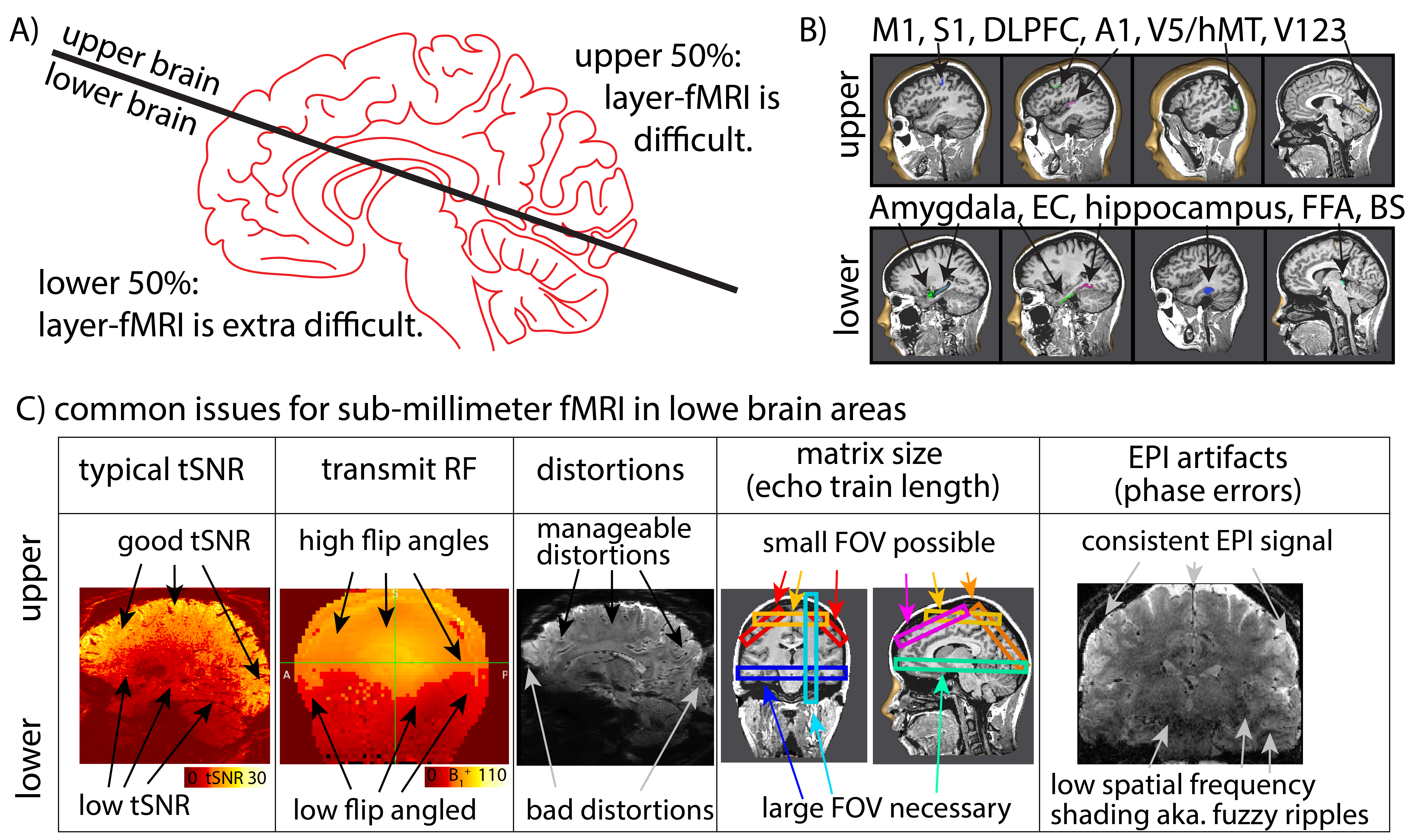
Fig. 1:
Layer-fMRI in lower brain areas (A, B) has the potential to inform questions of neural information of mesoscale circuits. However, it is under-investigated due to acquisition challenges related to low tSNR, limited transmit RF, geometric distortions, large echo train length and k-space trajectory imperfections (C). Only 3.4% of all layer-fMRI studies look at low brain structures (layerfmri.com/). In this abstract, we describe our experience from 5 studies evaluating the efficacy of various sequence approaches to address these challenges.
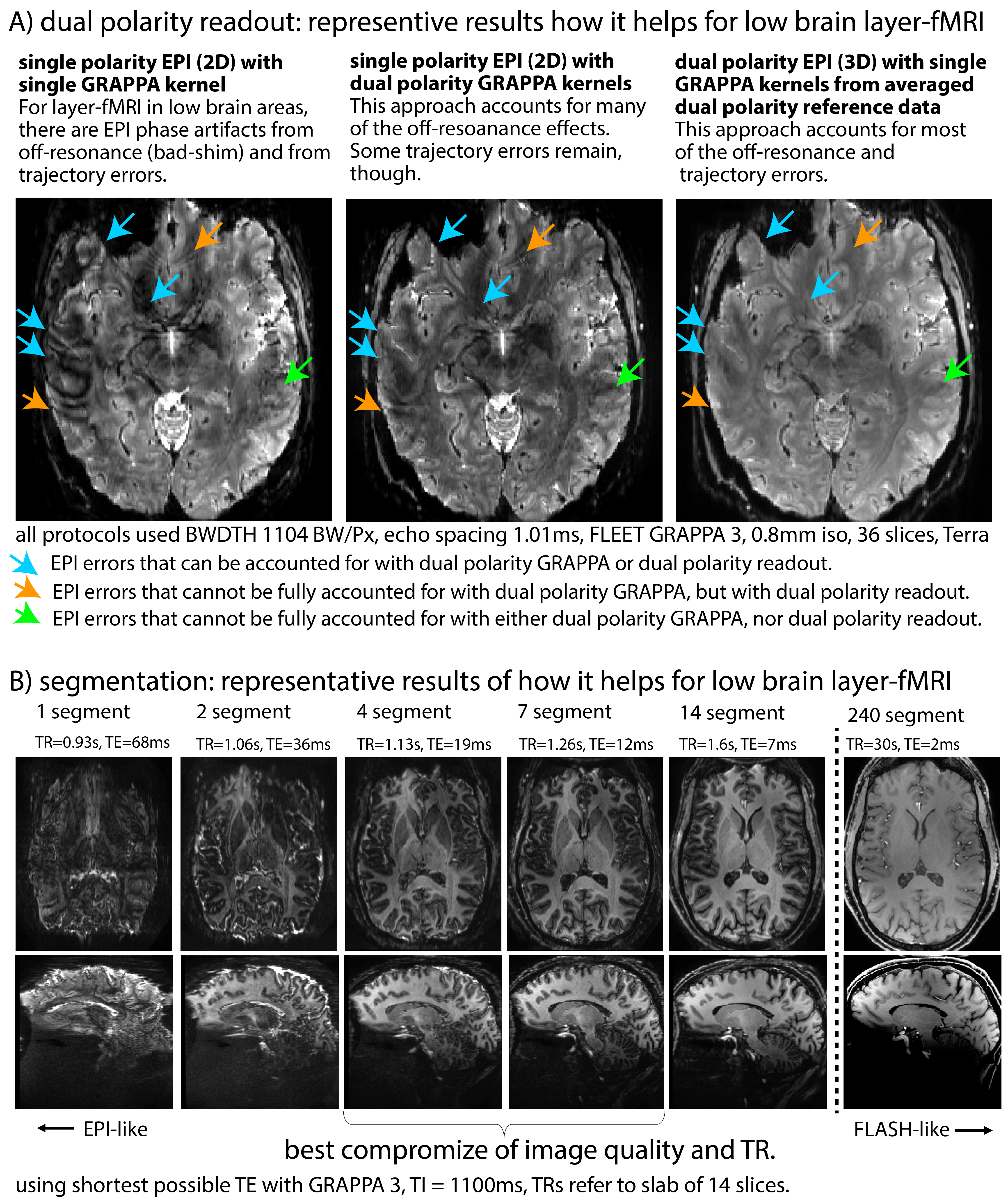
Fig. 2: Approaches that we found to be very helpful.
Representative results of investigating mitigation strategies for common EPI artifacts, when using low bandwidths (high resolution) with large matrix sizes (necessary in lower brain areas).
A) Dual polarity approaches are very helpful.
B) EPI segmentation improves data quality with respect to fuzzy ripples (low spatial-frequency shading) and distortions.
TRs increase only slightly, if TE is reduced with higher segmentation.
For dual polarity GRAPPA, see Houge & Polimeni 2016, for dual polarity readout see Huber 2023.
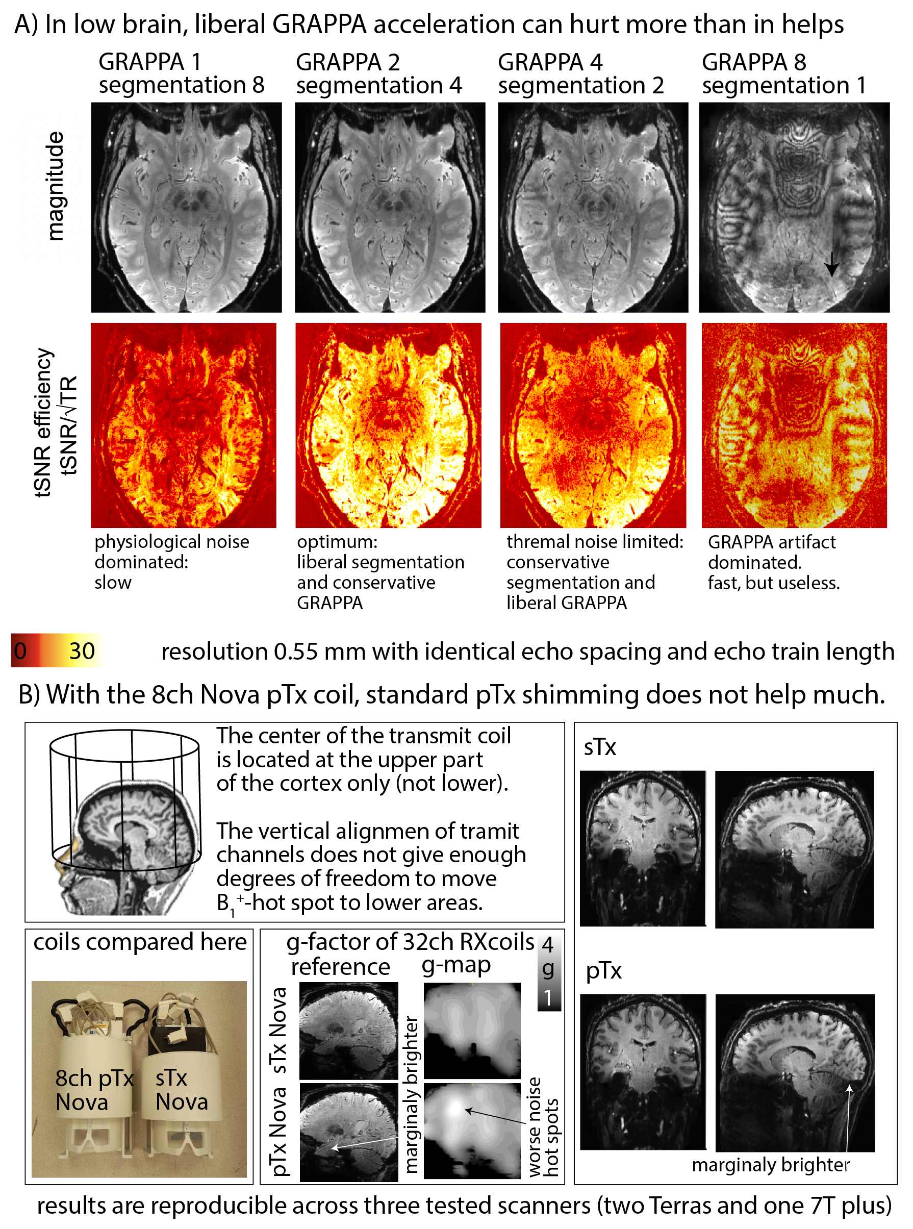
Fig. 3: Acquisition approaches that we found less helpful to facilitate layer-fMRI acquisition in lower brain areas.
A.) We found that aggressive GRAPPA acceleration beyond acceleration factors of 2-3 are counter indicative for layer-fMRI in lower brain areas.
B.) We found switching from the sTx Nova coil to pTx shimming with the 8ch Nova coil did not significantly help to make layer-fMRI data in lower brain areas more feasible.
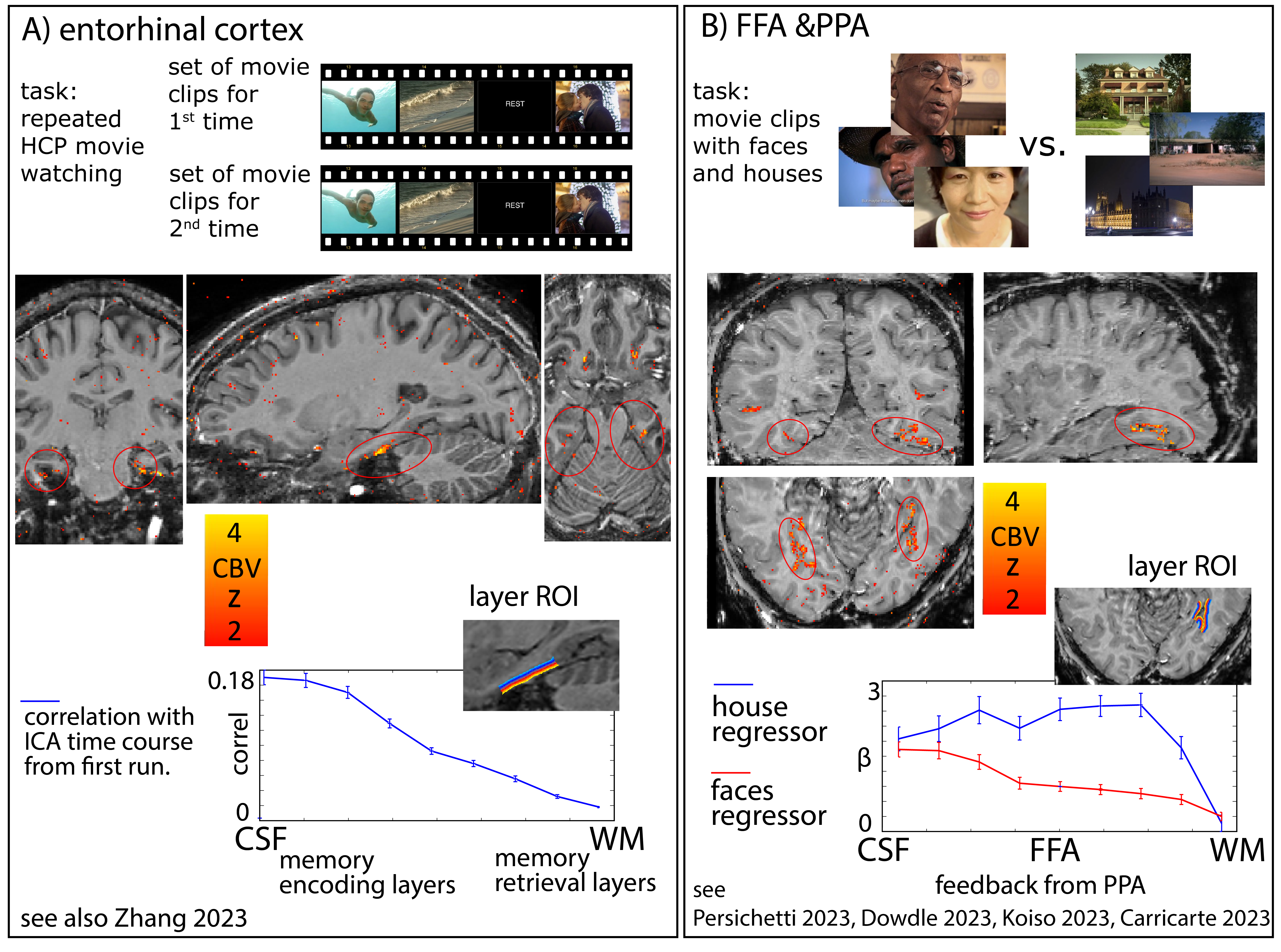
Fig. 4
We used five lower brain areas as ‘testbeds’ to confirm the feasibility of neuroscience application studies with the tested acquisition approach. Examples are in Figs. 4 and 5 use liberal segmentation factors (2-14 fold and conventional GRAPPA 3). Functional tasks in 4 were repeated movie watching (up to 50 times).
4A) For cross-run correlation (n->1), we expected memory encoding in upper layers.
4B) For face and house viewing in FFA, we expected feed-forward vs feedback signals, respectively.
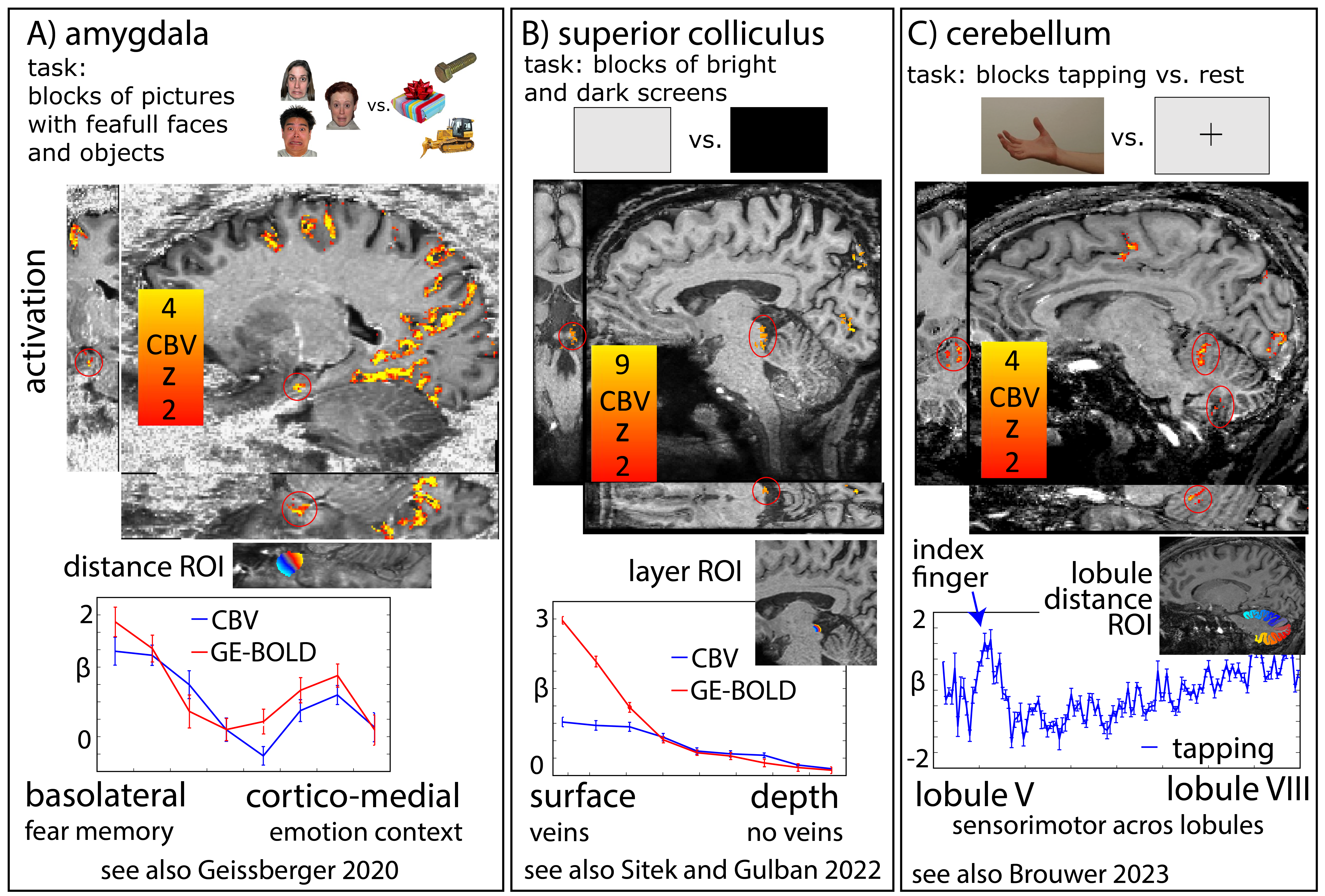
Fig. 5
Continuation of Fig. 4: Examples on the feasibility of the discussed approaches for neuroscience applications. Functional experiments were 12-14min (3runs), B1-shimming, segmentation, and dual-readouts.
A) We aimed to see activation distribution across sub-nuclei and whether they are differently biased by veins (VASO vs. BOLD).
B) We hypothesized that the surface voxels are more dominated by veins. C) For finger tapping, we expected focal responses in lobule V.