3125
NORDIC denoising on VASO data1Aarhus University, Aarhus, Denmark, 2University of Minnesota, Minneapolis, MN, United States, 3Maastricht University, Maastricht, Netherlands, 4National Institute of Mental Health, Bethesda, MD, United States
Synopsis
Keywords: fMRI Analysis, fMRI, NORDIC , VASO, laminar fMRI
Motivation: NORDIC denoising can effectively enhance the limited SNR in high-resolution fMRI. However, its application on VASO is yet to be validated.
Goal(s): We aimed to evaluate applications of NORDIC on VASO data and to offer recommendations for its execution.
Approach: We examined NORDIC’s capability to suppress noise while preserving the VASO signal across a wide parameter spectrum
Results: With a proper set of parameters, NORDIC effectively suppressed noise with minimal biases on the underlying signal.
Impact: NORDIC can substantially enhance the SNR in submillimeter VASO fMRI. We found the denoising performance to be sensitive to parameter choices and provide recommendations for safe execution.
Introduction
Functional MRI with submillimeter spatial resolution enables measurement of hierarchical information flow at the scale of cortical layers in the human brain. A common challenge in the field of layer-dependent fMRI is limited CNR, resulting from the small voxel sizes. This is particularly the case for VASO fMRI1, which is a popular supplement to BOLD in settings where layer-specificity is valued additionally to CNR. NORDIC denoising2 has shown promise in alleviating this challenge via thermal noise suppression. However, NORDIC has mainly been validated for BOLD acquisitions2-5 with limited attempts to apply it in VASO6-10. The data structure of VASO is notably different (alternating acquisitions with and without blood-nulling, different phase data, lower initial CNR, etc.), and it is thus unclear how NORDIC should be implemented.Here, we tested NORDIC applied to VASO across different strategies:
- combined or separately on nulled and not-nulled timeseries
- with or without an appended noise-volume
- on complex-valued versus magnitude-only data
- CNR
- spatiotemporal structure of removed components
- ability to retain response magnitudes and layer-profiles
Methods
NORDIC was tested on a range of field strengths and resolutions. This abstract highlight a 3T segmented VASO dataset (0.9 mm isotropic resolution), acquired from a single subject10. The generalizability across further datasets is described at: https://layerfmri.com/nordic/. The data shown here consist of six block-designed 12-minute runs of visuomotor stimulation alternating with rest. We reasoned this to be an ideal testbed as:- Submillimeter VASO at 3T is CNR limited and thermal noise dominated. It thus represents a setting in which NORDIC is most needed and presumably most effective.
- Low-rank denoising techniques are prone to bias in low-CNR domains. Hence, this data set provides a challenging test setting.
- The widespread brain activation associated with visuomotor stimulation and the excessive scan duration of 72 minutes facilitated reasonable ground-truth estimates through extensive spatiotemporal averaging of non-denoised data.
Relevant performance metrics (tSNR, t-values, PSC) were extracted from a large ROI (>7000 voxels). Laminar profiles were extracted from a small ROI placed in V1.
The data and NORDIC scripts are available at: https://github.com/LasseKnudsen1/NORDIC-VASO & https://layerfmri.page.link/ME_VASO3T.
Results and discussion
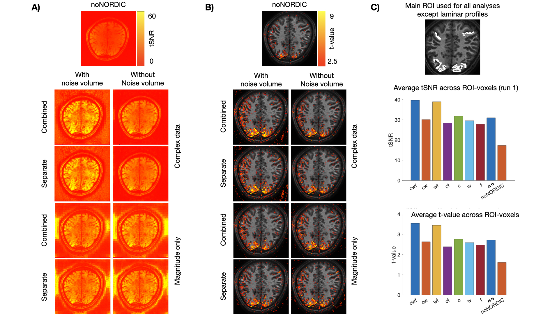
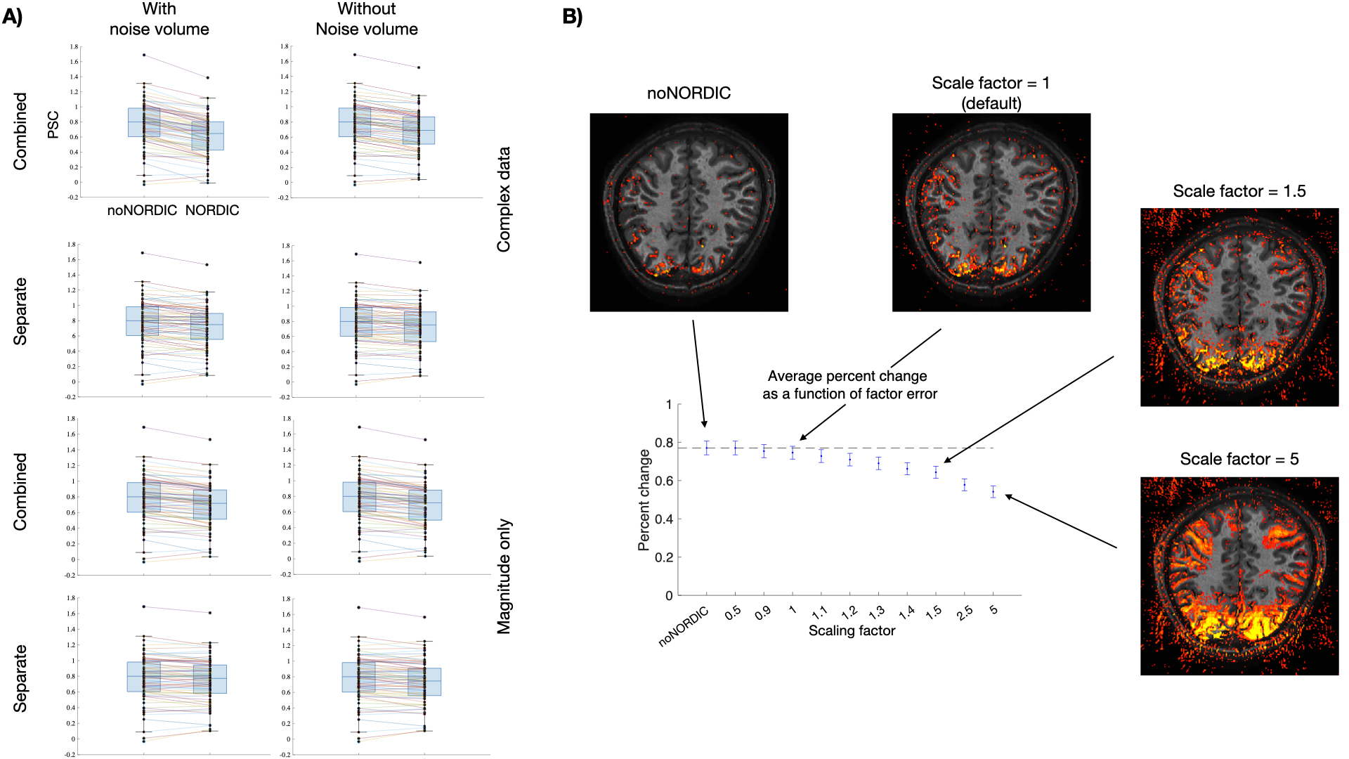
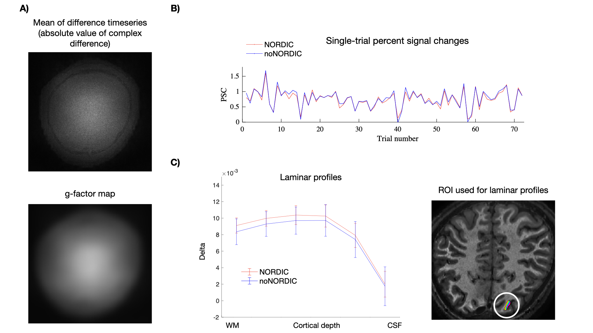
Figure 1 depicts tSNR- and t-value maps of each version. The largest gains in these quantities were observed when denoising was applied on combined and complex-valued nulled/not-nulled time series. However, combined versions were associated with a substantially reduced response magnitude (bias12) compared with the ground-truth estimate (Figure 2A), highlighting that denoising performance cannot be evaluated solely from CNR gains12-13. Figure 2B illustrates this CNR/bias tradeoff and shows how it can be manipulated by scaling the singular value threshold in NORDIC.Figure 3 depicts additional performance measures from the version “separate-withNoiseVol-MagnOnly” that best preserved the signal based on Figure 2A. Removed noise complied with that expected for g-factor-dependent, spatially varying thermal noise (3A), the temporal structure of single-trial PSC was unaltered (3B), and the laminar profile converged towards the ground-truth estimate (3C).
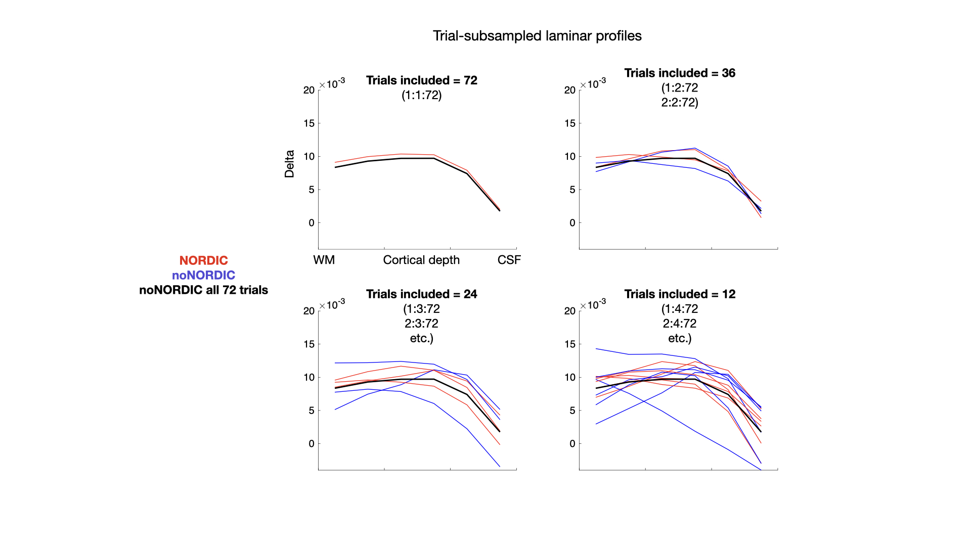
Figure 4 shows how the noise suppression mitigated the need for long scan durations, which could similarly facilitate higher resolution, lower field strength, small ROIs, etc.
VASO specific findings were:
- While denoising nulled/not-nulled time series combined provides more time points and PCs, with the potential to better separate signal from thermal noise, we find empirically that the strong contrast difference may ultimately introduce counter indications that make the separation harder for PCA.
- Like for conventional BOLD, we find that an appended noise volume in the right implementation improves the performance of NORDIC.
- We found the unique phase behavior in inversion-recovery VASO, with positive and negative phases (Mz-directions), calls for a conservative application of NORDIC with magnitude-only data. This is different from application of BOLD only, where complex-valued NORDIC is advised2. VASO users are advised to confirm the spatial integrity of their NORDIC data to rule out non-negligible spatial signal leakage compared to undenoised data.
Conclusion
We found that when NORDIC was implemented- on separate timeseries
- with an appended noise volume
- on magnitude-only data
While we show that NORDIC is sensitive to parameter choices, which limits generalizability (https://layerfmri.com/nordic/), we provide recommendations and scripts for a safe means of substantially improving the functional sensitivity of VASO fMRI with NORDIC.
Acknowledgements
This abstract is the outcome of a PhD secondment supported by Aarhus University and the NIH intramural program (#ZIAMH002783).References
1. Huber, L. et al. Slab-selective, BOLD-corrected VASO at 7 Tesla provides measures of cerebral blood volume reactivity with high signal-to-noise ratio. Magn. Reson. Med. 72, 137–148 (2014).
2. Vizioli, L. et al. Lowering the thermal noise barrier in functional brain mapping with magnetic resonance imaging. Nat. Commun. 12, (2021).
3. Dowdle, L. T. et al. Evaluating increases in sensitivity from NORDIC for diverse fMRI acquisition strategies. Neuroimage 270, 119949 (2023).
4. Knudsen, L. et al. Improved sensitivity and microvascular weighting of 3T laminar fMRI with GE-BOLD using NORDIC and phase regression. Neuroimage 271, 120011 (2023).
5. Fernandes, F. F., Olesen, J. L., Jespersen, S. N. & Shemesh, N. MP-PCA denoising of fMRI time-series data can lead to artificial activation “spreading”. Neuroimage 273, 120118 (2023).
6. Akbari, A., Gati, J. S., Zeman, P., Liem, B., & Menon, R. (2023). Layer Dependence of Monocular and Binocular Responses in Human Ocular Dominance Columns at 7T using VASO and BOLD. BioRxiv. https://doi.org/https://doi.org/10.1101/2023.04.06.535924
7. de Oliveira, Í. A. F., Siero, J. C. W., Dumoulin, S. O., & van der Zwaag, W. (2023). Improved Selectivity in 7 T Digit Mapping Using VASO-CBV. Brain Topography, 36(1), 23–31. https://doi.org/10.1007/s10548-022-00932-x
8. Huber, L., Krobichler, L., Stirnberg, R., Ehses, P., Stöcker, T., Fernandez-Cabello, S., Poser, B., & Kronbichler, M. (2021). Evaluating the capabilities and challenges of layer-fMRI VASO at 3T. ISMRM, #1229
9. Pizzuti et al., Laminar and columnar functional organization of human area MT using VASO at 7T, OHBM 2021, #1938.11.
10. Huber, L. et al. Evaluating the capabilities and challenges of layer-fMRI VASO at 3T. Aperture Neuro 3, (2023).
11. Huber, L. et al. LayNii: A software suite for layer-fMRI. Neuroimage 237, 118091 (2021).
12. Kay, K. The risk of bias in denoising methods: Examples from neuroimaging. PLoS One 17, 1–19 (2022).
13. Faes, L. et al., Mapping frequency preference in the auditory cortex using CBV-sensitive layer-fMRI, OHBM 2022. #2286
Figures