3124
Layer ReHo: Tool for characterizing mesoscale functional structure across layers and columns1NIMH, NIH, Bethesda, MD, United States, 2Faculty of Psychology and Neuroscience, Maastricht, Netherlands, 3Graduate School of Interdisciplinary Science and Engineering in Health Systems, Okayama University, Okayama, Japan
Synopsis
Keywords: Functional Connectivity, High-Field MRI, layer fMRI, ReHo
Motivation: High resolution fMRI is an emerging field, analysis pipelines are still under development. Local and short distanced time course similarities also named as Layer dependent regional homogeneity, can give us insights to map laminar dependent signal differences.
Goal(s): Using a simple metric to color code the depth dependent cortical activity.
Approach: Using mesoscale functional structure to predict and understand, time locked activity patterns in cortical ribbon without having necessity of specific model or predefined ROI
Results: Layer dependent regional homogeneity predicts local activity patterns, also can be used as a similarity matrix to rank different runs which potentially contributes to similar layer patterns.
Impact: This work can potentially be a publicly available analysis tool to observe and quality check, can also quantify laminar separability of the acquired depth dependent high resolution fMRI data.
Introduction
With the advance of high field scanners and the novel imaging methods, sub-millimeter fMRI acquisitions have become more common(Huber et al. 2018). Although a whole brain layer dataset(Koiso et al. 2023) recently became available, most of the high resolution fMRI studies are restricted to confined cortical regions like somatomotor(Yang et al. 2021; Yu et al. 2022) and visual areas(Akbari et al. 2021; Cho et al. 2022; Mourik et al. 2021). There is an increasing interest in confined activation patterns like layer dependent signals(Huber, Finn, et al. 2021; Müller et al. 2021). However investigating local and short distance connectivity structures at higher resolutions hasn't been studied and it could be a model free way to map and delineate confined functional structures. In this study we developed a simple data processing method called layer dependent regional homogeneity (Layer ReHo) to investigate above mentioned possibilites.Methods
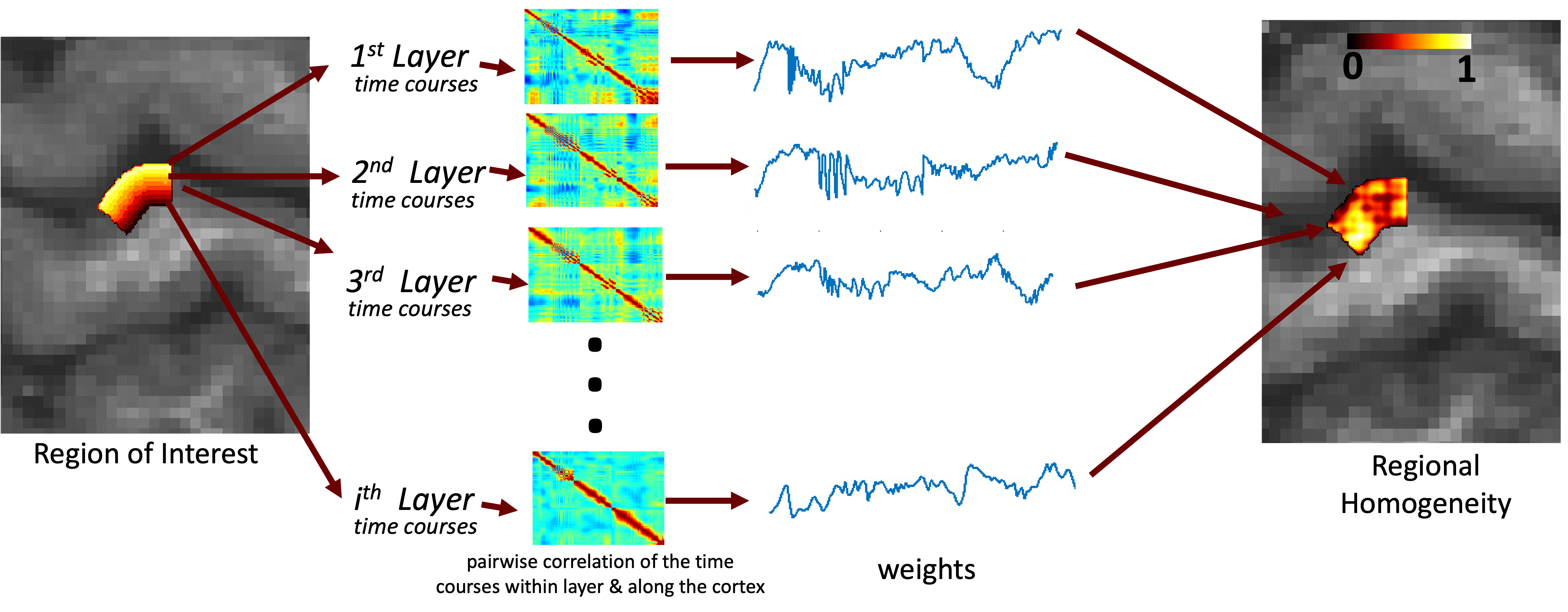
ReHo is defined and used previously in conventional fMRI resolutions(Jiang and Zuo 2016),also in one high resolution study(Pais-Roldán et al. 2020). Similarly we rank the time course similarity of the neighboring voxels. Differently, we restrict the neighbors to a certain cortical depth, a.k.a. layer bins calculated by open software called LayNii (Huber, Poser, et al. 2021). We calculated the similarity of each layer bins and values are given to corresponding voxels, see Fig1.Results & Conclusion
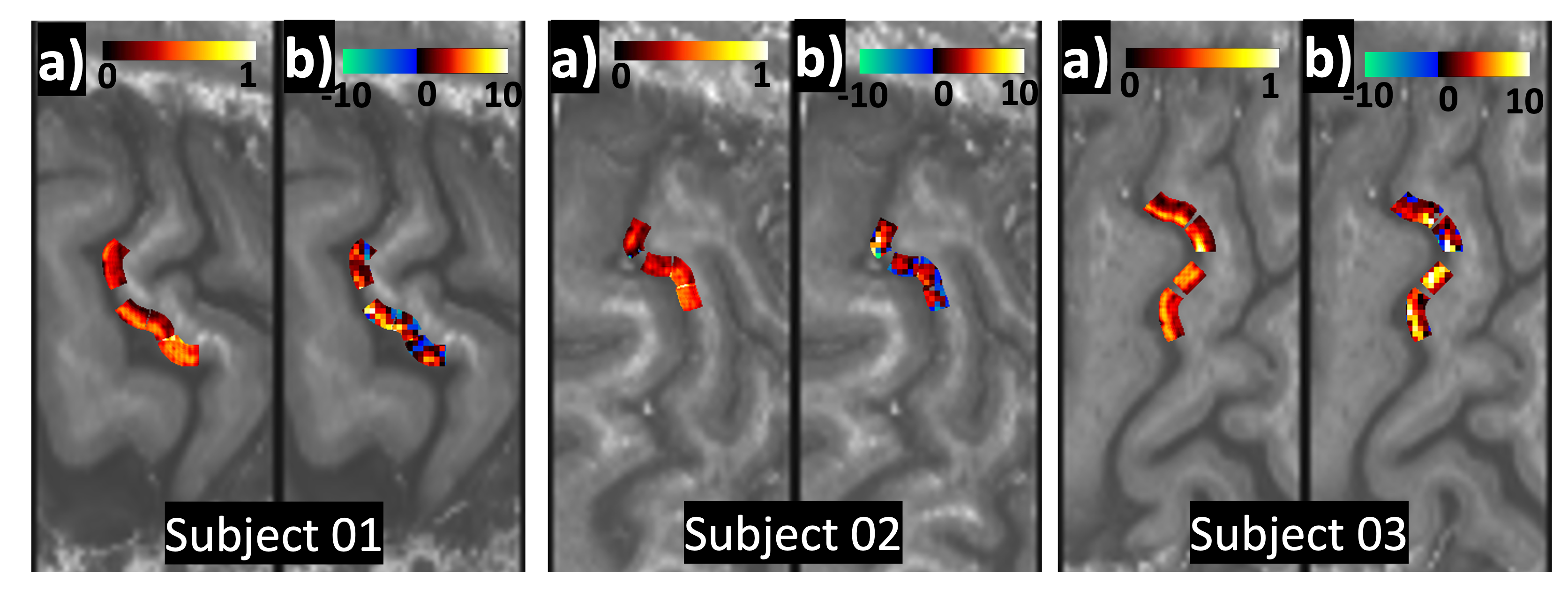
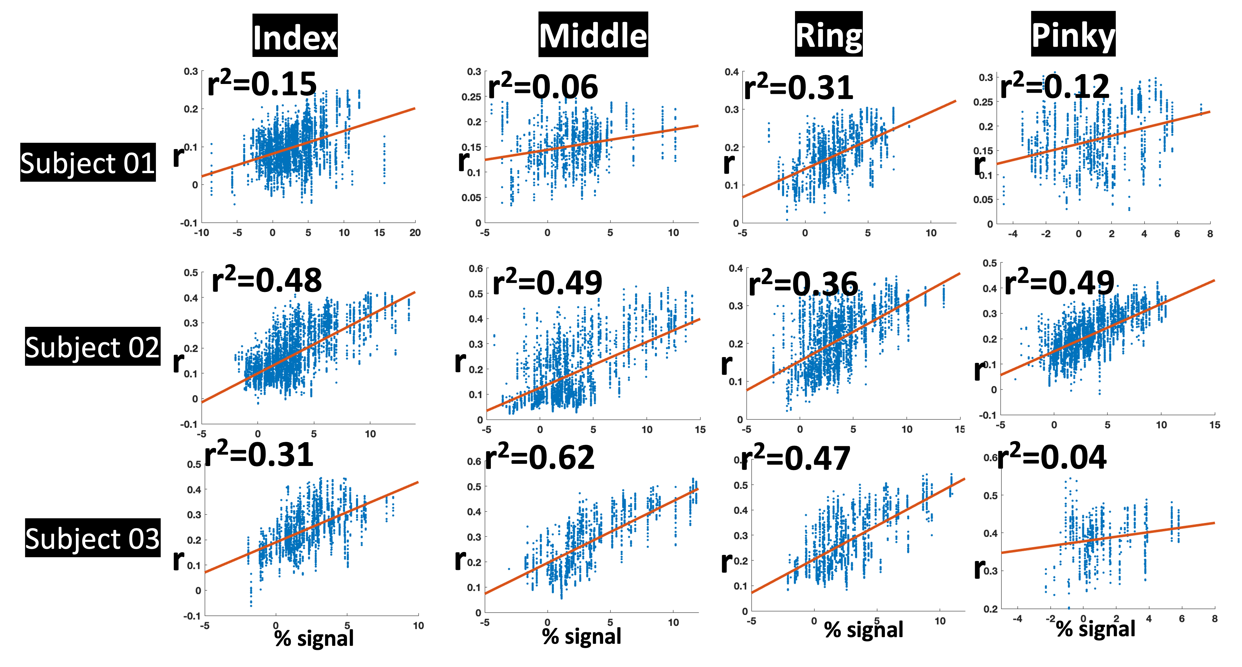
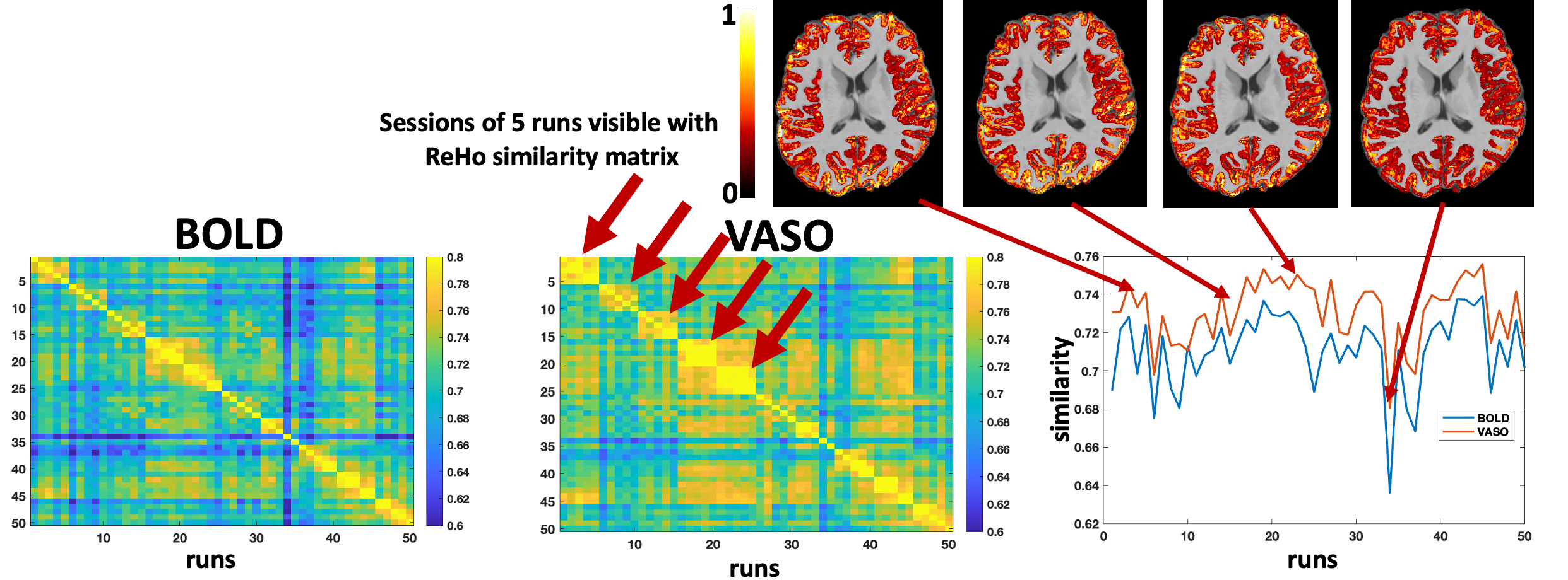
We’ve compared ReHo values calculated in somatomotor cortex with the activation pattern of fingers(index, middle, ring and pinky) stimulated vibrotactile setup described in the study(Yu et al. 2019) The percent activity change in finger areas show very high spatial resemblance with ReHo maps. See Fig2 & 3. We also calculate ReHo maps with publicly available whole brain dataset (https://openneuro.org/datasets/ds003216/versions/3.0.11) which consist of 50 individual runs of movie watching acquired in 10 different sessions, initial results have shown that, ReHo maps are highly similar across the runs. See Fig4. similar ReHo patterns across runs and days indicating that ReHo maps are picking up movie watching activity which are expected to be somewhat similar across runs.Acknowledgements
No acknowledgement found.References
- Akbari, Atena, Saskia Bollmann, Tonima S. Ali, and Markus Barth. 2021. “Modelling the Depth-Dependent VASO and BOLD Responses in Human Primary Visual Cortex.” https://doi.org/10.1101/2021.05.07.443052.
- Cho, Shinho, Arani Roy, Chao J. Liu, Djaudat Idiyatullin, Wei Zhu, Yi Zhang, Xiao-Hong Zhu, et al. 2022. “Cortical Layer-Specific Differences in Stimulus Selectivity Revealed with High-Field fMRI and Single-Vessel Resolution Optical Imaging of the Primary Visual Cortex.” NeuroImage 251 (May): 118978. https://doi.org/10.1016/j.neuroimage.2022.118978.
- Huber, Laurentius, Emily S. Finn, Yuhui Chai, Rainer Goebel, Rüdiger Stirnberg, Tony Stöcker, Sean Marrett, et al. 2021. “Layer-Dependent Functional Connectivity Methods.” Progress in Neurobiology 207 (101835). https://doi.org/10.1016/j.pneurobio.2020.101835.
- Huber, Laurentius, Dimo Ivanov, Daniel A. Handwerker, Sean Marrett, Maria Guidi, Kamil Uludag, Peter A. Bandettini, and Benedikt A. Poser. 2018. “Techniques for Blood Volume fMRI with VASO: From Low-Resolution Mapping towards Sub-Millimeter Layer-Dependent Applications.” NEUROIMAGE. 525 B ST, STE 1900, SAN DIEGO, CA 92101-4495 USA: ACADEMIC PRESS INC ELSEVIER SCIENCE. https://doi.org/10.1016/j.neuroimage.2016.11.039.
- Huber, Laurentius, Benedikt A. Poser, Peter A. Bandettini, Kabir Arora, Konrad Wagstyl, Shinho Cho, Jozien Goense, et al. 2021. “LayNii: A Software Suite for Layer-FMRI.” NeuroImage 237 (118091). https://doi.org/10.1016/j.neuroimage.2021.118091.
- Jiang, Lili, and Xi-Nian Zuo. 2016. “Regional Homogeneity.” The Neuroscientist 22 (5): 486–505. https://doi.org/10.1177/1073858415595004.
- Koiso, Kenshu, Anna K. Müller, Kazuaki Akamatsu, Sebastian Dresbach, Christopher J. Wiggins, Omer Faruk Gulban, Rainer Goebel, Yoichi Miyawaki, Benedikt A. Poser, and Laurentius Huber. 2023. “Acquisition and Processing Methods of Whole-Brain Layer-fMRI VASO and BOLD: The Kenshu Dataset.” Aperture Neuro 3 (September): 1–22. https://doi.org/10.52294/001c.87961.
- Mourik, Peter J.Koopmans Tim, Lauren J. Bains, David G. Norris, and Janneke F.M. Jehee. 2021. “Investigation of Layer Specific BOLD in the Human Visual Cortex during Visual Attention.” https://doi.org/10.1101/2021.02.07.430129.
- Müller, Anna Katharina, Miriam Heynckes, J. Wiggins, Gulban Christopher, Omer Faruk, Yuhui Chai, Benedikt A. Poser, and Laurentius Huber. 2021. “Whole Brain Layer-FMRI: An Open Dataset for Methods Benchmarking.”
- Pais-Roldán, Patricia, Seong Dae Yun, Nicola Palomero-Gallagher, and N.Jon Shah. 2020. “Cortical Depth-Dependent Human FMRI of Resting-State Networks Using EPIK.” https://doi.org/10.1101/2020.12.07.414144.
- Yang, Jiajia, Laurentius Huber, Yinghua Yu, and Peter A. Bandettini. 2021. “Linking Cortical Circuit Models to Human Cognition with Laminar FMRI.” Neuroscience & Biobehavioral Reviews 128: 467–78. https://doi.org/10.1016/j.neubiorev.2021.07.005.
- Yu, Yinghua, Laurentius Huber, Jiajia Yang, Masaki Fukunaga, Yuhui Chai, David C. Jangraw, Gang Chen, et al. 2022. “Layer-Specific Activation in Human Primary Somatosensory Cortex during Tactile Temporal Prediction Error Processing.” NeuroImage 248 (March): 118867. https://doi.org/10.1016/j.neuroimage.2021.118867.
- Yu, Yinghua, Laurentius Huber, Jiajia Yang, David C. Jangraw, Daniel A. Handwerker, Peter J. Molfese, Gang Chen, Yoshimichi Ejima, Jinglong Wu, and Peter A. Bandettini. 2019. “Layer-Specific Activation of Sensory Input and Predictive Feedback in the Human Primary Somatosensory Cortex.” SCIENCE ADVANCES. 1200 NEW YORK AVE, NW, WASHINGTON, DC 20005 USA: AMER ASSOC ADVANCEMENT SCIENCE. https://doi.org/10.1126/sciadv.aav9053.
Figures