3118
Cloud Computing Service Enabler for MR Workflows Based on Fluwiz1Biocliq Technologies private limited, Bangalore, India, 2GE HealthCare, Bangalore, India
Synopsis
Keywords: Software Tools, Data Processing, SaaS, PaaS, Cloud Computing, Cloud DICOM viewer
Motivation: Enable computationally expensive processing and dicom image review in cloud with low-cost solution amicable to low and unstable internet connectivity.
Goal(s): Utilize Cloud computing(SaaS) and cloud platform service(PaaS) to transfer data in secure and HIPPA compliant manner.
Approach: Fluwiz[5] enables HIPAA compliant data transfer to Cloud where MR workflows, reconstruction and post processing algorithms can be implemented. Dicom images are viewed on a cloud based DICOM viewer (Orthanc) or pushed back to the local PACS.
Results: The data was pushed to cloud using Rasberry PI 3 edge gateway device and image is viewed using Orthanc’s web based image viewer StoneViewer[6].
Impact: Low cost solution to transfer the raw MR data to the cloud in secure and HIPPA compliant manner would help latest advancements in MR imaging to remote parts of the world where internet bandwidth itself can be challenging
INTRODUCTION
AI has been increasing used in all aspects of MR imaging including prescription, reconstruction, and post-processing[1]. This has placed an increased burden on the limited compute resource availability on the MR Scanner. Recent surge in AI algorithms for data acquisition acceleration and image quality enhancement has propelled the need for additional compute units. Some clinical research groups[2-4] have developed dedicated cloud or local compute clusters and software platform to support such recon intensive applications but successful integration to the scanner environment within a secure hospital network has been a challenge. In addition to security, the bandwidth required for such transmission is also a challenge. In this work, we present a secure and scalable solution using Fluwiz[5] that enables transfer of Health Insurance Portability and Accountability Act (HIPAA) compliant data to Cloud where MR workflows, reconstruction and post processing algorithms can be implemented, and results viewed on a cloud based DICOM viewer or pushed back to the local PACS. Here, cloud computing service was used as Software as a service (SaaS) and Platform as a service (PaaS).METHODS
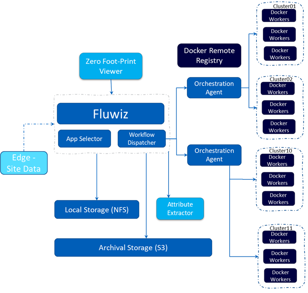
Low-cost gateway device: Small footprint low-cost edge device like a Raspberry PI3 that can either act as a gateway device for reliable data transfer or act as an additional node for compute.Transfer of HIPAA compliant data: Server/client framework powered by Fluwiz platform was used to transfer data from the MR scanner to local or a public cloud based on the organization’s security needs (Figure 1). Such data includes raw data, machine logs and DICOM images from the scanner. Given the sensitive nature of such data, we conform to HIPAA standards and remove patient sensitive information from the data streams before data transfer. Data is compresses by zipping DICOM images, raw data, and uses REST API services for data management of all relevant data streams.
Data Processing: The server platform carries out several commands to co-ordinate execution of different applications on the transferred data such as image reconstruction and image-enhancment. Dedicated orchestration agents (Figure 1) handle running multiple applications simultaneously and co-ordinate parallel resource requests. Output DICOM images and other data is stored locally on the cloud.
Image Viewing/Storage: The platform architecture is integrated to standard DICOM communication protocols and QIDO/WADO web services. This helps install images back to the PACS or to a secure Cloud based image viewer.
RESULTS
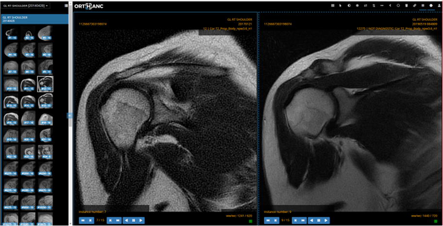
Figure 2 shows the original and denoised and super-resolved MR images of shoulder acquired on a commercial 1.5T MRI scanner (Signa HDxt, GE HealthCare, Waukesha, WI, USA). The data was pushed to cloud using Rasberry PI 3 edge gateway device and image is viewed using Orthanc’s web based image viewer StoneViewer[6].DISCUSSION AND CONCLUSION
A framework powered by Fluwiz utilizing low-cost edge gateway, Raspberry PI3, and cloud computing service as SaaS and PaaS was presented and shown to transfer DICOM images, raw data and system logs to the AWS cloud in compressed zip format in an HIPPA compliant manner. The reconstructed and post processed DICOM image on the cloud were then viewed using a web-based cloud viewer. The proposed solution could enable widespread use of AI without the need for installing additional compute units at the scanner/local institution thereby enabling shorter MR scans with high quality diagnostic clinical MR images.Acknowledgements
No acknowledgement found.References
[1] Hosny, Ahmed et al. “Artificial intelligence in radiology.” Nature reviews. Cancer vol. 18,8 (2018): 500-510. doi:10.1038/s41568-018-0016-5
[2] Hansen MS and Sorensen TS, "Gadgetron: An open source framework for medical image reconstruction", MRM,2013, 69(6), 1768-1776.
[3]Yarra Framework – A toolbox for clinical MRI research https://yarra-framework.org.
[4] Uecker, M.,et.al. Berkeley ad vanced reconstruction toolbox. ISMRM 2015:2486. https://mrirecon.github.io/bart/
[5]Fluwiz : https://www.fluwiz.com/
[6] Orthanc Inc. https://www.orthanc-server.com/
Figures