3109
Robust generation of tract-wise myelination measurements from infant T1- and T2-weighted MRI using synth based deep learning methods1Centre for Medical Image Computing, University College London, London, United Kingdom, 2Department of Paediatrics and Child Health, University of Cape Town, Cape Town, South Africa, 3Department of Neuroimaging, King's College London, London, United Kingdom, 4Department of Informatics, University of Sussex, Brighton, United Kingdom, 5Radiology, MGH & Harvard Medical School, Charlestown, MA, United States, 6Martinos Center for Biomedical Imaging, MGH & Harvard Medical School, Boston, MA, United States, 7CUBRIC, School of Psychology, Cardiff University, Cardiff, United Kingdom
Synopsis
Keywords: Data Processing, Data Processing, Infant myelination
Motivation: While T1/T2-weighted ratio maps are important for the study of myelination, existing reconstruction tools can fail in infants and present difficulty to automated segmentation.
Goal(s): To provide a pipeline for obtaining accurate regional myelination measures of whole brain regions and white matter tracts.
Approach: We adapted existing T1/T2-weighted ratio pipelines to incorporate deep learning methods for segmentation and registration as well as a high-quality tract atlas.
Results: Our pipeline showed reduced errors and improved differentiation between 3- and 6-month-old infants from a South African longitudinal birth cohort study.
Impact: The improved T1/T2-weighted ratio contrast and detailed segmentations provided by our pipeline will enable study of specific myelination patterns during neurodevelopment, especially in populations exposed to risk factors for altered white matter maturation.
Introduction
Accurate tract-wise measurement of T1/T2-weighted ratio maps in infants can provide valuable insight into neurodevelopment using the sensitivity of T1- and T2-weighted (T1w & T2w) scans to myelin content1. Myelination begins early in the development of the central nervous system, insulating axonal connections. This happens non-uniformly across white matter regions, proceeding rapidly over the first eight postnatal months and more gradually thereafter2. Disruptions in myelination are associated with neurological disorders, and are seen in relation to pre-term birth, malnutrition, and adverse experiential factors3.Regional T1/T2-weighted ratio measurements are hampered by difficulties in both map generation and segmentation with off-the-shelf tools. Time-varying non-uniform contrast changes, higher variability in head pose, and smaller global intracranial volume, all increase difficulty for registration and segmentation in developmental populations. Meanwhile, ratio generation pipelines developed for study of demyelinating disease in adults4 involve: 1) alignment of T1w and T2w images to an adult template, 2) non-linear registration between contrasts, 3) harmonisation of contrasts, 4) ratio calculation. Hence applications of such pipelines either run into issues with infant-to-adult registration, or substitute age-based templates5 increasing difficulty for automated segmentation.
Here we adapt existing pipelines to generate T1/T2-weighted ratio maps for infants. To do this we use emerging synth based deep learning methods, trained on synthetic contrasts to substantially increase robustness and generalisability of registration6,7 and segmentation8. We then refine segmentations with cerebral white-matter tract labels, enabling tract-wise measurements of myelination. We demonstrate this pipeline on a South African longitudinal birth cohort study with a high prevalence of infections, prenatal substance use and maternal stress.
Methods
1. Input data: We require T1w and T2w images with accompanying 2D ROI labels in the eyes and facial muscles.2. Alignment to template: We perform an alignment of the T1w image to the MNI space template using FreeSurfer’s EasyReg7 algorithm based on the centroids of cortical parcellations.
3. Non-linear registration: We register the T2w image to the MNI-aligned T1w using EasyReg7. Here, EasyReg applies a non-linear deformation field generated by a U-net pretrained for contrast agnostic registration.
4. Harmonisation and ratio map: We apply a model-based bias field correction method and normalise by linearly rescaling intensities to match eye and muscle measurements with MNI space template values. We then divide the T1w by the T2w to produce a ratio Image.
5. Segmentation: We apply synthseg8 to obtain whole brain labels. In the absence of diffusion MRI for tractography, we parcellate the white matter by registering whole brain segmentations of 16 training subjects from TRACULA9 and transfer the manually reviewed streamlines to our synthseg segmentations.
Results
We applied an existing SPM ratio generation toolbox4 as well as our ratio generation and segmentation methods to scans of 69 infants from the Khula study (28 at age 3 months, 37 at age 6 months, and 4 at both 3 and 6 months). While the SPM toolkit failed to reconstruct one subject and showed variable registration and segmentation errors in others, our method consistently oriented the scans and segmented all regions successfully as demonstrated in Figure 1.Figure 2 shows the mean T1/T2-weighted ratio measurement for each method across all whole brain segmentation labels. Our method shows greater contrast than SPM, both between ROI measurements and between age groups within target ROIs. For example, in discrimination between age groups thresholding of the white matter obtained an area under the ROC curve (AUC) of 97.64% with our method compared to 91.39% with SPM.
Figure 3 shows the mean measurements for each tract from our method. Each tract shows an individual pattern of myelin increase, corresponding to non-uniform myelination of white matter in infants. Combining these measures using linear discriminant analysis for discrimination between age groups we see an increased AUC of 99.39% highlighting possible utility of such measures.
Discussion and Conclusion
This work introduces a new pipeline to construct T1/T2-weighted ratio maps aligned to adult templates, allowing segmentation by existing tools and the detailed study of myelination in white-matter regions corresponding to expected tracts. An area for possible future work is removing the requirement of manual ROIs using methods similar to synthSR10, which is trained to take any adult MRI contrast and generate an equivalent, harmonised, T1w MPRAGE. Training tools such as synthSR and synthSeg using infant specific modelling could improve segmentation, registration, and harmonisation of scans in such a pipeline.The current work produces more accurate segmentation and sharper myelination maps than off the shelf tools. This allows us to provide tract wise myelination measurements in a unique dataset from a comparatively underreported population with substantial exposure risks affecting white matter maturation.
Acknowledgements
We would like to thank the mothers, families, and infants who made this work possible. We would also like to thank members of the Khula South Africa recruitment team (R. Gulwa and P. Madikane) and data collection team (M. Miles, D. Herr, L. Davel, R. Samuels, S. Williams, C. A. Jacobs, N. Mlandu, T. Pan, Z. Madi, T. Mhlakwaphalwa, B. Methola, C. Knipe, and K. Nkubungu) who contributed to authorship of this work. This research is supported by funding from the Wellcome Leap 1kD Program and the Bill & Melinda Gates Foundation. Wellcome Trust grant 221915 and the NIHR UCLH Biomedical Research Centre support DCA’s work on this topicReferences
1. Glasser, M.F. and Van Essen, D.C., 2011. Mapping human cortical areas in vivo based on myelin content as revealed by T1-and T2-weighted MRI. Journal of neuroscience, 31(32), pp.11597-11616. https://doi.org/10.1523/JNEUROSCI.2180-11.2011
2. Kinney, H.C., Brody, B.A., Kloman, A.S. and Gilles, F.H., 1988. Sequence of central nervous system myelination in human infancy: II. Patterns of myelination in autopsied infants. Journal of Neuropathology & Experimental Neurology, 47(3), pp.217-234. https://doi.org/10.1097/00005072-198805000-00003
3. Kinney, H.C. and Volpe, J.J., 2018. Myelination events. In Volpe's neurology of the newborn (pp. 176-188). Elsevier. https://doi.org/10.1016/B978-0-323-42876-7.00008-9
4. Ganzetti, M., Wenderoth, N. and Mantini, D., 2014. Whole brain myelin mapping using T1-and T2-weighted MR imaging data. Frontiers in human neuroscience, 8, p.671. https://doi.org/10.3389/fnhum.2014.00671
5. Soun, J.E., Liu, M.Z., Cauley, K.A. and Grinband, J., 2017. Evaluation of neonatal brain myelination using the T1‐and T2‐weighted MRI ratio. Journal of Magnetic Resonance Imaging, 46(3), pp.690-696. https://doi.org/10.1002/jmri.25570
6. Hoffmann, M., Billot, B., Greve, D.N., Iglesias, J.E., Fischl, B. and Dalca, A.V., 2021. SynthMorph: learning contrast-invariant registration without acquired images. IEEE transactions on medical imaging, 41(3), pp.543-558. https://doi.org/10.1109/TMI.2021.3116879
7. Iglesias, J.E., 2023. A ready-to-use machine learning tool for symmetric multi-modality registration of brain MRI. Scientific Reports, 13(1), p.6657. https://doi.org/10.1038/s41598-023-33781-0
8. Billot, B., Greve, D.N., Puonti, O., Thielscher, A., Van Leemput, K., Fischl, B., Dalca, A.V. and Iglesias, J.E., 2023. SynthSeg: Segmentation of brain MRI scans of any contrast and resolution without retraining. Medical image analysis, 86, p.102789. https://doi.org/10.1016/j.media.2023.102789
9. Maffei, C., Lee, C., Planich, M., Ramprasad, M., Ravi, N., Trainor, D., Urban, Z., Kim, M., Jones, R.J., Henin, A. and Hofmann, S.G., 2021. Using diffusion MRI data acquired with ultra-high gradient strength to improve tractography in routine-quality data. Neuroimage, 245, p.118706. https://doi.org/10.1016/j.neuroimage.2021.118706
10. Iglesias, J.E., Billot, B., Balbastre, Y., Tabari, A., Conklin, J., González, R.G., Alexander, D.C., Golland, P., Edlow, B.L., Fischl, B. and Alzheimer’s Disease Neuroimaging Initiative, 2021. Joint super-resolution and synthesis of 1 mm isotropic MP-RAGE volumes from clinical MRI exams with scans of different orientation, resolution and contrast. Neuroimage, 237, p.118206. https://doi.org/10.1016/j.neuroimage.2021.118206
Figures
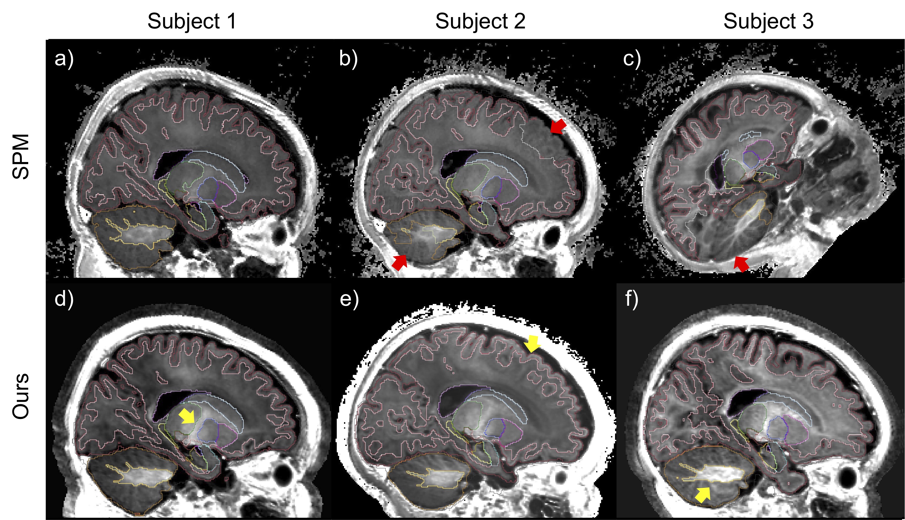
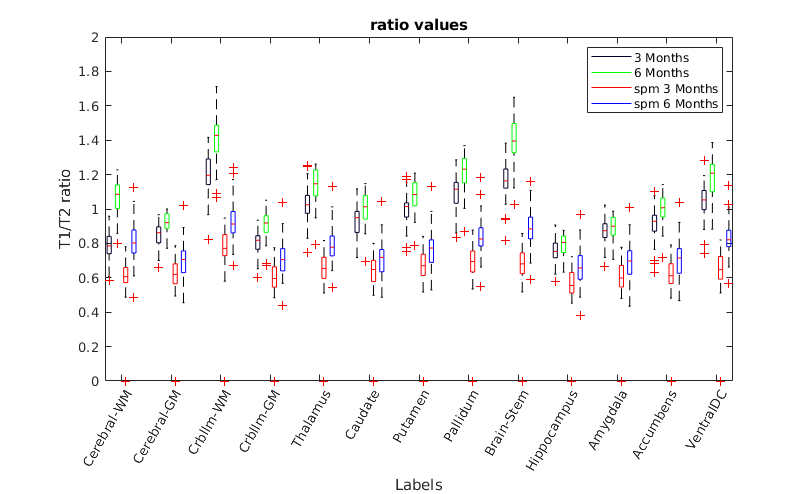
Figure 2. Comparison of regional T1/T2 ratio measurements between 3- and 6-month-old subjects, obtained using our proposed pipeline and the existing SPM toolkit. Our method improves the contrast between different ROIs and provides better separation between 3- and 6-month-old subjects.
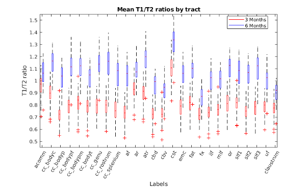
Figure 3. Comparison of tract-wise mean T1/T2 ratio values between age groups. Values shown are generated using our reconstruction and segmentation pipeline and are averaged across hemispheres. It is these white matter regions that most directly reveal myelination in the first year of life making such measures important to the understanding of neurodevelopment.