3106
Machine Learning-Based Classification of IDH Mutant Gliomas Using VASARI and Radiomics Features1Radiology, Yale School of Medicine, New Haven, CT, United States, 2Radiology, University of Leipzig, Leipzig, Germany, 3Neuroradiology, The Ohio State University School of Medicine, Columbus, OH, United States
Synopsis
Keywords: Analysis/Processing, Cancer, VASARI
Motivation: Diagnosis of molecular subtypes of IDH-mutant gliomas on MRI has presented a challenge in clinical practice.
Goal(s): To classify IDH-mutant gliomas we compared different ML models using qualitative and quantitative features from preoperative MRI.
Approach: Three models were compared, using only qualitative VASARI features as scored by two blinded neuroradiologists, the other used quantitative features from FLAIR and T1Gd and finally combining both in a third model.
Results: The VASARI feature based model showed moderate diagnostic accuracies for different tumor entities, which was higher than the Radiomics only model. Combining both features improved results, emphasizing the importance of feature selection in clinical applications.
Impact: This study demonstrates the potential of machine learning models in enhancing the accuracy of IDH-mutant glioma classification on preoperative MRI images.
BACKGROUND OR PURPOSE
Accurate diagnosis and classification of IDH mutant glioma subtypes are crucial for guiding treatment decisions and improving patient outcomes [1-3]. In this study, we investigated the performance of various machine learning models in classifying these tumor entities based on two distinct feature sets: VASARI features and Radiomics features derived from FLAIR and T1Gd MRI images. Additionally, we explored the impact of combining these feature sets on classification accuracy. The purpose of this study was to assess the performance of various machine learning models in classifying brain tumor entities using two distinct feature sets: the VASARI (Visually Accessible Rembrandt Images) feature set and Radiomics features derived from FLAIR and T1Gd MRI images. In a third step, we investigated the impact of combining these feature sets on classification accuracy.METHODS
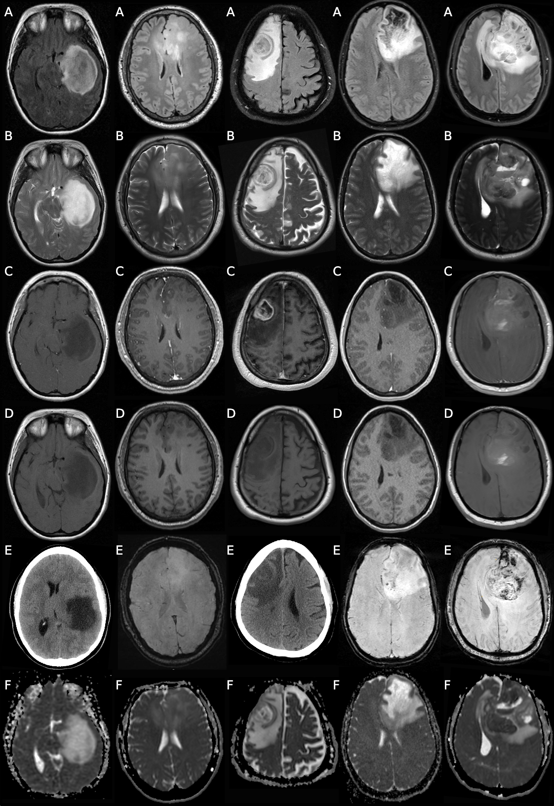
In this retrospective study, a total of 168 patients who received surgical and/or radiotherapeutic treatment for primary brain tumors at the study institution between January 2000 and December 2023 were reviewed. The inclusion criteria for this study were as follows: (1) Patients with IDH-mutant astrocytomas of grade 2 to 4 and IDH-mutant, 1p/19q co-deleted oligodendrogliomas of grade 2 to 3, as confirmed by histopathology according to the 2021 WHO CNS5 classification criteria; (2) Inclusion required the availability of pretreatment MRI scans, which included conventional MRI sequences. For the assessment of the VASARI features a board-certified neuroradiologist with 4 years of experience conducted independent assessments of preoperative imaging to diagnose glioma subtype. The included sequences were FLAIR, T1-weighted images after contrast enhancement. The radiologist conducting the assessments was blinded to the clinical data and the diagnosis of each case. For the classification tasks, we selected various machine learning models to compare performance: XGBoost, Logistic Regression, Random Forest, SVM, K-Nearest Neighbors, Decision Tree, and Neural Network. We evaluated the accuracy and area under the curve (AUC) of each model in predicting the IDH-mutant glioma tumor subtype, codeletion status and T2/FLAIR mismatch sign.RESULTS
Our findings reveal that diagnostic accuracy, as assessed by two readers (F.M., B.S.), varies across different tumor entities: 37.6% of grade 2 astrocytomas, 20% of grade 3 astrocytomas, 37.1% of grade 4 astrocytomas, 41.3% of grade 2 oligodendrogliomas, and 32% of grade 3 oligodendrogliomas were correctly diagnosed. When applying a classification model on the set of 25 VASARI features, Random Forest demonstrated strong predictive power, achieving the highest accuracy in predicting codeletion status (91.18%). The XGBoost model demonstrated very good discriminative ability amongst classes, achieving AUC values ranging from .86 to .91 across the different subtypes. Notably, Random Forest also exhibited strong performance with AUC values between .80 and .92. On the other hand, classification based on radiomics features exhibited lower accuracy in predicting the tumor subtypes. As an example XGBoost AUCs ranged .38 to .58. The best performing model was the Logistic Regression model with AUC of .88 for astrocytoma, grade 4. Combining VASARI and Radiomics features did not improve with AUC of .57 for Random Forest and evidence of overtraining for XGBoost.CONCLUSIONS
We demonstrate that machine learning models can be used as an adjunct for classifying molecular subtype of IDH-mutant gliomas. Due to significant imaging feature similarity amng these tumors differentiation of imaging alone is difficult. Further analysis and expanded datasets may refine our understanding and inform more precise model selection for specific diagnostic tasks and address the potential issue of class imbalance.Acknowledgements
No acknowledgement found.References
- van den Bent MJ, Chang SM. Grade II and III Oligodendroglioma and Astrocytoma. Neurol Clin. 2018;36(3):467-484. doi:10.1016/j.ncl.2018.04.005
- White ML, Zhang Y, Kirby P, Ryken TC. Can Tumor Contrast Enhancement Be Used as a Criterion for Differentiating Tumor Grades of Oligodendrogliomas? AJNR Am J Neuroradiol. 2005;26(4):784-790.
- Juratli TA, Tummala SS, Riedl A, et al. Radiographic assessment of contrast enhancement and T2/FLAIR mismatch sign in lower grade gliomas: correlation with molecular groups. J Neurooncol. 2019;141(2):327-335. doi:10.1007/s11060-018-03034-6
Figures