3105
Subtype classification of Functional Pituitary Adenomas based on MRI Radiomics.1Department of Biomedical Imaging and Radiological Sciences, National Yang Ming Chiao Tung University, Taipei, Taiwan, 2Department of Radiology, Taipei Veterans General Hospital, Taipei, Taiwan
Synopsis
Keywords: Radiomics, Radiomics
Motivation: Pituitary adenomas (PAs) are a rare but clinically diverse group of tumors with varying hormone secretion profiles and clinical characteristics, comprising 15% of intracranial tumors. Typical classification of PAs relies on blood hormone levels as gold standard test, with a limited exploration into assessing hormone status using neuroimaging biomarkers.
Goal(s): We aim to offer a practical MRI-based classification model, improving clinical PA management.
Approach: Our study developed a machine learning model using MRI radiomics as image biomarkers for the classification of PAs focusing on six subtypes.
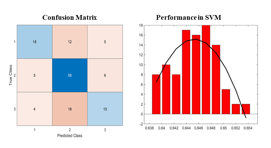
Results: Our SVM model showed an accuracy of 0.65 based on MRI images.
Impact: Our radiomics classification model promises to revolutionize MRI PA classification and diagnosis, enhancing clinical management and benefiting scientists, clinicians, and patients by enabling more accurate and efficient diagnostics and treatments.
Background and Purpose
Pituitary adenomas (PAs) are tumors of the anterior lobe of the pituitary gland. The pituitary gland is responsible for helping to regulate the functions of other endocrine glands, such as the thyroid gland, adrenal glands, ovaries, and testes. Therefore, any disruption or growth in the pituitary, such as a tumor, can have a profound impact on a patient's overall well-being [1]. Although PAs are usually considered benign, their prevalence is currently increasing. PAs can be classified into various subtypes based on their cell type and hormone secretion patterns. Those that secrete hormones are known as functioning adenomas, and those that do not secrete hormones are known as nonfunctioning adenomas [2]. Functioning adenomas are further divided into different subtypes depending on the type of hormone they secrete. Each subtype has distinct clinical features and requires specialized treatment approaches tailored to the specific hormone abnormalities and tumor characteristics. Traditionally, the classification of PAs has relied on the expertise of skilled pathologists by basing it on the hormone level in the blood, which is subjective to human errors and is time-consuming, leading to misdiagnosis and delay in treatment [3]. The differentiation of hormone patterns using neuroimaging biomarkers was less explored. To address this problem, we aim to develop a PA subtypes classification model to precisely classify PA subtypes using a machine learning approach as image biomarkers.Materials and Methods
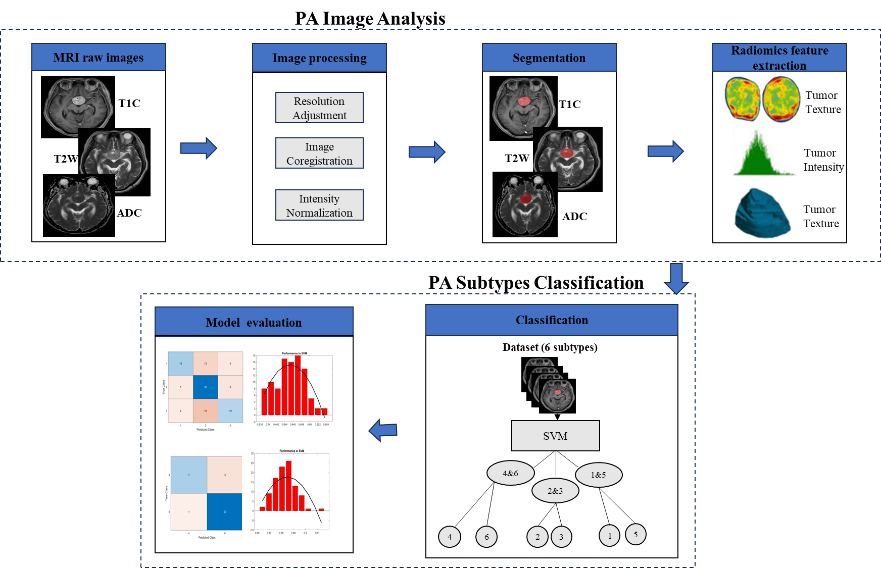
We retrospectively recruited 705 patients with PA and hormone pattern proved by immunohistology at Taipei Veterans General Hospital. MRI data include contrast-enhanced T1-weighted (CET1), T2-weighted images (T2W), and apparent diffusion coefficient (ADC) derived from diffusion-weighted images. Several postprocessing steps on the MR images were applied to reduce the discrepancy of imaging parameters using our previously published MRI radiomics platform (MRP, http://www.ym.edu.tw/~cflu/MRP_MLinglioma.html) with a graphic user interface built on MATLAB programming environment [4]. The adjustment of image resolution was first performed to resample all voxel size to isotropic spatial resolution (1.00 x 1.00 x 1.00 mm3) for each MRI contrast according to Image biomarker standardization initiative (IBSI). The PA lesions were automatically segmented (using 3D U-Net with an average dice coefficient of 0.88) followed by the extraction of 1178 radiomic features, including the gray-scale intensity patterns, lesion shape/size geometry, and texture features. One-way analysis of variance (One -way ANOVA) was performed to identify the significantly different features (p <0.05) between six types of hormone pattern in PA. Radiomics features were used to train support vector machine (SVM). In view of the inherent complexity, the simultaneous classification of all six subtypes is challenging. Consequently, we employed a pairwise merging approach, grouping the six subtypes into three categories and performing the classification. Subsequently, binary classification was executed to assign each instance to the appropriate subtype. The dataset was randomly separated into training dataset and testing dataset with a ratio of 8:2. To assess the reproducibility of this model, we tested modeling performance with bootstrap 100 times and attained accuracy. Figure 1 shows the overall workflow of the study.Results and Discussion
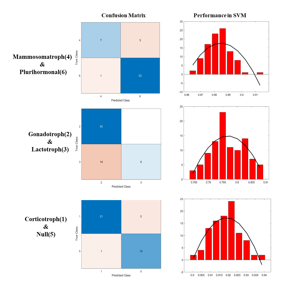
A total of 705 patients’ MRI data was used to identify radiomic features for classification. Our preliminary results for the merged subtypes of our SVM model are shown in Figure 2 with overall accuracy of 65% in the test group respectively. The performance of binary classification between the merged subtypes, (Mammosomatroph & Plurihormonal), (Gonadotroph & Lactotroph) and (Corticotroph & Null) showed an accuracy of 88%, 80% and 92% respectively as shown in Figure 3. By analyzing our confusion matrix, we observed that the model's performance exhibited notable disparities across different subtypes and there were a few instances where the model misclassified the subtypes or failed to classify them. This study is not only committed to the creation of precise classifiers but also includes a comprehensive evaluation framework to identify the most suitable model for practical clinical use. In this abstract, we present our initial models, and we are committed to ongoing refinement and optimization. Our future work will focus on enhancing the model's performance, exploring additional features, and fine-tuning hyperparameters to achieve higher accuracy and robustness.Conclusions
This study developed MRI radiomics based Machine learning model for PAs subtype classification. The SVM model showed an accuracy of 0.65 . The MRI radiomic features could be helpful to identify the PA subtypes and benefit diagnosis and clinical treatment.Acknowledgements
This work was supported by the Ministry of Science and Technology, Taiwan (MOST 109-2314-B-010-022-MY3).References
- Thapar, K., et al., Diagnosis and management of pituitary tumors. 2000: Springer Science & Business Media.
- Mete, O. and M.B. Lopes, Overview of the 2017 WHO classification of pituitary tumors. Endocrine Pathology, 2017. 28: p. 228-243.
- Dai, C., et al., How to classify and define pituitary tumors: recent advances and current controversies. Frontiers in endocrinology, 2021. 12: p. 604644.
- Lu, C.-F., et al., Machine learning–based radiomics for molecular subtyping of gliomas. Clinical Cancer Research, 2018. 24(18): p. 4429-4436.
Figures