3094
Differentiation of IDH-mutant Glioma Subtypes Using Unsupervised Dimensionality Reduction of MRI Biomarkers1Radiology, Yale School of Medicine, New Haven, CT, United States, 2Radiology, University of Leipzig, Leipzig, Germany, 3Biomedical Engineering, Yale University, New Haven, CT, United States, 4Pathology, Yale School of Medicine, New Haven, CT, United States, 5Therapeutic Radiology, Yale School of Medicine, New Haven, CT, United States
Synopsis
Keywords: Radiomics, Brain, Diagnosis/Prediction, Data Analysis
Motivation: Radiomic features can potentially help distinguish subtypes of IDH-mutant gliomas that appear similar on MRI.
Goal(s): The aim of this study was to evaluate whether imaging-based clustering of radiomic biomarkers of IDH-mutant gliomas may identify patterns or subgroups based on the 2021 CNS WHO classification.
Approach: Dimensionality reduction techniques were applied to radiomic features of 179 patients of different sequence combinations to analyze the high dimensional feature space.
Results: FLAIR and T1 post-contrast imaging revealed unique clusters, and survival analysis suggested potential differences amongst clusters. However, further research with a larger dataset is needed to determine whether the observed differences are significant.
Impact: This study analyzed quantitative imaging biomarkers to differentiate IDH-mutant gliomas according to the 2021 WHO classification. The findings suggest that radiomic features may hold insights into potential survival differences among subtypes. Larger-scale research is required to further investigate these findings.
INTRODUCTION
IDH-mutant glioma share many similarities on MRI and histopathology but have distinct differences in their survival based on their molecular subtypes [1-3]. Radiomic biomarkers provide insights into glioma imaging phenotypes that may not appear in clinical evaluation [4-6]. This study aimed to evaluate whether MRI-based biomarkers of IDH-mutant gliomas can differentiate 2021 CNS WHO classification molecular subtypes [7] and identify distinct patterns of overall survival [8-10].METHODS
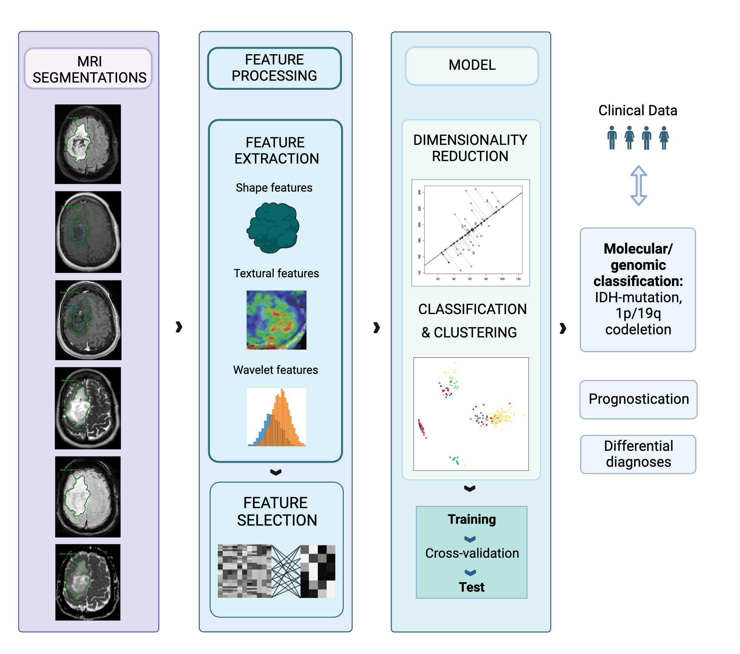
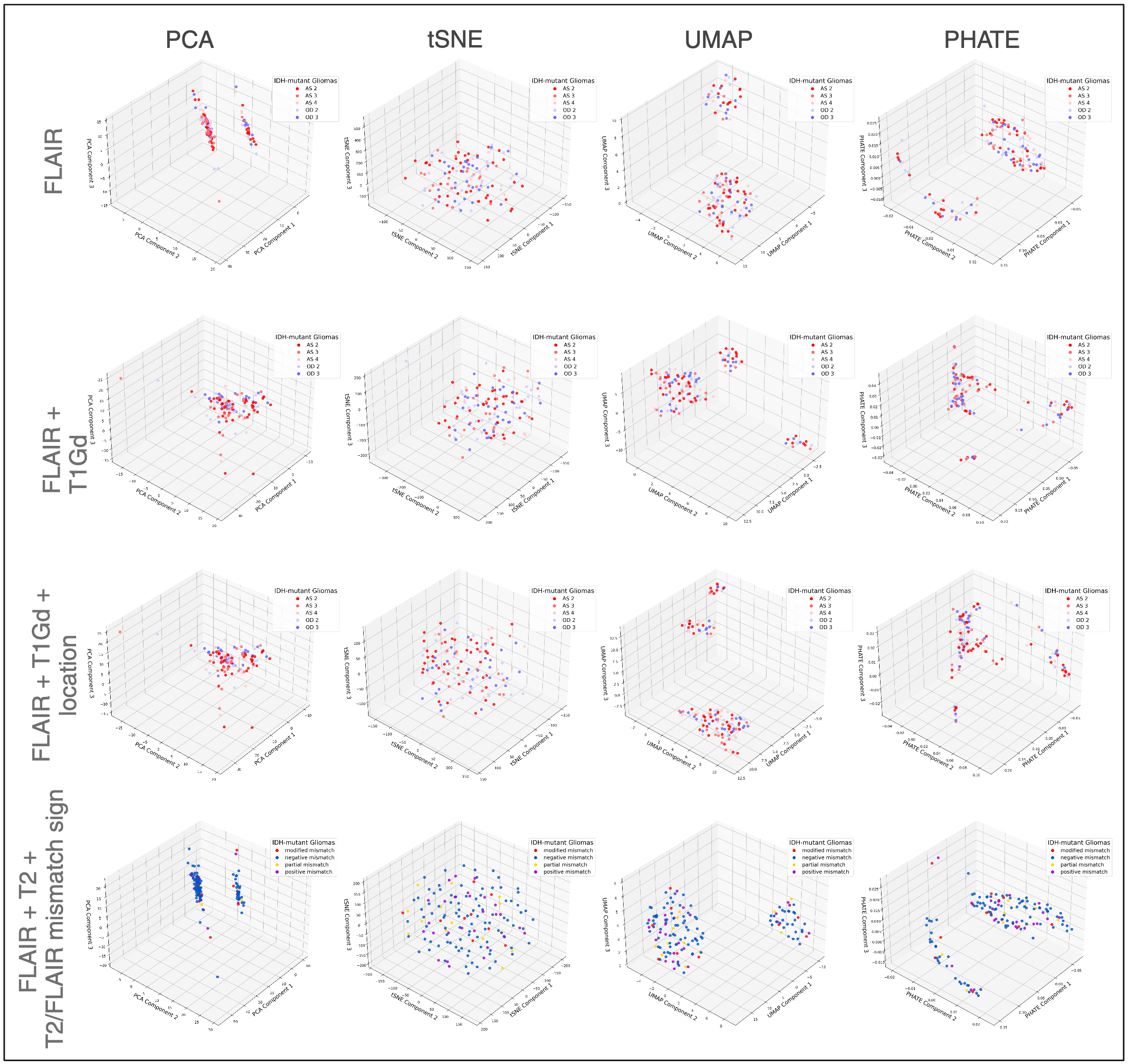
We included n=179 adult patients with IDH-mutant, 1p/19q co-deleted oligodendrogliomas (WHO CNS5 grade 2-3) and IDH-mutant, non co-deleted astrocytoma (WHO CNS5 grades 2-4) with pre-treatment MRI on FLAIR (n=179 cases), T1 post-contrast (n=129 cases) and T2 (n=144 cases). Segmentations were performed using a semiautomatic workflow with a UNETR algorithm trained on the BRaTS 2021 dataset, which were corrected by medical student research fellows (M.R., J.L., N.T.) or segmented manually and checked by a board-certified neuroradiologist (M.A.). MRI scans were independently resampled to a common voxel spacing (0.7x0.7x5mm) and their voxel intensity values were normalized. M=94 texture biomarkers were extracted from the segmentation region of each scan using the PyRadiomics 3.1.0 pipeline (s. Figure 1) [6]. Dimensionality reduction techniques (PCA, t-SNE, UMAP, and PHATE) were applied to the radiomic biomarkers of (i) FLAIR; (ii) FLAIR and T1 post-contrast imaging (PGGE/PGSE); (iii) incorporating one-hot encoded lesion location information; and (iv) FLAIR and T2 imaging. Subsequently, K-Means clustering was used, with k based on visually analyzing the dimensionality-reduced features to identify relevant clinical clusters in the reduced space.RESULTS
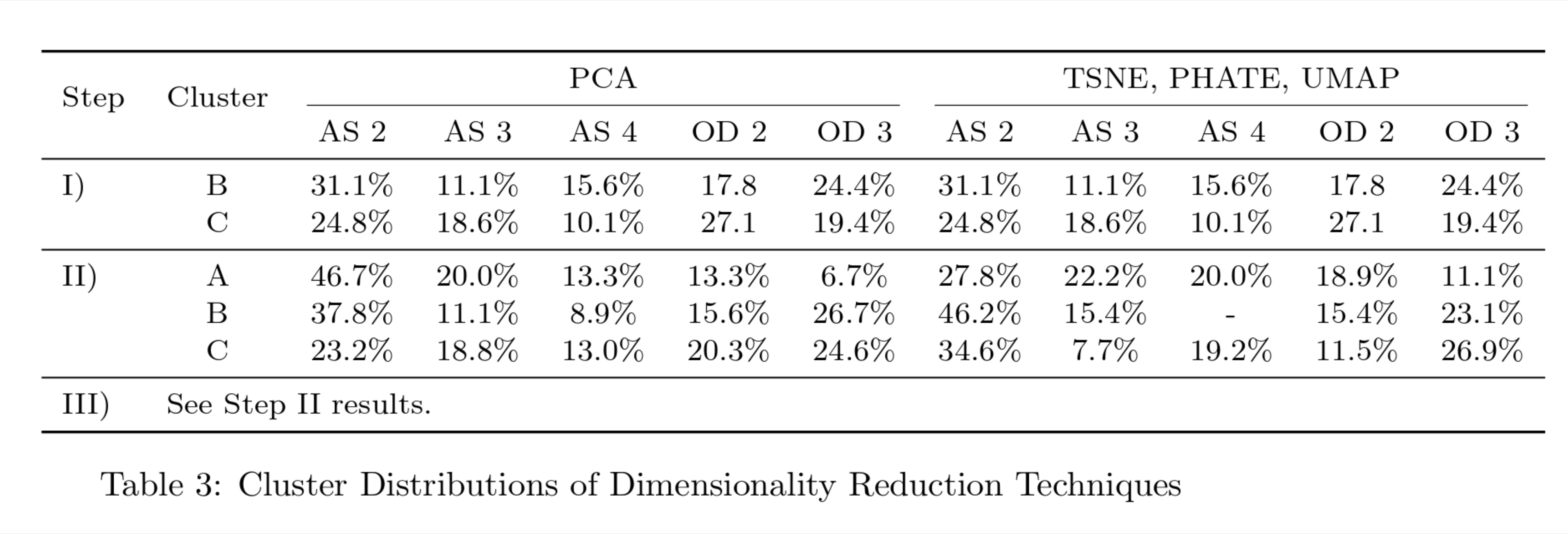
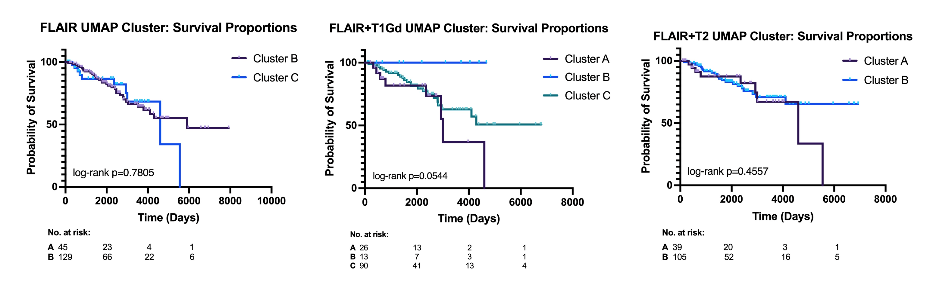
Figure 2 shows the distribution of subtypes across K-Means clusters. (i) For FLAIR-based biomarkers, all four dimensionality reduction techniques yielded the same distribution among clusters B and C, with the highest representation being 27.1% of oligodendroglioma, grade 2, 24.8% of astrocytoma, grade 2, and 19.4% of oligodendroglioma in one cluster, and 31.1% of astrocytoma, grade 2, 24.4% oligodendroglioma, grade 3, and 17.8% oligodendroglioma, grade 2 in the other cluster. (ii) When adding radiomic features from T1 post-contrast only PCA showed different distributions: Cluster A had 46.7% of astrocytoma, grade 2, whereas cluster B had 37.8% and cluster C 23.2% within the clusters. TSNE, PHATE, and UMAP presented 46.2% of astrocytomas, grade 2 in Cluster B, and 34.6% in Cluster C (s. Table 1). When adding T2 in (iv), the distribution of T2/FLAIR mismatch sign showed no significant differences: 20.9% positive mismatch in Cluster A, and 12.8% in Cluster B. Kaplan Meier survival analysis of the three clusters in UMAP using log-rank test (Mantel-Cox) indicated no significant differences in survival between clusters with p=0.781, p=0.054, p=0.455 for FLAIR, FLAIR + T1Gd, and FLAIR+T2 respectively (s. Figure 3).CONCLUSIONS
We show that there is a spectrum of imaging features of IDH-mutant gliomas that have significant overlap on FLAIR and T1 post-contrast imaging, but cluster into two groups on FLAIR and three clusters when adding T1WI post-gadolinium radiomic features. This suggests that MRI radiomic features provide additional information to molecular based classification of IDH-mutant gliomas and shows that imaging features do not directly correspond to molecular based classifications and may need to be used together. Additional analyses or a larger sample size may be needed to determine whether the observed differences in survival per cluster are significant.Acknowledgements
The author would like to acknowledge the generous support provided by the BMEP stipend (Biomedical Education Program).References
- References 1. van den Bent MJ, Chang SM. Grade II and III Oligodendroglioma and Astrocytoma. Neurol Clin. 2018;36(3):467-484. doi:10.1016/j.ncl.2018.04.005
- White ML, Zhang Y, Kirby P, Ryken TC. Can Tumor Contrast Enhancement Be Used as a Criterion for Differentiating Tumor Grades of Oligodendrogliomas? AJNR Am J Neuroradiol. 2005;26(4):784-790.
- Juratli TA, Tummala SS, Riedl A, et al. Radiographic assessment of contrast enhancement and T2/FLAIR mismatch sign in lower grade gliomas: correlation with molecular groups. J Neurooncol. 2019;141(2):327-335. doi:10.1007/s11060-018-03034-6
- Bera K, Braman N, Gupta A, Velcheti V, Madabhushi A. Predicting cancer outcomes with radiomics and artificial intelligence in radiology. Nature Reviews Clinical Oncology. Published online October 18, 2021:1-15. doi:https://doi.org/10.1038/s41571-021-00560-7
- Kumar, V., Gu, Y., Basu, S., Berglund, A., Eschrich, S. A., Schabath, M. B., Forster, K., Aerts, H. J., Dekker, A., Fenstermacher, D., Goldgof, D. B., Hall, L. O., Lambin, P., Balagurunathan, Y., Gatenby, R. A., & Gillies, R. J. (2012). Radiomics: the process and the challenges. Magnetic resonance imaging, 30(9), 1234–1248. https://doi.org/10.1016/j.mri.2012.06.010
- van Griethuysen, J. J., Fedorov, A., Parmar, C., Hosny, A., Aucoin, N., Narayan, V., Beets-Tan, R. G., Fillion-Robin, J.-C., Pieper, S., & Aerts, H. J. (2017). Computational Radiomics System to Decode the Radiographic Phenotype. Cancer Research, 77(21), e104–e107. https://doi.org/10.1158/0008-5472.CAN-17-0339
- Louis, D. N., Perry, A., Wesseling, P., Brat, D. J., Cree, I. A., Figarella-Branger, D., Hawkins, C., Ng, H. K., Pfister, S. M., Reifenberger, G., Soffietti, R., von Deimling, A., & Ellison, D. W. (2021). The 2021 WHO Classification of Tumors of the Central Nervous System: a summary. Neuro-oncology, 23(8), 1231–1251. https://doi.org/10.1093/neuonc/noab106
- Kocher, M., Ruge, M. I., Galldiks, N., & Lohmann, P. (2020). Applications of radiomics and machine learning for radiotherapy of malignant brain tumors. Strahlentherapie und Onkologie : Organ der Deutschen Rontgengesellschaft ... [et al], 196(10), 856–867. https://doi.org/10.1007/s00066-020-01626-8
- Li, Y., Ammari, S., Lawrance, L., Quillent, A., Assi, T., Lassau, N., & Chouzenoux, E. (2022). Radiomics-Based Method for Predicting the Glioma Subtype as Defined by Tumor Grade, IDH Mutation, and 1p/19q Codeletion. Cancers, 14(7), 1778. https://doi.org/10.3390/cancers14071778
- Pashmina Kandalgaonkar, Sahu, A., Ann Christy Saju, Joshi, A., Mahajan, A., Thakur, M., Sahay, A., Sridhar Epari, Sinha, S., Dasgupta, A., Chatterjee, A., Shetty, P., Aliasgar Moiyadi, Agarwal, J., Gupta, T., & Jayant Sastri Goda. (2022). Predicting IDH subtype of grade 4 astrocytoma and glioblastoma from tumor radiomic patterns extracted from multiparametric magnetic resonance images using a machine learning approach. Frontiers in Oncology, 12. https://doi.org/10.3389/fonc.2022.879376
Figures