3091
Enhanced Lateralization of Mesial Temporal Lobe Epilepsy through Vascular Territory Analysis: Insights from Arterial Spin Labeling MRI1Research Center for Molecular and Cellular Imaging, Tehran University of Medical Sciences, Tehran, Iran (Islamic Republic of), 2Biomedical Engineering and Medical Physics, Shahid Beheshti University of Medical Sciences, Tehran, Iran (Islamic Republic of), 3Bioinformatics and Biophysics, Tarbiat Modares University, Tehran, Iran (Islamic Republic of), 4Medical School, Tehran University of Medical Sciences, Tehran, Iran (Islamic Republic of), 5Pars Advanced and Minimally Invasive Medical Manners Research Center, Pars Hospital, Iran University of Medical Sciences, Tehran, Iran (Islamic Republic of), 6Isfahan Neuroscience Research Center, Isfahan University of Medical Sciences, Isfahan, Iran (Islamic Republic of)
Synopsis
Keywords: Epilepsy, Perfusion, ASL Perfusion MRI, Lateralization of TLE, Blood Teritorry
Motivation: To enhance lateralization of drug-resistant mesial temporal lobe epilepsy (mTLE) for improved surgery decisions. Using arterial spin labeling (ASL) MRI, this study explores vascular territories to differentiate left and right mTLE.
Goal(s): To assess ASL MRI's potential in distinguishing between mTLE types by studying cerebral blood flow changes.
Approach: It involves comparing mTLE groups to controls using ASL MRI and vascular territory analysis, identifying specific brain regions affecting lateralization.
Results: They highlight vascular territory changes crucial in distinguishing mTLE types, emphasizing the clinical significance of ASL MRI.
Impact: This research may significantly impact treatment decisions, benefitting individuals with challenging mTLE and guiding future epilepsy research.
Introduction:
Epilepsy is a prevalent neurological disorder affecting millions globally, with mesial temporal lobe epilepsy (mTLE) being one of the most common forms. For individuals with drug-resistant mTLE, epilepsy surgery becomes necessary, with a key consideration being the localization of the seizure focus in the left or right temporal lobe [1]. Recent advancements in neuroimaging, particularly arterial spin labeling (ASL) MRI, a noninvasive method for perfusion analysis, provide an opportunity to improve this critical aspect of epilepsy treatment [2-4]. ASL MRI, which measures blood flow, can be enhanced by utilizing a vascular territory atlas to aid in determining the lateralization of mTLE. This study seeks to explore the potential of vascular territory analysis with ASL MRI in distinguishing between left and right mTLE and its relevance in clinical practice.Methods:
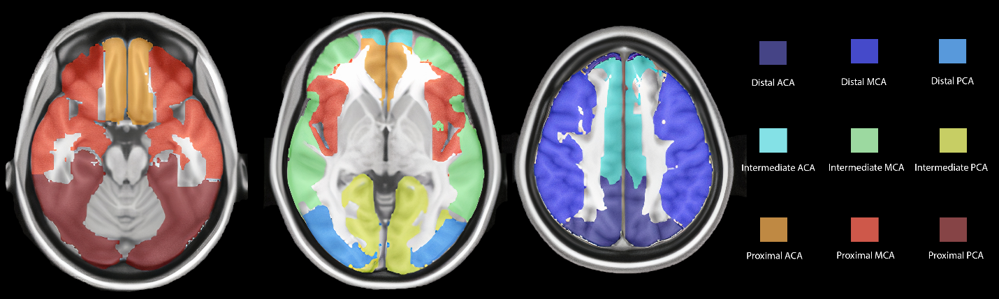
In this research, Pulsed ASL MRI data from 15 healthy volunteers and 23 left mTLE, 22 right mTLE were collected using a 3 T Siemens Prisma MRI scanner. The analysis was conducted using the Bayesian Inference for Arterial Spin Labeling MRI (BASIL) toolbox, providing pre-processing and post-processing procedures for CBF mapping. The vascular territory brain template is employed to compute CBF (cerebral blood flow) in each vascular territory, encompassing the entire gray matter nourished by the ACA, MCA, and PCA, with subdivisions into proximal, intermediate, and distal areas, resulting in a total of 18 regions of interest (ROIs) [5].Result:
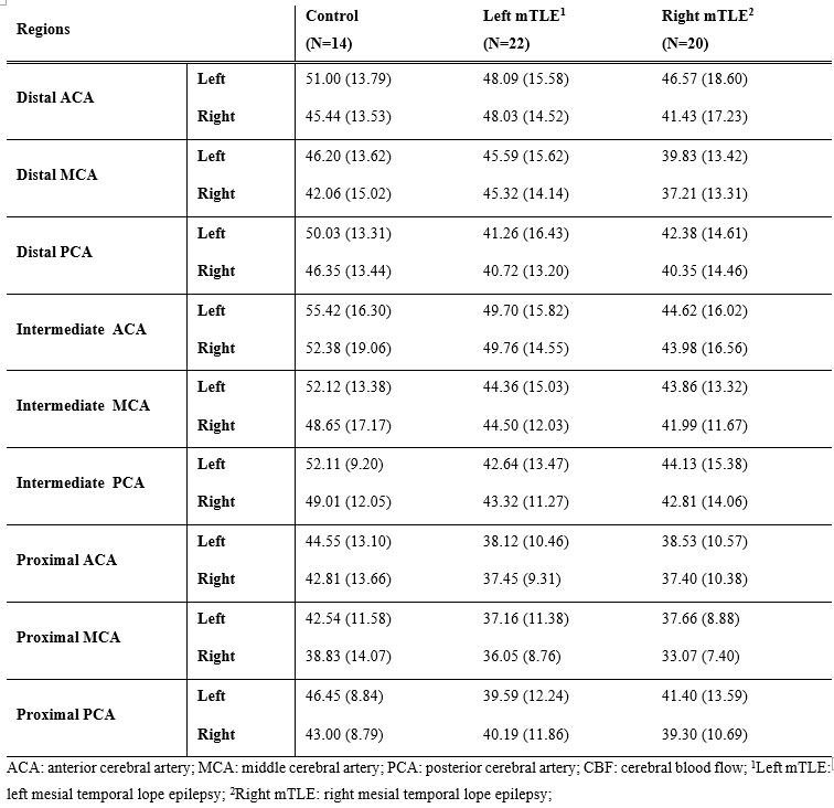
Intra-cohort comparison using paired T-test between left and right brain hemispheres showed significant differences for control cohort in left and right distal ACA, distal MCA, proximal ACA, proximal MCA, and proximal PCA. There was a significant difference between bilateral regions of right mTLE in distal ACA, intermediate PCA, proximal ACA, proximal MCA, and proximal PCA. In patients with left mTLE, the comparison of bilateral regions did not reveal any significant difference in any of the ROIs. Inter-cohort comparison of ROIs was performed using SPSS’s independent T-Test. left mTLE vs. control data comparison resulted in a significant difference in left intermediate PCA. Right mTLE cohort was different in left intermediate ACA, regions compared to control cohort. Statistical comparison of right mTLE vs. left mTLE showed a significant difference in right distal MCA.Discussion:
The use of vascular territories has proven valuable in studying various cerebrovascular and neurological diseases, including Moyamoya disease and Alzheimer's disease [6, 7]. While certain regions of interest (ROIs) were effective in distinguishing right and left mTLE from control cohorts, differentiation between left and right mTLE was challenging. Notably, the right distal anterior cerebral artery (ACA) vascular territory's significance in separating the two mTLE subcohorts suggests that hypoperfusion in this region could serve as a potential imaging biomarker for lateralization in temporal lobe epilepsy. Comparing left mTLE with the control group revealed significant differences in the left intermediate posterior cerebral artery (PCA) territory. This region is considered highly susceptible to minor vessel disease or autoregulation issues [8]. The observed drop in cerebral blood flow (CBF) can be attributed to its supply to the left parahippocampus and the left occipital lobe. This aligns with findings of hypoperfusion in the left inferior occipital and left parahippocampus regions in both voxel-wise and ROI analyses. In the comparison between right mTLE and control cohorts, significant differences were observed in the left intermediate anterior cerebral artery (ACA) territory, which supplies various brain regions including the superior frontal gyrus and frontal pole. These findings support previous research [9, 10] indicating cortical thinning in the superior frontal lobe in right mTLE and disruptions in cerebrocerebellar functional networks, emphasizing the relevance of CBF changes in the left intermediate ACA to existing studies. Conclusion: The results of this study underscore the importance of considering vascular territory analysis in the assessment of mTLE lateralization. While previous research often focused on structural changes in the gray matter, the significance of vascular territory changes cannot be underestimated. By considering vascular territory changes, clinicians may be better equipped to make informed decisions regarding surgical interventions, especially when faced with challenging cases of mTLE.Acknowledgements
No acknowledgement found.References
1. Sheikh SR, Nair D, Gross RE, Gonzalez‐Martinez J. Tracking a changing paradigm and the modern face of epilepsy surgery: a comprehensive and critical review on the hunt for the optimal extent of resection in mesial temporal lobe epilepsy. Epilepsia. 2019;60(9):1768-93.
2. Rahimzadeh H, Kamkar H, Hoseini-Tabatabaei N, Mobarakeh NM, Habibabadi JM, Hashemi-Fesharaki S-S, et al. Alteration of intracranial blood perfusion in temporal lobe epilepsy, an arterial spin labeling study. Heliyon. 2023;9(4).
3. Yoganathan K, Malek N, Torzillo E, Paranathala M, Greene J. Neurological update: structural and functional imaging in epilepsy surgery. Journal of Neurology. 2023;270(5):2798-808.
4. Fallahi A, Pooyan M, Habibabadi JM, Nazem-Zadeh M-R. Comparison of multimodal findings on epileptogenic side in temporal lobe epilepsy using self-organizing maps. Magnetic Resonance Materials in Physics, Biology and Medicine. 2022;35(2):249-66.
5. Mutsaerts H, Van Dalen J, Heijtel D, Groot P, Majoie C, Petersen E, et al. Cerebral perfusion measurements in elderly with hypertension using arterial spin labeling. PloS one. 2015;10(8):e0133717. 6. Huang C-W, Hsu S-W, Chang Y-T, Huang S-H, Huang Y-C, Lee C-C, et al. Cerebral perfusion insufficiency and relationships with cognitive deficits in Alzheimer’s disease: a multiparametric neuroimaging study. Scientific reports. 2018;8(1):1-14.
7. Dounavi M-E, Low A, McKiernan EF, Mak E, Muniz-Terrera G, Ritchie K, et al. Evidence of cerebral hemodynamic dysregulation in middle-aged APOE ε4 carriers: The PREVENT-Dementia study. Journal of Cerebral Blood Flow & Metabolism. 2021;41(11):2844-55.
8. Huang C-W, Hsu S-W, Chang Y-T, Huang S-H, Huang Y-C, Lee C-C, et al. Cerebral perfusion insufficiency and relationships with cognitive deficits in Alzheimer’s disease: a multiparametric neuroimaging study. Scientific reports. 2018;8(1):1541.
9. Zhou X, Zhang Z, Liu J, Qin L, Pang X, Zheng J. Disruption and lateralization of cerebellar–cerebral functional networks in right temporal lobe epilepsy: a resting-state fMRI study. Epilepsy & Behavior. 2019;96:80-6.
10. Jber M, Habibabadi JM, Sharifpour R, Marzbani H, Hassanpour M, Seyfi M, et al. Temporal and extratemporal atrophic manifestation of temporal lobe epilepsy using voxel-based morphometry and corticometry: clinical application in lateralization of epileptogenic zone. Neurological Sciences. 2021;42(8):3305-25.
Figures