3087
A clinical study of the hippocampus subregion in temporal lobe epilepsy patients with neurite directional dispersion and density imaging1Department of Radiology, General Hospital of Ningxia Medical University, Yinchuan 750004, China, Yinchuan, China, 2GE HealthCare MR Research, Beijing, China, Beijing, China
Synopsis
Keywords: Epilepsy, Diffusion/other diffusion imaging techniques
Motivation: To explore the microstructural changes of hippocampal subregions in temporal lobe epilepsy(TLE) patients.
Goal(s): The microstructure of hippocampal subregion in TLE patients was significantly changed, which would provide imaging basis for the diagnosis of MRI negative TLE patients.
Approach: Using neurite orientation dispersion and density imaging (NODDI) combined with automatic segmentation technology to explore TLE changes in microstructure and volume of hippocampal subregion in patients.
Results: This study demonstrated the ability of NODDI technique to detect the changes of hippocampal microstructure in TLE patients. NDI may be more able to highlight neuronal damage and fiber recombination in TLE patients.
Impact: This study demonstrated the ability of NODDI technique to detect the changes of hippocampal microstructure in TLE patients. NDI may be more able to highlight neuronal damage and fiber recombination in TLE patients.
Temporal lobe epilepsy (TLE) is the most common drug-resistant epilepsy in adult focal epilepsy1. Its main pathology is hippocampal sclerosis (HS), but about 20% - 30% of epilepsy patients have no obvious abnormality on routine MRI. Neurite orientation dispersion and density imaging (NODDI), an advanced diffusion technique that quantifies neurite density and orientation. Many previous diffusion studies have shown abnormal microstructure of the entire hippocampus in TLE patients2. In fact, the hippocampus is composed of several subregions, such as the hippocampal horn (CA1-CA4), the subiculum (Sub) and the granular cell layer of the dentate gyrus (GC-DG). We used NODDI combined with automatic segmentation technique to explore the changes of the volume and NODDI indexes of the hippocampal subregion.
Methods
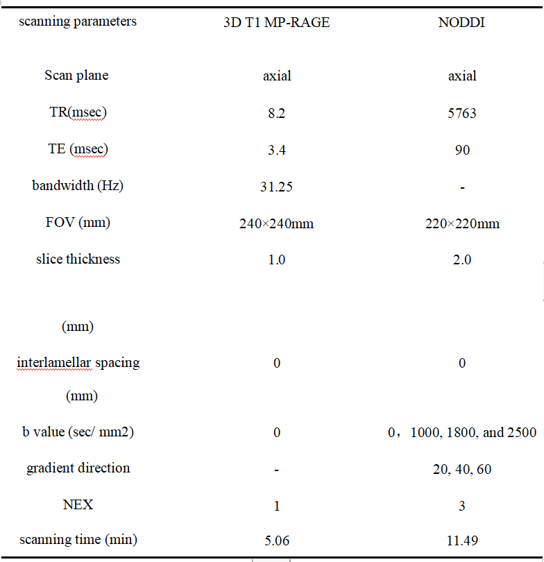
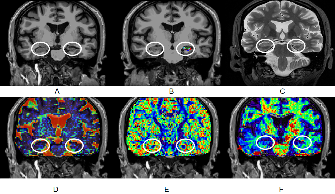
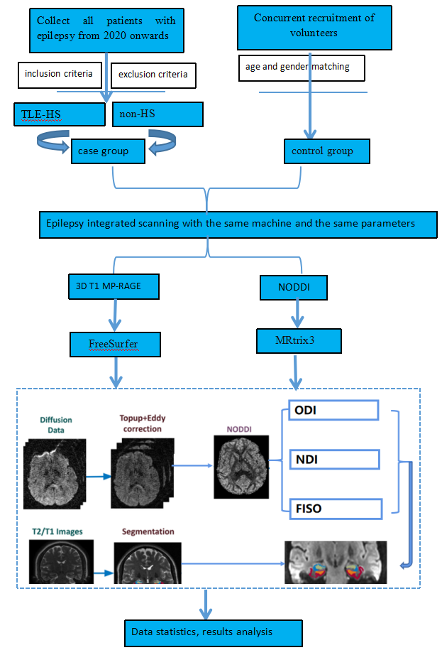
According to the diagnostic criteria of the International League Against Epilepsy(ILAE), 20 TLE patients with hippocampal sclerosis (TLE-HS) and 12 MRI negative TLE patients (non-HS) were included. 15 matched healthy controls (con) were recruited at the same time. TI structure images and multi-b-value NODDI images were collected. FreeSurfer software3 was used to segment the hippocampus on T1 images. MRtrix34 was used for post-processing and NODDI index maps: ODI, NDI and fiso maps were calculated.The volume and diffusion indexes of hippocampal subregions were calculated respectively.
Results
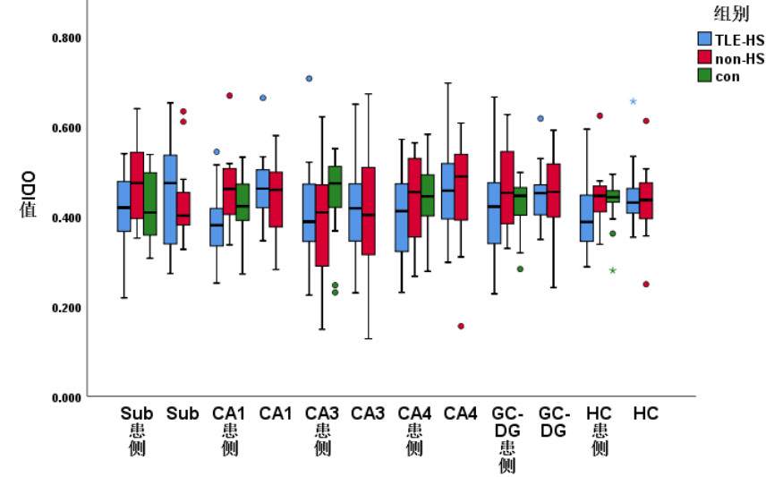
In TLE-HS group, the whole hippocampal, CA1, CA3, CA4 and GC-DG subregions of the pathologic side were significantly smaller than the opposite side. In TLE-HS group, the fiso values of CA3 subregion of the pathologic side were significantly higher than the contralateral hippocampus, the ODI values of CA1 and CA4 subregions were significantly lower than the contralateral hippocampal subregions, and the NDI values of CA1, CA3, CA4 and GC-DG subregions were significantly lower than the contralateral hippocampus. The fiso value of GC-DG pathologic subregion in TLE-HS group was significantly higher than con group, the ODI value of CA1 pathologic subregion in TLE-HS group was significantly lower than con and non-HS group, the NDI values in Sub, CA1, CA4 and GC-DG subregions in TLE-HS group were significantly lower than con group, and the NDI value in TLE-HS group was significantly lower than control group. The NDI values of CA1, CA4 and GC-DG hippocampus subregions in Non-HS group were significantly lower than con group, but there was no significant difference among other groups. In TLE-HS group, the NDI value of pathologic hippocampal CA4 subregion was negatively correlated with the course of disease.
Discussion
TLE is a chronic disease caused by the abnormal firing of neurons in the hippocampus. However, changes in the microstructure of specific regions of the brain are uncertain. Fiso stands for the volume fraction of free water in an organization. In theory, due to neuronal degeneration and apoptosis, the extracellular space increases and the extracellular fluid increases. Our results were consistent with theoretical speculation. The ODI value of CA1 subregion in TLE-HS group was significantly lower than con and non-HS group. Meanwhile, in the intra-group comparison, ODI values in the affected hippocampal subregions of the TLE-HS group were all decreased compared to the contralateral, but the differences were significant in the CA1 and CA3 subregions, which was consistent with the ILAE classification. This may indicate its potential to provide pathological types. Compared with fiso and NDI indicators, ODI showed significant differences in more hippocampal subregions. The decline in NDI results is attributed to the gradual decrease in nerve axon density and myelin content5, which also implies significant neuronal damage. NDI can detect the change of microstructure more sensitively than other indexes. When the whole hippocampus was studied, only the NDI value of the affected hippocampus was found to be lower than the con group, suggesting that the microstructure characteristics of the tissue could be better captured at the hippocampal subregion level.For the non-HS group, the conventional MRI sequence could not detect abnormal findings. However, NODDI technology was applied to find that the NDI values of the pathologic hippocampus in the CA1, CA4 and GC-DG subregions in the non-HS group were significantly lower than the con group. It provides the imaging basis for the diagnosis of non-HS. In addition, in the correlation analysis it was found that the NDI value of CA4 pathologic subregion in the TLE-HS group was negatively correlated with the course of disease, indicating that the neurons gradually lost with the progression of the disease.
Conclusion
We combined NODDI and automatic segmentation to study the microstructure of hippocampal subregion in TLE patients, and found the abnormal microstructure in TLE patients, providing image information for the diagnosis of patients with negative MRI.
Acknowledgements
No acknowledgement found.References
[1] Thijs RD, Surges R, O'Brien TJ, et al. Epilepsy in adults. Lancet, 2019 Feb 16,393(10172):689-701. [2] Giachetti I, Padelli F, Aquino D, et al. Role of NODDI in the MRI Characterization of Hippocampal Abnormalities in Temporal Lobe Epilepsy: Clinico-histopathologic Correlations. Neurology,2022 Apr 26,98(17):e1771-e1782. [3] Fischl B, Salat DH, Busa E, et al. Whole brain segmentation: automated labeling of neuroanatomical structures in the human brain. Neuron, (2002) 33:341–55. [4] Tournier JD, Smith R, Raffelt D, et al. MRtrix3: A fast, flexible and open software framework for medical image processing and visualisation. Neuroimage, (2019) 202:116137. [5] Alotaibi A, Podlasek A, AlTokhis A, et al. Investigating Microstructural Changes in White Matter in Multiple Sclerosis: A Systematic Review and Meta-Analysis of Neurite Orientation Dispersion and Density Imaging. Brain Sci, 2021 Aug 29,11(9):1151.Figures