3085
A potential mechanism of neurological impairment in children with infantile spasm:a structure analysis employing voxel-based morphometry1Radiology, Shenzhen Children's Hospital, Shenzhen, China, 2Radiology, Longhua District Shenzhen People’s Hospital, Shenzhen, China, 3Philips Healthcare, Guangzhou, China, Shenzhen, China, 4China Medical University, Shenyang, China, Shenzhen, China
Synopsis
Keywords: Epilepsy, Brain Connectivity, Spasms, Infantile; Magnetic Resonance Imaging; Imaging; Three-Dimensional; Cerebral Cortex
Motivation: Whether there is micro-structural alteration in the temporal lobe of Infantile epileptic spasms syndrome (IESS) patients is yet to be clarified, of which resulting in severe language function impairment.
Goal(s): To explore the potential mechanism of neurological impairment in children with IESS.
Approach: We conducted a study with BSID-II for clinical evaluation, meanwhile voxel-based morphometry (VBM) for MRI data analysis.
Results: In IESS group, the most significant volume loss of gray matter in right fusiform (Broadman area 21) and middle temporal gyrus (Wernicke's area) were key node for voice recognition, language processing, semantic retrieval, memory, and understanding network.
Impact: The findings of this study light up an anatomical basis for language impairment in children with IESS.
Introduction
IESS is a refractory age-dependent epileptic syndrome in infants, with peak onset age ranging 3 to 12 months1.A long-term follow-up study found language function impairment in children with IESS2. Neuropsychology and neuroimaging study showed that abnormal temporal and frontal connectivity was responsible for language function impairment3. The temporal lobe played a key role in auditory processing, including perception of sounds, giving meaning to sounds, and remembering sounds. However, whether there is micro-structural alteration in the temporal lobe of IESS patients is yet to be clarified. We conducted a study to investigate the alteration of micro-structure of IESS patients, focusing on linguistic and visual function and their networks. The objective was to explore the potential mechanism of neurological impairment in children with IESS by using the self-made brain template and tissue probability map (TPM) based on diffeomorphic anatomical registration through exponential lie algebra (DARTEL)4.Methods
IESS group had 21 males and 13 females (mean age: 17.7±15.6 months), while HC group had 22 males and 10 females (mean age: 29.4±18.7 months). Three-dimensional magnetization prepared rapid acquisition gradient echo sequences were used to acquire high-resolution 3D structural images (matrix size: 256×256; voxel size: 1.0×1.0×1.0 mm3). Computational Anatomy Toolbox implemented in Statistical Parametric Mapping 12 was used to measure the gray matter and white matter volume, and the cortical thickness separately. Independent sample t test was used to assess between-group differences. IESS group was assessed using the Bayley Scales of Infant Development.Results
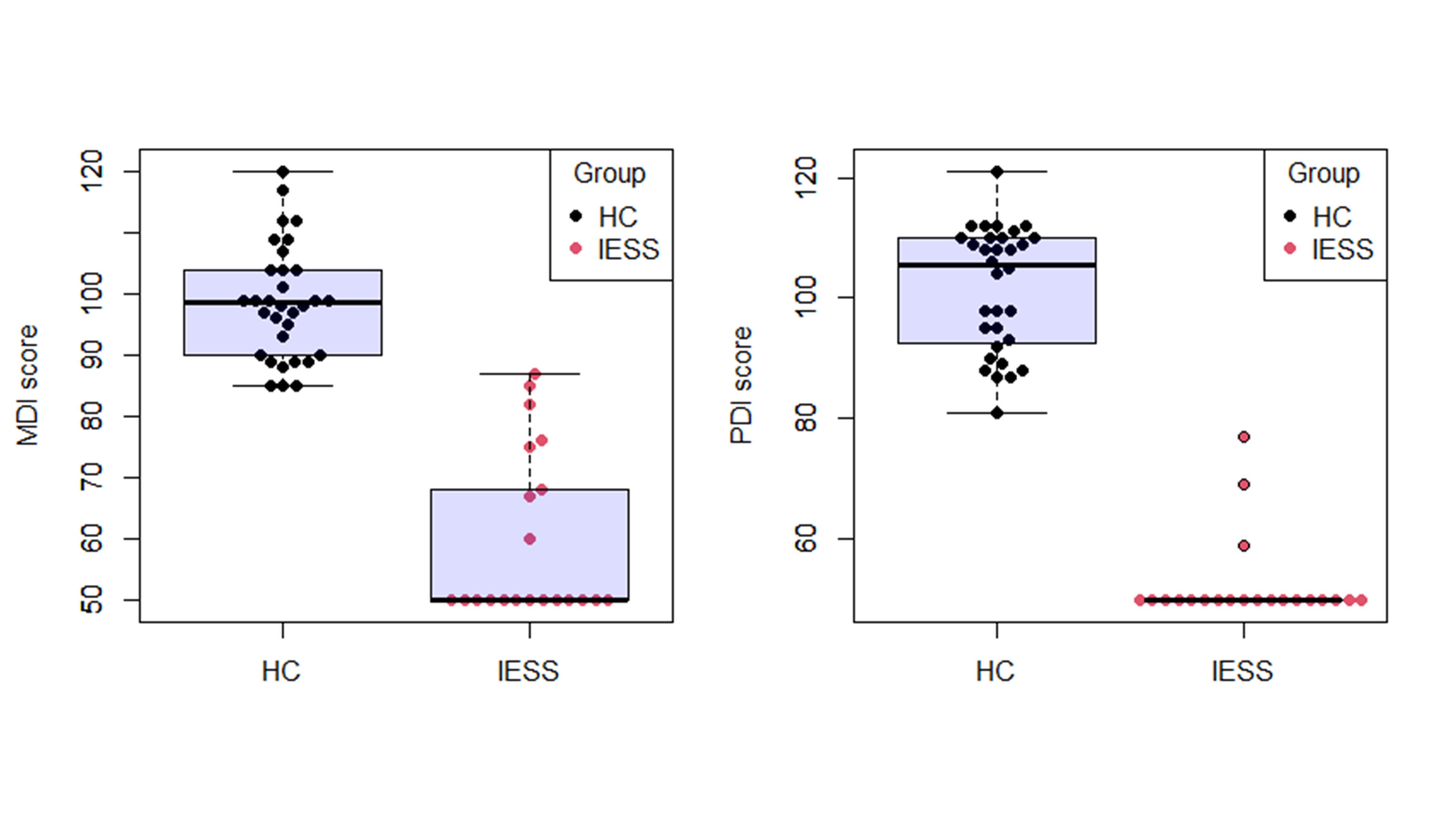
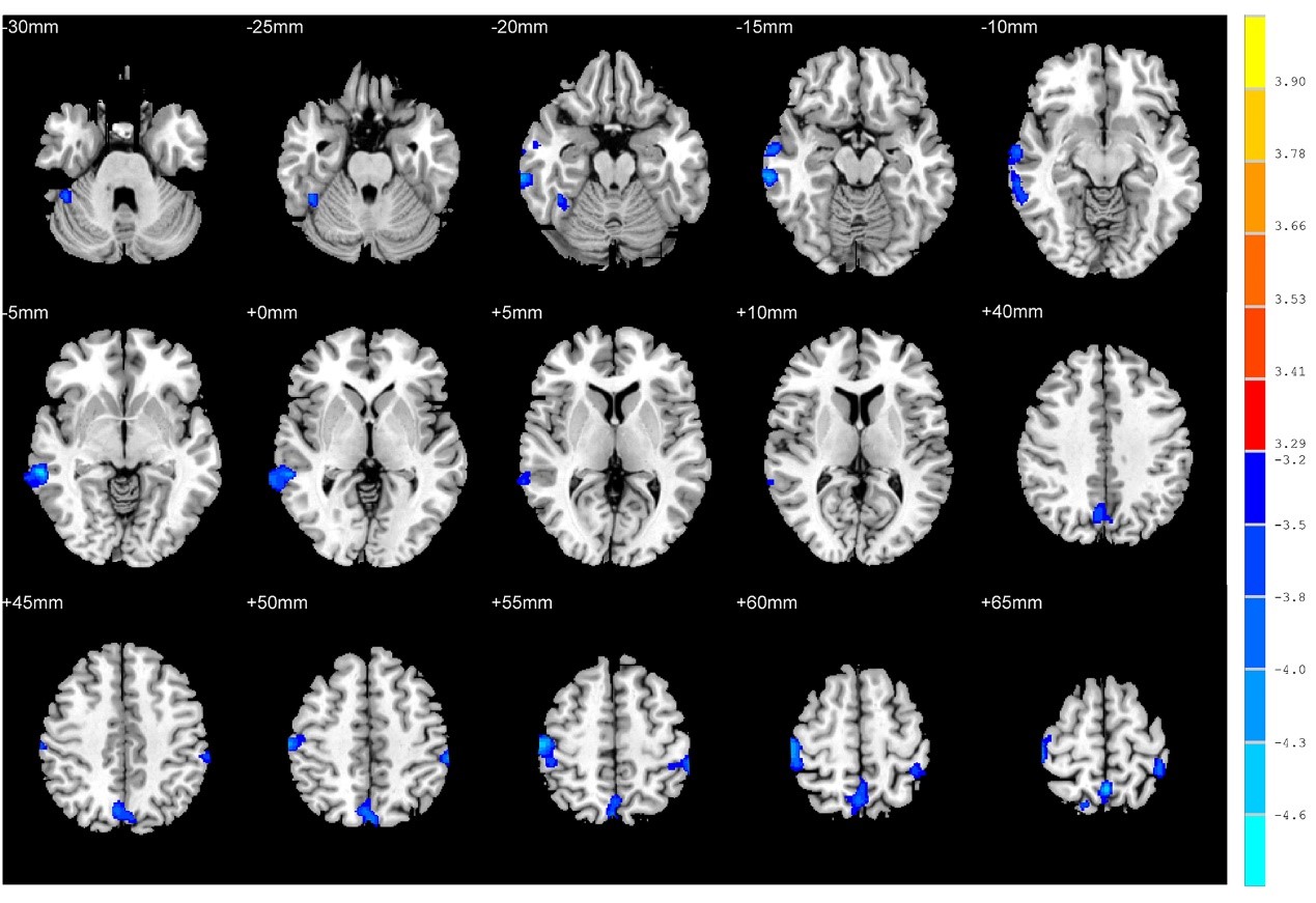
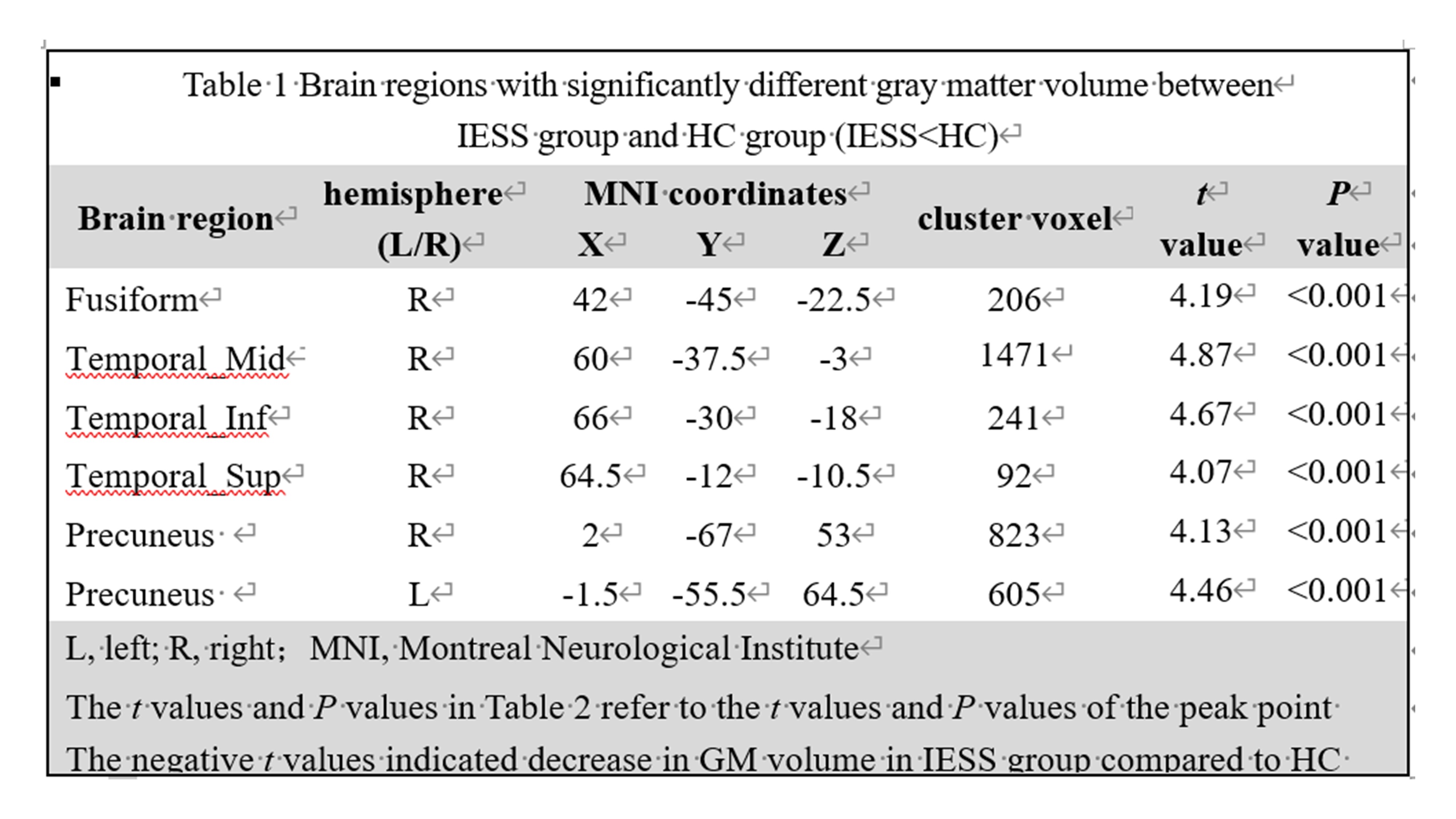

Compared to HC group, IESS group showed a significantly decreased volume of gray matter in right middle temporal gyrus, inferior temporal gyrus, superior temporal gyrus, right fusiform, and bilateral precuneus (P<0.001). There were no significant between-group differences with respect to white matter volume or cortical thickness (P>0.001). The results of Bayley Scales of Infant Development showed that the mental development scale (MDI) and psychomotor development scale (PDI) scores of children with IESS were almost concentrated in the range of <70. MDI score showed a positive correlation with gray matter reduction area in IESS group.Discussion
In this study, the IESS group exhibited decreased gray matter volume in three important functional areas (the right inferior temporal gyrus, fusiform, and bilateral precuneus) closely related to the visual pathway, suggesting impaired visual processing and attention, resulting in corresponding clinical development retardation, thus affecting the overall development of cognitive function5-7. Meanwhile, significantly decreased GM volume in the right superior temporal gyrus and middle temporal gyrus might lead to impaired sound processing, thus causing impaired language function of different types, which further affected the cognitive function development8-10. In this study, we found significantly decreased gray matter volume in the right middle and inferior temporal gyrus in IESS group, reflected the anatomical basis of language function impairment. Because the ventral pathway (one of the neural pathways of language function) projected to the ventrolateral through the superior temporal sulcus cortex, and finally reached the middle temporal gyrus and the inferior temporal gyrus, which acted as a transit point between sound-based language comprehension and conceptual expression in the language development11. Combined with the clinical language deficiency (MDI scores), it reflected the anatomical basis of language function impairment in children with IESS.Conclusion
Children with IESS had impaired cognitive and delayed motor development. Decreased volume of gray matter in the right temporal lobe, fusiform, and bilateral precuneus might be the potential anatomic basis for impaired hearing, visual and language function in children with IESS. In IESS group, the most significant volume loss of gray matter in right fusiform (Broadman area 21) and middle temporal gyrus (Wernicke's area), which were key node for voice recognition, language processing, semantic retrieval, memory, and understanding network.Acknowledgements
This project was supported by Sanming Project of Medicine in Shenzhen (SZSM202011005) from Shenzhen Medical and Health Project, Guangdong High-level Hospital Construction Fund (ynkt2021-zz46).References
[1] Zuberi SM, Wirrell E, Yozawitz E, Wilmshurst JM, Samia P, Hirsch E, et al. ILAE classification and definition of epilepsy syndromes with onset in neonates and infants: Position statement by the ILAE Task Force on Nosology and Definitions |. 23 | Solomon L Moshé 2022;63:26. [2] Benn EKT, Hesdorffer DC, Levy SR, Testa FM, DiMario FJ, Berg AT. Parental report of behavioral and cognitive diagnoses in childhood-onset epilepsy: A case-sibling-controlled analysis. Epilepsy and Behavior 2010;18:276–9. [3] Valente KD, Caciagli L, Ratcliffe C, Wandschneider B, Baxendale S, Thompson P, et al. Cognitive Function in Genetic Generalized Epilepsies: Insights From Neuropsychology and Neuroimaging. Frontiers in Neurology | WwwFrontiersinOrg 2020;1:144. [4] Kim GW, Kim YH, Jeong GW. Whole brain volume changes and its correlation with clinical symptom severity in patients with schizophrenia: A DARTEL-based VBM study. PLoS ONE 2017;12. https://doi.org/10.1371/journal.pone.0177251. [5] Sebastian R, Gomez Y, Leigh R, Davis C, Newhart M, Hillis AE. The roles of occipitotemporal cortex in reading, spelling, and naming. Cogn Neuropsychol 2014;31:511–28. [6] Pehere N, Chougule P, Dutton GN. Cerebral visual impairment in children: Causes and associated ophthalmological problems. Indian J Ophthalmol 2018;66:812–5. [7] Harroud A, Boucher O, Tran TPY, Harris L, Hall J, Dubeau F, et al. Precuneal epilepsy: Clinical features and surgical outcome. Epilepsy and Behavior 2017;73. [8] Binder JR. Current Controversies on Wernicke’s Area and its Role in Language. Current Neurology and Neuroscience Reports 2017 17:8 2017;17:1–10. [9] Fosi T, Werner K, Boyd SG, de Haan M, Scott RC, Neville BG. Auditory processing following infantile spasms: An event-related potential study. Epilepsia 2017;58:872–81. [10] Smith AB, Dawes P, Smith S, Pal DK. A specific deficit of auditory processing in children with Rolandic Epilepsy and their relatives. Epilepsy & Behavior 2017;72:135–9. [11] DeWitt I, Rauschecker JP. Wernicke’s area revisited: Parallel streams and word processing. Brain and Language 2013;127:181–91.Figures