3084
Altered brain structural and functional network in children and adolescents with drug-resistant epilepsy1Department of Radiology, Affiliated Hospital of Zunyi Medical University, Zunyi, China
Synopsis
Keywords: Epilepsy, fMRI (resting state), drug-resistant,structural, functional, networks
Motivation: Epilepsy is widely regarded as a brain network disorder. However, few studies have investigated the interaction between brain structural networks and functional networks in epilepsy.
Goal(s): To investigate the topological alterations of brain networks in children and adolescents with drug-resistant epilepsy.
Approach: Global graph theoretic network measures of global properties, nodal properties and rich club organizational properties were computed.
Results: Aberrant topological attributes and connectivity patterns have been found in DRE. Rich-club connectivity was lower in patients with DRE than that in controls.
Impact: Altered Dc in the left pallidum may provide a new theoretical basis for clinical treatment of DRE. The present findings enhanced our understanding of the neurophysiologic mechanisms associated with DRE.
Purpose
This study aims to investigate the topological changes of structural and functional networks in children and adolescents with drug-resistant epilepsy(DRE) by combining resting-state functional magnetic resonance imaging (rs-fMRI) and diffusion tensor imaging(DTI), and to analyze the correlation between topological changes of brain networks and disease duration and intelligence quotient(IQ), providing new insights into the underlying neuropathophysiological mechanism of behavioral and cognitive impairments observed in DRE.Methods
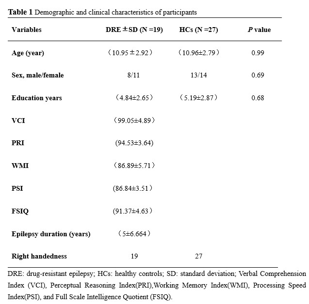
Nineteen children with DRE and 27 healthy controls (HCs) underwent rs-fMRI and DTI scans. Neuropsychological assessment of IQ was performed. Graph theoretical analyses were chosen to compare the global properties (small-world parameters, and network efficiency), nodal properties, and rich club organizational properties in DRE patients and HCs.Results
DRE and HCs both exhibited small-world attributes. Compared with HCs, DRE showed decreased global efficiency(Eg) local efficiency(Eloc), and the nodal efficiency(Ne) of the left inferior temporal gyrus in the structural network(Figure 1). Rich nodes include left orbital superior frontal, superior occipital gyrus, middle occipital gyrus, right supplementary motor area, putamen, insula, bilateral precuneus, and bilateral medial cingulate gyri(Figure 2B). DRE exhibited decreased rich club connectivity, feeder connectivity and local connectivity (Figure 2A). Rich-club connectivity positively correlated with the Ne in left inferior temporal gyrus((Figure 2C). Feeder connectivity positively correlated with Eg, Eloc, and Ne in left inferior temporal gyrus(Figure 3). DRE patients showed reduced Dc in the left pallidum in the functional network compared with controls(Figure 4A). The Dc in the left pallidum were negatively correlated with processing speed index (PSI)( Figure 4B).Discussion
Epilepsy is widely regarded as a brain network disorder. According to the neural network hypothesis, reduced ASD efficacy can be caused by an abnormal (epileptic) neural network formed by epilepsy-induced structural alterations such as neurodegeneration, axonal sprouting,neurogenesis, gliosis, and synaptic reorganization[1]. There is broad experimental evidence that the modulation of part of the epileptic network by targeted intracranial drug delivery is sufficient to control focal seizure initiation, propagation, or generalization[2].However, few studies have investigated the interaction between brain structural networks and functional networks in epilepsy. To our knowledge, this was the first study to investigate the topological changes of structural and functional networks in children and adolescents with DRE combining DTI and rs-fMRI. The rich-club topology plays a key role in the integrative capacity among regions of the whole-brain network[3]. In our study, we showed that the existence of a rich-club organization of structural brain networks in DRE, but the rich-club connectivity in DRE patients was significantly reduced, reflecting a lower level of connectivity among hub brain regions. Interrupted brain network topology affects the dissemination and integration of information between distant brain regions. This study found that the decrease in global and local efficiency of structural networks is closely related to the decrease in connectivity. The main reason for the lower global and local efficiency in DRE patients is the abnormal interruption of global connectivity.Therefore,the destruction of Rich Club had a serious impact on the global communication efficiency level of the network[4]. Abnormal rich-club region can also adversely affect on the brains higher cognitive function due to its central role in the network. DRE patients showed that Dc in left pallidum were negatively correlated with PSI. The processing speed depends on various functional abilities, such as attention, planning,visuospatial and auditory skills.These functions are supported by complex brain network that provides the physiological basis for information processing.[5, 6].The pallidum, as part of the basal ganglia, is not only important for the motor system but also plays a role in cognitive functions, including working memory, executive function, reward, and learning[7]. We speculated that decreased Dc in the left pallidum was closely related to cognitive impairment in DRE. Altered Dc in the left pallidum may provide a new theoretical basis for clinical treatment of DRE.Conclusion
Combining DTI and rs-fMRI provided a more comprehensive reflection of brain network changes in patients with DRE. And Dc in the left pallidum may serve as a therapeutic target for DRE in the future.The present findings enhanced our understanding of the neurophysiologic mechanisms associated with DRE.Acknowledgements
We are grateful to all of the individuals who participated in the current study.References
[1] Bastos F, Cross JH. Epilepsy. Handb Clin Neurol. 2020. 174: 137-158.
[2] Gernert M, Feja M. Bypassing the Blood-Brain Barrier: Direct Intracranial Drug Delivery in Epilepsies. Pharmaceutics. 2020. 12(12): 1134.
[3] van den Heuvel MP, Sporns O. Rich-club organization of the human connectome. J Neurosci. 2011. 31(44): 15775-86.
[4] Cao R, Wang X, Gao Y, et al. Abnormal Anatomical Rich-Club Organization and Structural-Functional Coupling in Mild Cognitive Impairment and Alzheimer's Disease. Front Neurol. 2020. 11: 53.
[5] Takeuchi H, Kawashima R. Effects of processing speed training on cognitive functions and neural systems. Rev Neurosci. 2012. 23(3): 289-301.
[6] Bassett DS, Sporns O. Network neuroscience. Nat Neurosci. 2017. 20(3): 353-364.
[7] Schroll H, Horn A, Gröschel C, et al. Differential contributions of the globus pallidus and ventral thalamus to stimulus-response learning in humans. Neuroimage. 2015. 122: 233-45.
Figures
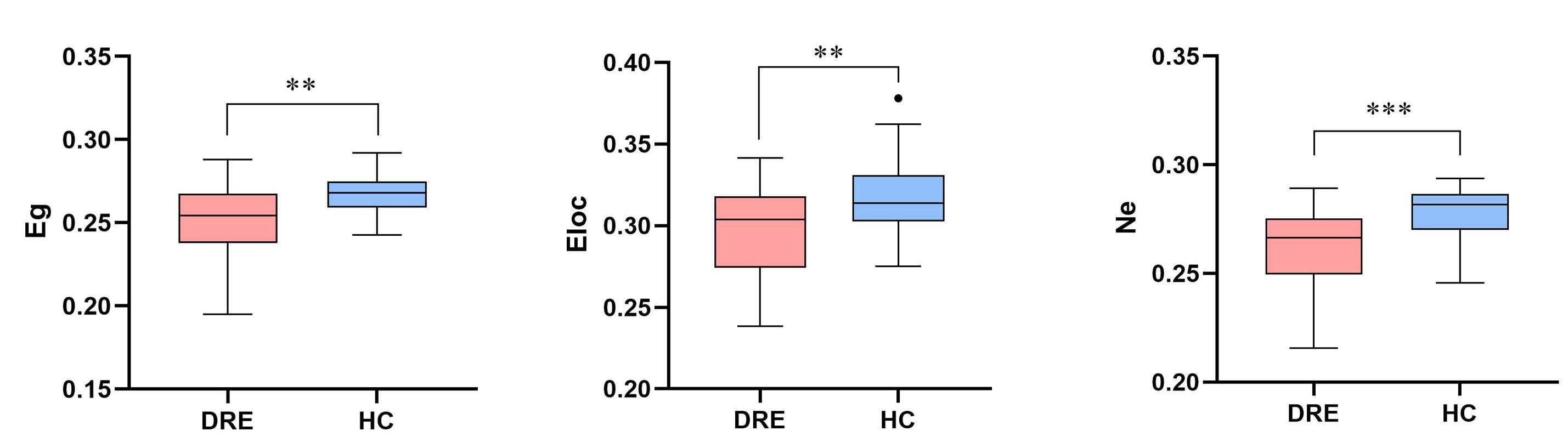
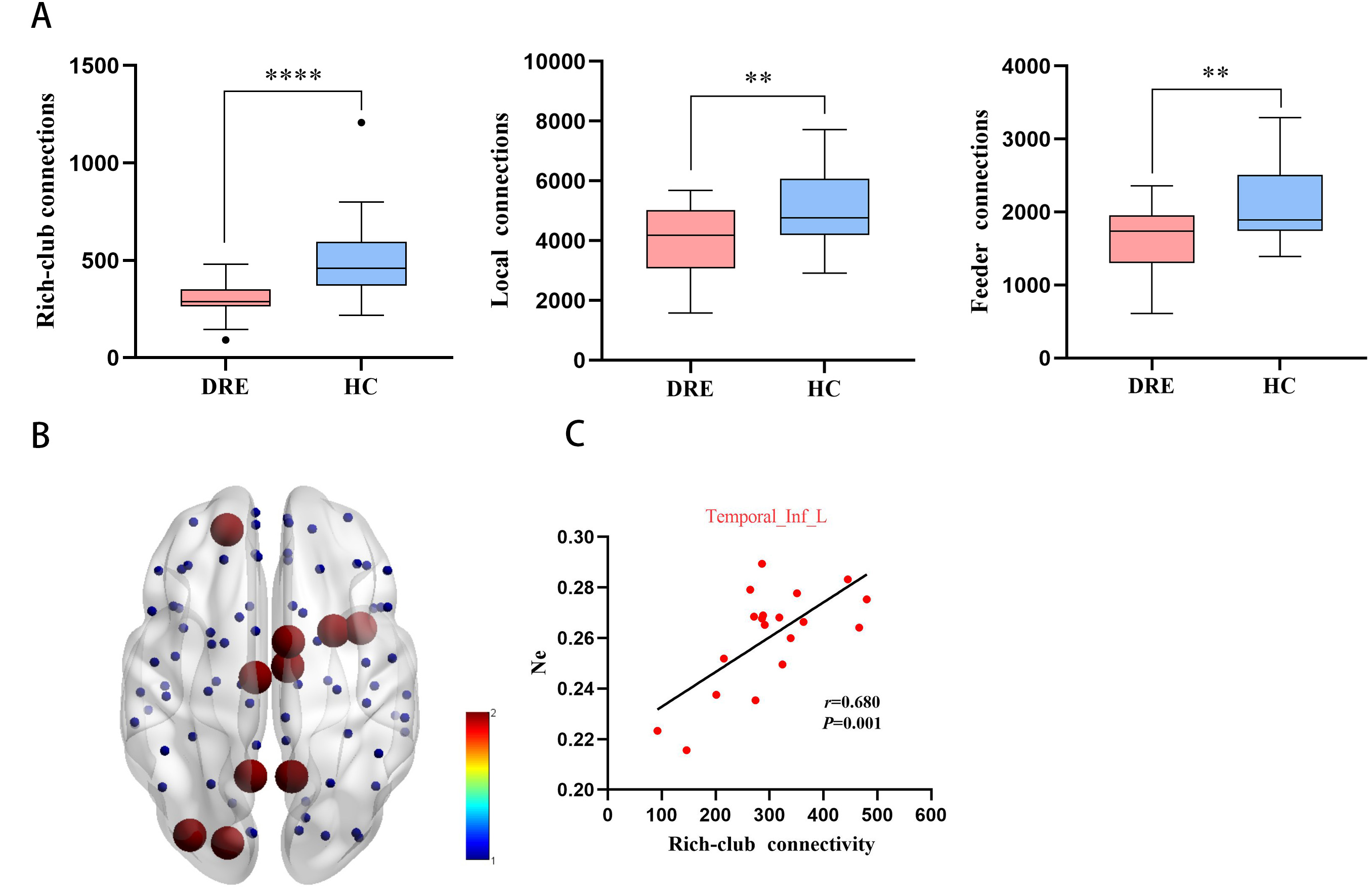
Figure 2A The different kinds of connections in structural networks.
Figure 2B The rich-club nodes (red nodes) are shown across all groups.
Figure 2C Rich-club connectivity correlated with the Ne in left inferior temporal gyrus.
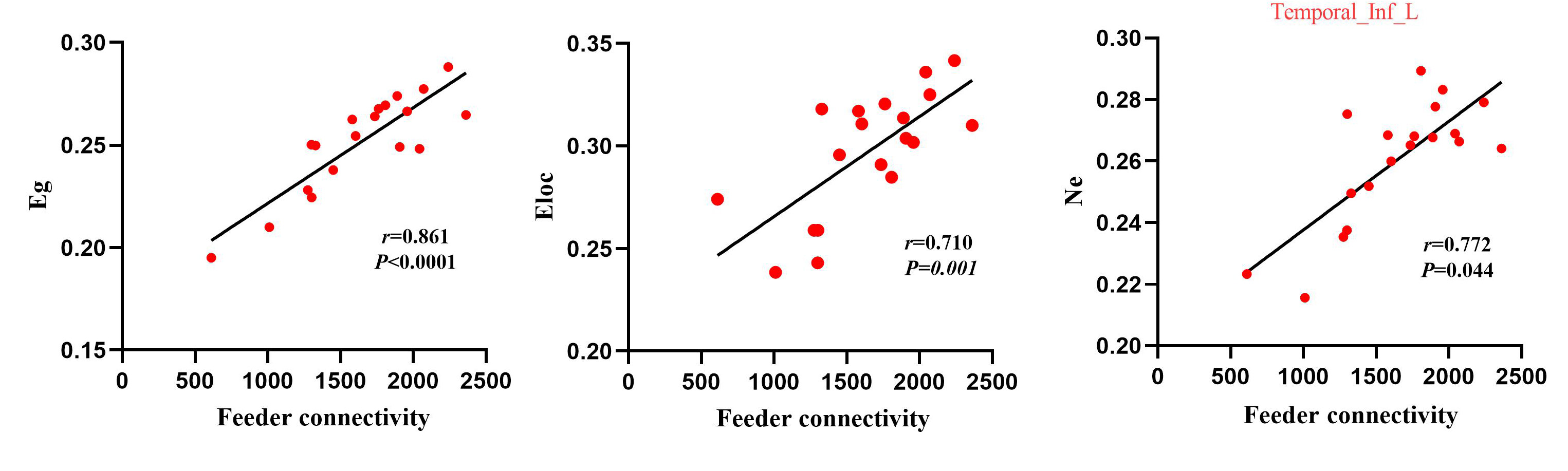
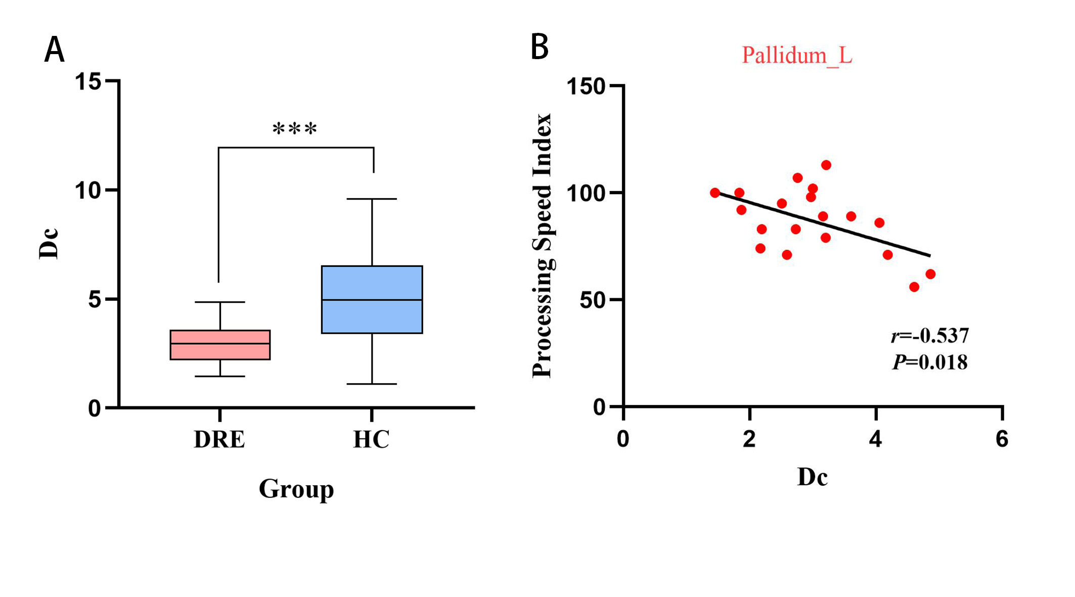
Figure4A Reduced Dc in the left pallidum in DRE.
Figure 4B The Dc in the left pallidum were negatively correlated with processing speed index (PSI).