3081
Study of brain network alternations in sleep-related hypermotor epilepsy by resting-state functional magnetic resonance imaging1Radiology, West China Hospital of Sichuan University, Chengdu, China, 2Department of Radiology and Huaxi MR Research Center (HMRRC), West China Hospital of Sichuan University, Chengdu, China, 3Department of Neurology, West China Hospital of Sichuan University, Chengdu, China, 4Department of Radiology, The First Affiliated Hospital, College of Medicine, Zhejiang University, Hangzhou, China, 5Department of Radiology, Huashan Hospital, Fudan University, Shanghai, China, 6Department of Radiology, West China Hospital of Sichuan University, Chengdu, China, 7Radiology, West China Xiamen Hospital of Sichuan University, Xiamen, China
Synopsis
Keywords: Epilepsy, fMRI (resting state), Sleep-related hypermotor epilepsy, graph theory analysis, functional brain network.
Motivation: To investigate the topological properties of brain functional connectomes in patients with sleep-related hypermotor epilepsy (SHE).
Goal(s): Little is known about the topological organization changes of brain functional networks in SHE patients.
Approach: 57 SHE patients and 54 healthy controls underwent resting-state functional MRI examination. Topological properties of brain networks were identified using graph-based theoretical analysis.
Results: Compared to controls, SHE patients showed longer characteristic path length (Lp) and lower global efficiency (Eglob), decreased nodal centralities in several regions, and connectivity aberrations. Lp and normalized Lp had positive correlations while Eglob and nodal centralities of thalamus had negative correlations with epilepsy duration.
Impact: The aberrant topological properties in brain functional networks of SHE may provide insights into the pathophysiology of epileptogenesis. The identification and characterization of network changes may contribute to clinical treatment through the disruption or inhibition of these epileptogenic networks.
Introduction
Sleep-related hypermotor epilepsy (SHE) is a focal epilepsy characterized by hyperkinetic seizures occurring mainly during non-rapid eye movement sleep[1-3]. Recent advances in graph-based theoretical approaches have allowed for noninvasive characterization of the topological properties of brain networks[4]. However, little is known about the topological organization changes of whole-brain networks in SHE based on resting-state functional MRI (rs-fMRI), which may provide further insight into the neuropathophysiological mechanisms underlying SHE.Methods
SubjectsThe study was approved by the Ethics Committee of our hospital, and written informed consent was obtained from each subject. Fifty-seven SHE patients with normal routine MRI findings and fifty-four healthy controls (HCs) were enrolled in this study.
Image acquisition and processing
MRI data were acquired using a Siemens 3 T scanner (Trio Tim, Erlangen, Germany) with an 8-channel phased array head coil. The scanning parameters were as follows: (1) T1WI: Magnetization Prepared Rapid Gradient Echo (MPRAGE) sequence, TR/TE = 2250/2.6 ms, flip angle = 9°, slice thickness = 1 mm, matrix size= 256 × 256, and FOV = 256 × 256 mm2; (2) rs-fMRI: gradient echo-planar imaging (EPI) sequence, TR/TE = 2000/35 ms, flip angle = 68°, voxel size = 3.3 × 3.3 × 3.0 mm3, slice thickness = 3 mm, matrix size = 64 × 64, FOV = 208 × 208 mm2, time points = 220, and acquisition time = 7.20 min. Preprocessing, brain network construction, and calculation of topological properties metrics were performed using the GRETNA toolbox[5].
Statistical analysis
Group differences were tested using two-sample t-test. False discovery rate (FDR) (P < 0.05) was applied to correct for multiple comparisons in nodal properties. The network-based statistic (NBS) was used to control the error rate for between-group comparisons in internodal connections (Edge p = 0.001, Component p = 0.05, Number of iterations = 5000). Pearson or Spearman correlation test was performed to determine the relationship between network matrices and clinical data.
Results
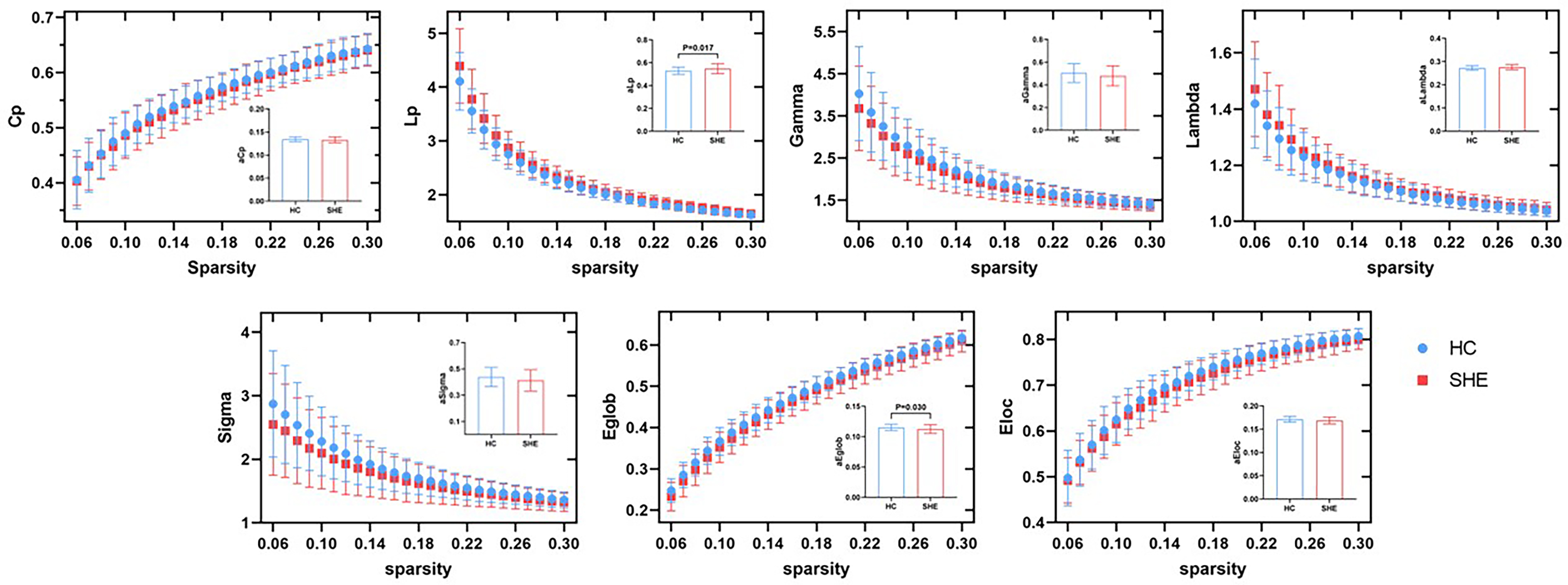
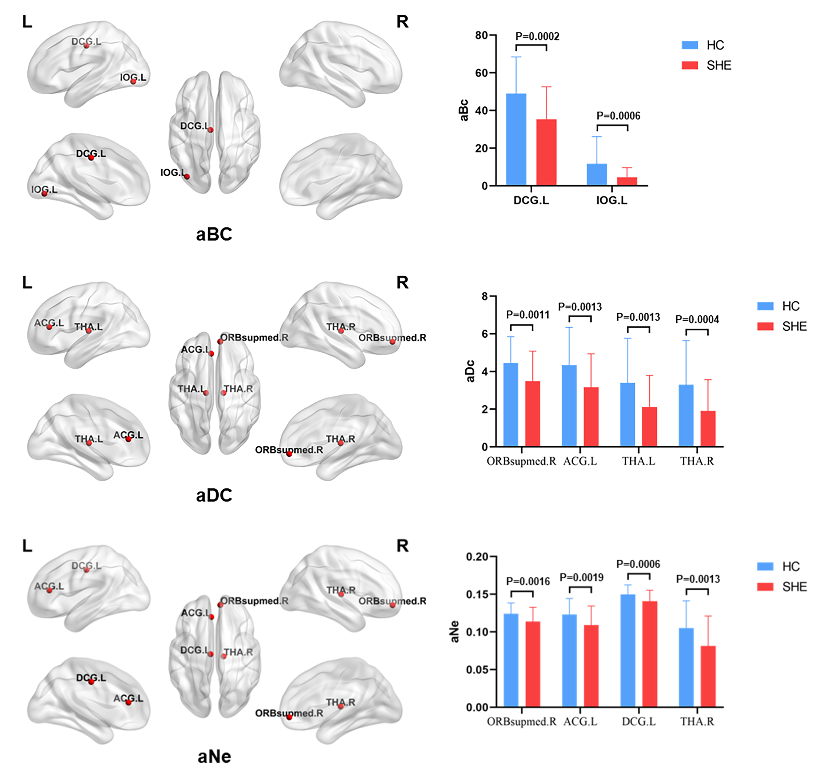
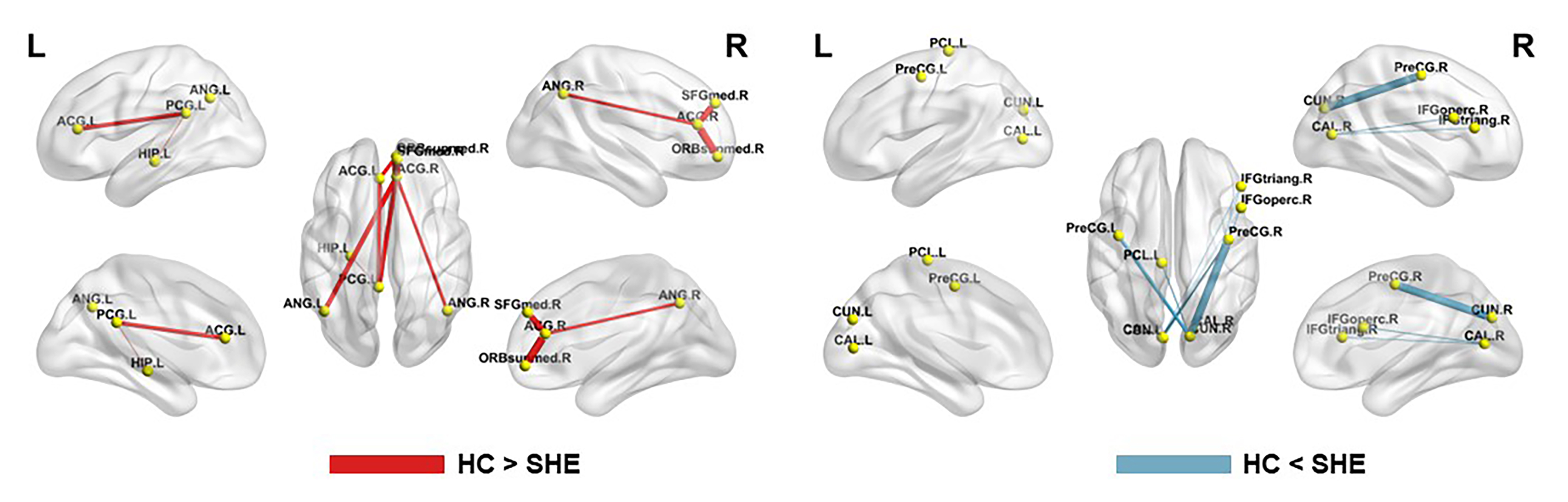
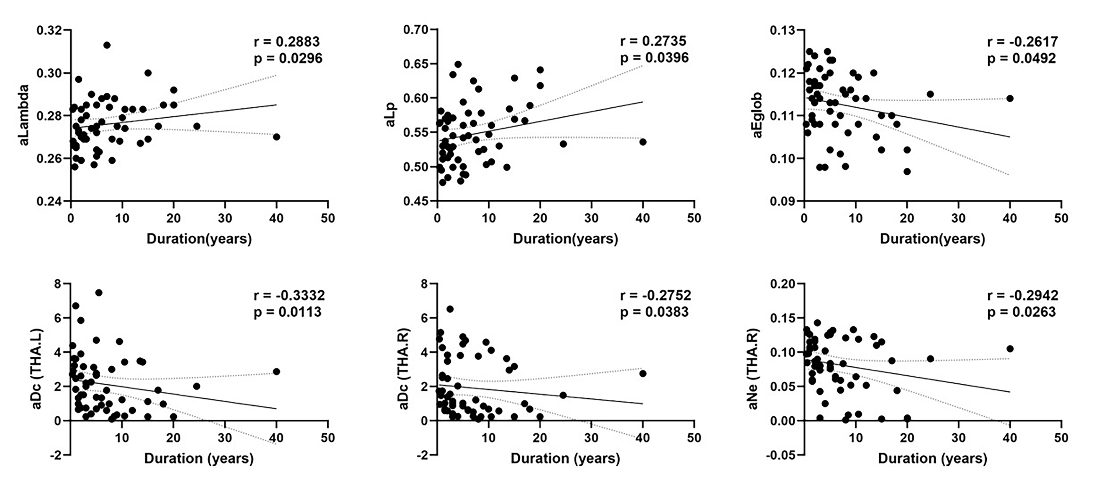
Compared with HCs, SHE showed significantly longer characteristic path length (Lp) and lower global efficiency (Eglob) (P=0.017-0.030); decreased betweenness centrality in left median cingulate and inferior occipital gyrus, decreased degree centrality in right superior frontal gyrus, left anterior cingulate gyrus and bilateral thalamus, and decreased nodal efficiency in right superior frontal gyrus, left anterior and median cingulate gyrus and right thalamus (all P<0.05). The NBS method identified two altered subnetworks in SHE group (PHCs>SHE = 0.043 and PHCs<SHE = 0.044). Correlations analysis showed that Lp and normalized Lp (lambda) had positive correlations (r = 0.288-0.274, P = 0.030-0.040) while Eglob and nodal centralities of thalamus had negative correlations with illness duration (r = -0. 262 – -0.333, P = 0.011–0.049).Discussion
Our study showed the brains of SHE patients with significantly longer Lp and lower Eglob compared with controls, which indicates decreased global integration. Surprisingly, both Lp and lambda had positive correlations with illness duration, while Eglob had a negative correlation with illness duration, illustrating that the alterations of brain network topology are progressive in the course of epilepsy.Patients with SHE exhibited impaired nodal centralities in frontal and some subcortical regions, suggesting regional functional decline, possibly due to the destructive effects of repeated epileptiform activities on the functional coordination of emotional/memory/cognitive-related networks[6]. The altered topological centralities in bilateral thalamus were negatively correlated with disease duration. The thalamic nuclei may play a pivotal role in seizure development and propagation in patients with SHE.
We found significantly decreased connectivity involved in the important components of DMN, including the medial prefrontal cortex, posterior cingulate gyrus, angular gyrus, and hippocampus in SHE group[7]. The reduced functional connections between the DMN and the anterior cingulate located in salience network (SN) indicate poor efficiency between each other and might be associated with abnormal cognitive and executive functions. Additionally, we observed enhanced functional connectivity among precentral gyrus, paracentral lobule, calcarine, cuneus, and inferior frontal gyrus in SHE patients, probably indicating compensatively increased coupling and integrated communication. Involvement of precentral gyrus and paracentral lobule of sensorimotor network (SMN) was consistent with the clinical “hypermotor” semiology of SHE[8]. The cuneus and calcarine regions of visual network (VN) had enhanced connectivity with SMN, which might facilitate visuo-motor control[9]. Combined with the results on decreased and increased connectivity, the generation and maintenance of seizures may be associated with disequilibrium and remodeling between these networks.
Conclusion
Overall, our results provide empirical evidence for the disrupted topological organization of functional brain network in SHE patients with normal MRI. These topological abnormalities could be potential neuroimaging biomarkers for SHE, such as localization of epileptic seizure or biomarkers related to AEDs response.Acknowledgements
This work was supported by the National Natural Science Foundation of China (Grant Nos. 82271961, 82302160, 81820108018 and 82027808), and the Sichuan Provincial Foundation of Science and Technology (Grant No. 2022YFS0073).References
[1] Tinuper P, Bisulli F, Cross JH, et al. Definition and diagnostic criteria of sleep-related hypermotor epilepsy. Neurology. 2016;86(19):1834-42.
[2] Menghi V, Bisulli F, Tinuper P, et al. Sleep-related hypermotor epilepsy: prevalence, impact and management strategies. Nat Sci Sleep. 2018;10:317-326.
[3] Vignatelli L, Bisulli F, Giovannini G, et al. Prevalence of nocturnal frontal lobe epilepsy in the adult population of Bologna and Modena, Emilia-Romagna region, Italy. Sleep. 2015;38(3):479-85.
[4] Zhang Z, Lu G, Zhong Y, et al. Altered spontaneous neuronal activity of the default-mode network in mesial temporal lobe epilepsy. Brain Res. 2010;1323:152-60.
[5] Blümcke I, Coras R, Busch RM, et al. Toward a better definition of focal cortical dysplasia: An iterative histopathological and genetic agreement trial. Epilepsia. 2021;62(6):1416-1428.
[6] Tong X, An D, Xiao F, et al. Real-time effects of interictal spikes on hippocampus and amygdala functional connectivity in unilateral temporal lobe epilepsy: An EEG-fMRI study. Epilepsia. 2019;60(2):246-254.
[7] McCormick C, Quraan M, Cohn M, et al. Default mode network connectivity indicates episodic memory capacity in mesial temporal lobe epilepsy. Epilepsia. 2013;54(5):809-18.
[8] Liu W, Yue Q, Wu X, et al. Abnormal blood oxygen level-dependent fluctuations and remote connectivity in sleep-related hypermotor epilepsy. Acta Neurol Scand. 2021;143(5):514-520.
[9] Wan X, Zhang P, Wang W, et al. Abnormal brain functional network dynamics in sleep-related hypermotor epilepsy. CNS Neurosci Ther. 2023;29(2):659-668.
Figures