3080
GABA+ and Glutamate Metabolites Correlates with Clinical Semeiology of Lateralization in Unilateral Temporal Lobe Epilepsy: MEGA-PRESS Study.1Neuroimaging and Interventional Radiology, National Institute of Mental Health and Neurosciences, Bengaluru, India, 2Neuology, National Institute of Mental Health and Neurosciences, Bengaluru, India, 3Radiology, Center for Magnetic Resonance Research (CMRR),, University of Minnesota, Minneapolis, Minneapolis, MN, United States, 4Biostatistic, National Institute of Mental Health and Neurosciences, Bengaluru, India
Synopsis
Keywords: Epilepsy, Epilepsy, MRS, Neurometabolites, Neurotransmission
Motivation: TLE is a common epileptic syndrome. Potential dysregulation in GABAergic and glutamatergic mechanisms in epilepsy include neuronal, glial, and/or neuronal-glial interaction dysfunction, leading to increased seizure risks.
Goal(s): The study aimed to explore the utility of MEGA-PRESS MRS in patients with drug-resistant temporal lobe epilepsy for seizure lateralization.
Approach: In-vivo MRS to assess GABA and Glu levels and video-EEG in drug-resistant unilateral TLE patients for seizure localization.
Results: Concordance between neurometabolites with video-EEG for lateralization demonstrates that the correct classification percentage for GABA was 86.7%, indicating an 86.7% chance that GABA will be able to lateralize the unaffected side as detected by VEEG.
Impact: Clinical utility of MEGA-PRES as a presurgical tool for assessing in-vivo neurometabolic profiles and adding knowledge of the role of GABA and Glu in epilepsy and its interplay
Introduction
Temporal lobe epilepsy (TLE) is a common epileptic syndrome and a most typical form of drug-refractory focal epilepsy1. TLE patients demonstrate hippocampal sclerosis, and recurrent seizures are often accompanied by neuronal damage with altered neurometabolite levels2,3. A potential dysregulation in glutamatergic mechanisms in epilepsy includes neuronal, glial, and/or neuronal-glial interaction dysfunction4, and a decrease in inhibitory GABA levels would also increase the seizure risks5. Hence, it is paramount to investigate metabolic changes and modulation of both GABA and Glu concentrations in TLE patients. This study aimed to explore the role of GABA and Glu concentration in seizure lateralization and also look for the association between alterations in neurometabolites concentration with clinical parameters, including seizure profile, history, and disease duration.Material and Methods
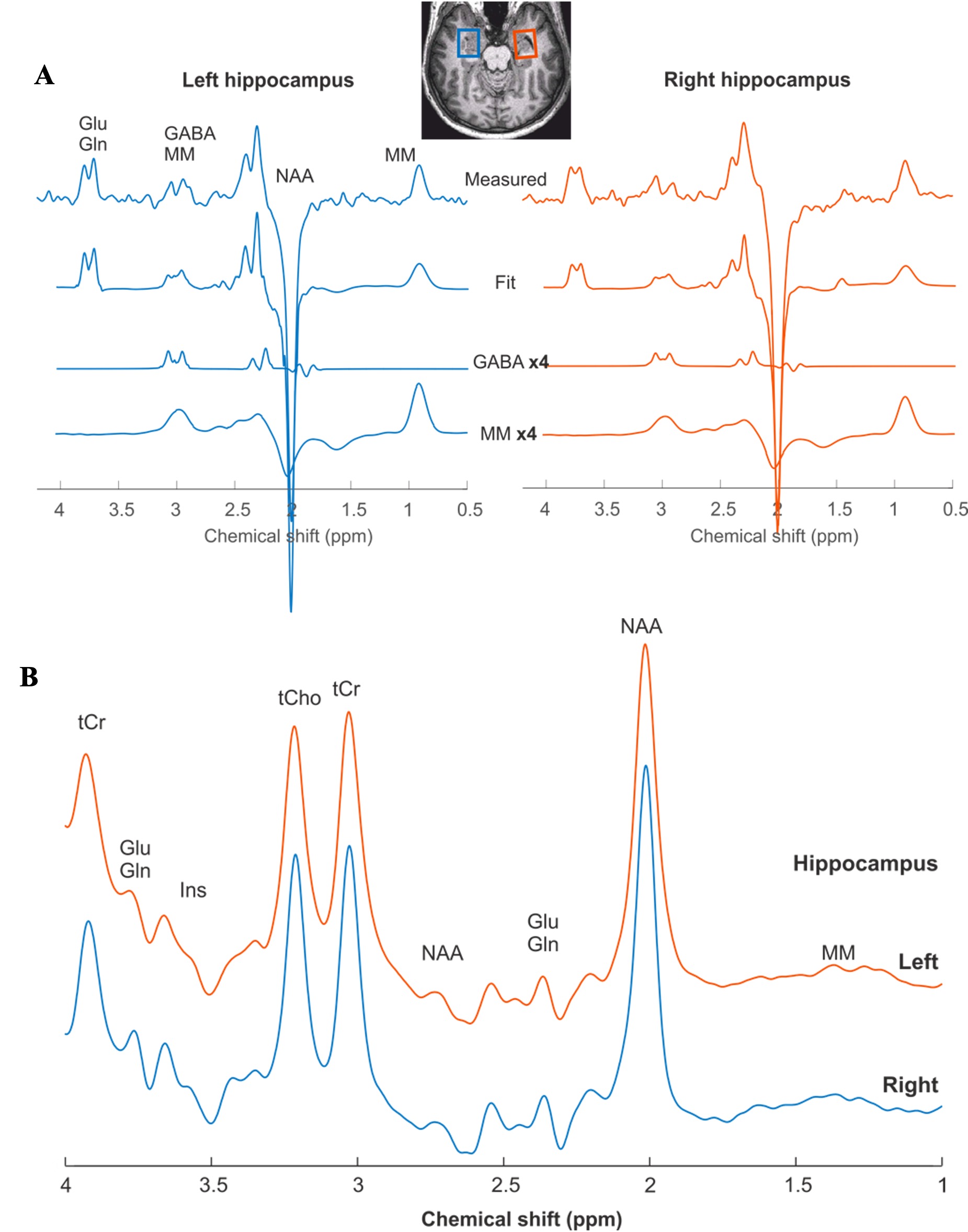
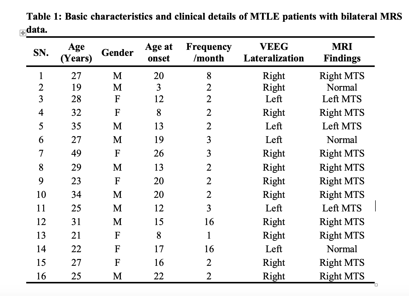
Drug-resistant unilateral TLE patients (n=25) diagnosed based on clinical features, MRI Brain, and Video-EEG and aged between 18-50 years were included in the study (Table 1). Bilateral single-voxel MEGA-PRESS (Figure 1) data was acquired from MTL on 3.0T MRI (Siemens Biograph).In-vivo MRI and MEGA-PRESS Spectroscopy Data Acquisition
Structural anatomical images were acquired using a T1-MPPRAGE to place spectral voxel. MRS data was acquired using a MEGA-PRESS edited pulse involving the collection of two interleaved datasets that differ in their treatment of the GABA spin, and MEGA-editing was achieved with 20-ms Gaussian editing pulses applied at 1.9 ppm and 7.5 ppm in an interleaved fashion6,7, and following acquisition parameters: were used: TR/TE=2260/68ms, Flip-angle =900, voxel-size=25x25x20mm, number of averages=128. VAPOR technique8 for water suppression, interleaved OVS modules to remove unwanted coherences and minimize signal outside the VOI, and FAST(EST) MAP for 1st and 2nd order B0 shimming were applied9. An unsuppressed water spectrum (n=16) was acquired as a water reference for eddy current correction and water scaling for absolute neurometabolites quantification.MRI and MRS Post-processing
MRSpa10 and LC-Model11-12 were used for postprocessing and neurometabolites quantification (Figure 1). SPM/FreeSurfer13,14 was used for tissue segmentation from the hippocampus and spectroscopic voxel. Segmented volumes were used for tissue metabolite, CSF correction, and volumetric measurement15-17.Statistical Analysis
Descriptive statistics were expressed using mean, standard deviation, percentages, and frequencies. Measurements of metabolite peaks, alterations, and metabolite ratios from the volume of interest were compared between seizure lateralized sides with opposite sides. Data were compared statistically using Mann-Whitney U, Wilcoxon signed-rank, and Kruskal-Wallis tests to analyze differences in neurometabolic between affected and normal sides and correlations with clinical parameters, including seizure profile, history, and disease duration. Spearman's correlation coefficient was performed between metabolites and ratio correlated with hippocampal volume. Hosmer and Lemeshow's model fits data for concordance analysis. Linear regression was done to assess correct classification for lateralization. A p-value of <0.05 was considered to be statistically significant.Results
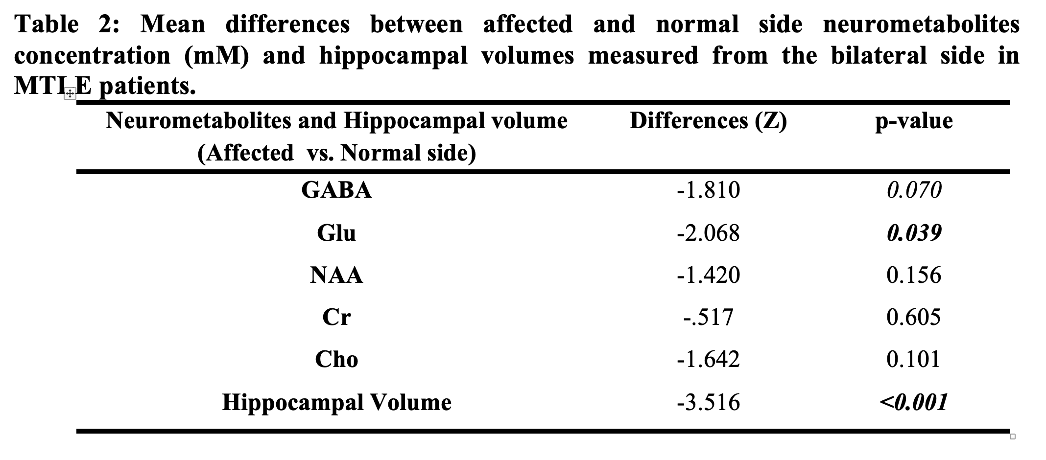
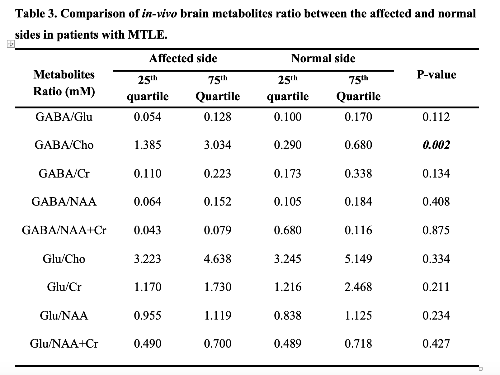
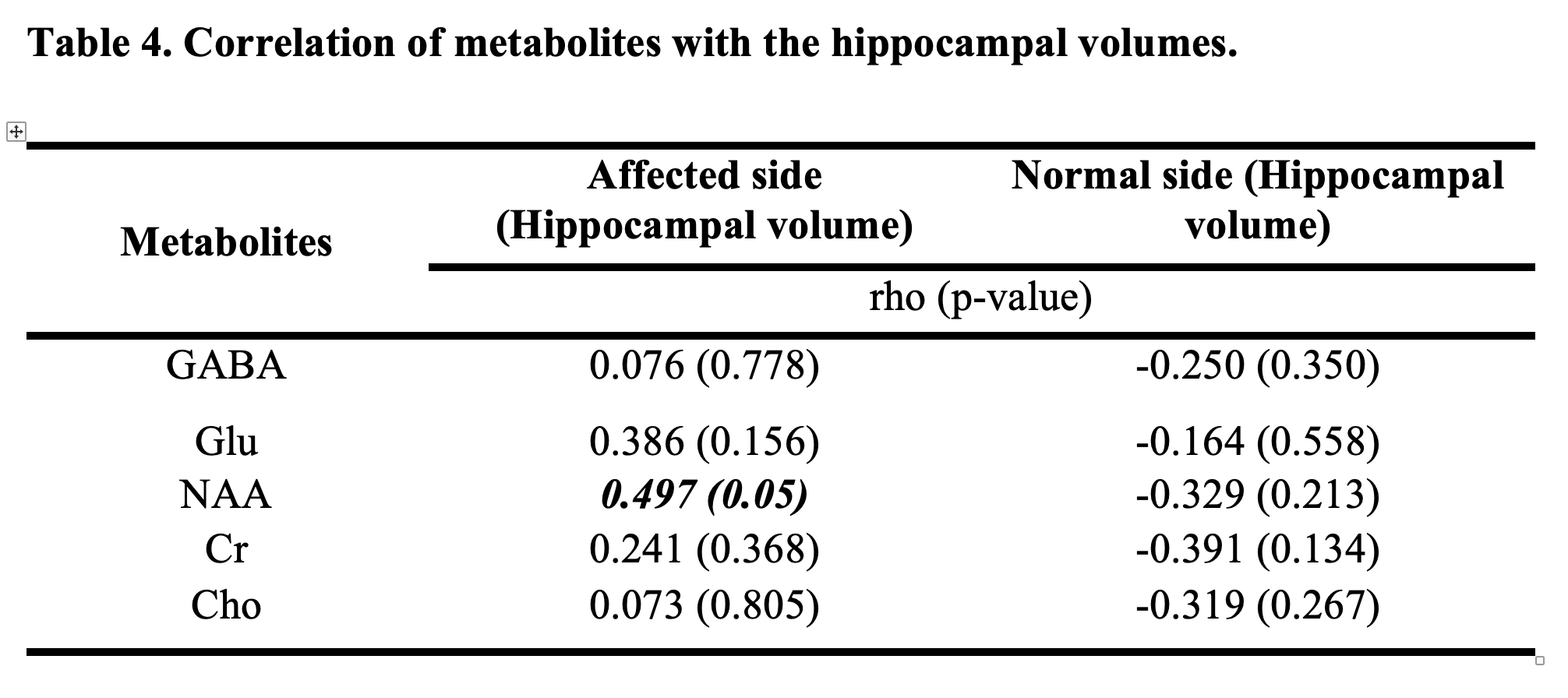
GABA, Glu, NAA, Cr, and Cho neurometabolites were computed. The group-wise statistical comparison revealed a significantly reduced NAA level from the affected compared to the normal side of the hippocampus (p-value=0.039). We observed reduced GABA (p-value=0.07) values and hippocampal volume (p-value<0.001) from the affected side (Table 2). A significant negative correlation was noted between affected-side hippocampal volume and NAA concentration (rho=-0.497, p-value=0.05). Glu/NAA ratios from both affected (rho=-0.692, p-value=0.012) and normal-sides (rho=-0.608, p-value=0.016) of the hippocampus were negatively correlated with age (Tables 3 and 4). Concordance between neurometabolites with video-EEG for lateralization demonstrates that the correct classification percentage for GABA was 86.7%, indicating an 86.7% chance that GABA will be able to lateralize the unaffected side as detected by VEEG.Discussion
This study explored the role of GABA, Glu, and their ratio in the temporal lobe of drug-resistant TLE patients and evaluated its concordance with other presurgical modalities like Video-EEG, MRI, and PET. Multiple studies have demonstrated that neuronal loss reduces the intracellular glutamate concentration associated with mTLE in various pathological conditions18-20. The GABA, Glu concentration, and hippocampal volume were lower in the ipsilateral temporal lobe compared to the contralateral temporal lobe. Authors have reported an increased GABA/Cr ratio in a subgroup of patients with a long duration of epilepsy and high seizure frequency21. Similarly, Simister et al. reported alteration in neurometabolites following a seizure episode, significantly increasing the Glx/Cr ratio22. The GABA/Glu ratio was lower in patients with a higher frequency of seizures/month observed in the current study. However, hippocampal volume showed better predictability for lateralizing an epileptic zone in TLE than GABA, Glu, and GABA/Glu ratios.Conclusion
A novel exploratory study demonstrates an alternation of in-vivo GABA and Glu levels and hippocampal volume. This study may provide a direction for utilizing MEGA-PRES as a presurgical tool for assessing in-vivo neurometabolic profiles. This study also added knowledge on the role of GABA and Glu in epilepsy and its interplay.Acknowledgements
Acknowledgments: We thank all the participants and their family members for their time and willingness to participate in the study. We also thank our MRI technologists for their extended technical support in MRS data acquisition.References
1. Kwan P, Sander JW. The natural history of epilepsy: an epidemiological view. J Neurol Neurosurg Psychiatry 2004;75:1376–81.
2. Schmidt D, Loscher W. Drug Resistance in Epilepsy: Putative Neurobiologic and Clinical Mechanisms. Epilepsia 2005;46:858–77.
3. Zhao F et al. Neuropsychological deficits in temporal lobe epilepsy: A comprehensive review. Ann Indian Acad Neurol 2014;17:374–82.
4. Thom M. Review: Hippocampal sclerosis in epilepsy: a neuropathology review. Neuropathol Appl Neurobiol 2014;40:520–43.
5. Curia G, et al. Pathophysiogenesis of mesial temporal lobe epilepsy: is prevention of damage antiepileptogenic? Curr Med Chem 2014;21:663–88.
6. Deelchand DK, Marjańska M, Henry P, et al. MEGA‐PRESS of GABA+: Influences of acquisition parameters. NMR in Biomedicine 2021;34.
7. Branzoli F, Deelchand DK, Sanson M, et al. In vivo 1 H MRS detection of cystathionine in human brain tumors. Magn Reson Med 2019;82:1259–65.
8. Tkác I, et al. In vivo 1H NMR spectroscopy of rat brain at 1 ms echo time. Magn Reson Med 1999;41:649–56.
9. Gruetter R, Tk I. Field mapping without reference scan using asymmetric echo-planar techniques. Magn Reson Med 2000;43:319–23.
10. MRspa: Magnetic Resonance signal processing and analysis.(https://www.cmrr.umn.edu/downloads/mrspa/).
11. Provencher SW. Automatic quantitation of localized in-vivo1H spectra with LCModel. NMR Biomed 2001;14:260–4.
12. Provencher SW. Estimation of metabolite concentrations from localized in vivo proton NMR spectra. Magn Reson Med 1993;30:672–9.
13. Fischl B. FreeSurfer. NeuroImage 2012;62:774–81.
14. Sämann PG, et al. FreeSurfer-based segmentation of hippocampal subfields: A review of methods and applications, with a novel quality control procedure for ENIGMA studies and other collaborative efforts. Hum Brain Mapp 2022;43:207–33.
15. Peek AL, et al. A comprehensive guide to MEGA-PRESS for GABA measurement. Anal Biochem 2023;669:115113.
16. Van Veenendaal TM, et al. Glutamate quantification by PRESS or MEGA-PRESS: Validation, repeatability, and concordance. Magn Reson Imaging 2018;48:107–14.
17. Deelchand DK, McCarten JR, Hemmy LS, et al. Changes in the intracellular microenvironment in the aging human brain. Neurobiology of Aging 2020;95:168–75.
18. Bartnik-Olson BL, Alger JR, Babikian T, et al. The clinical utility of proton magnetic resonance spectroscopy in traumatic brain injury: recommendations from the ENIGMA MRS working group. Brain Imaging and Behavior 2021;15:504–25.
19. Muhlert N, Atzori M, De Vita E, et al. Memory in multiple sclerosis is linked to glutamate concentration in grey matter regions. J Neurol Neurosurg Psychiatry 2014;85:833–9.
20. Kaiser LG, Schuff N, Cashdollar N, et al. Age-related glutamate and glutamine concentration changes in normal human brain: 1H MR spectroscopy study at 4 T. Neurobiology of Aging 2005;26:665–72.
21. He C, Liu P, Wu Y, et al. Gamma-aminobutyric acid (GABA) changes in the hippocampus and anterior cingulate cortex in patients with temporal lobe epilepsy. Epilepsy Behav 2021;115:107683.
22. Simister RJ, McLean MA, Salmenpera TM, et al. The effect of epileptic seizures on proton MRS visible neurochemical concentrations. Epilepsy Res 2008;81:36–43.
Figures