3077
Employ diffusion kurtosis imaging in conjunction with the XGBoost model to unveil white matter abnormalities in pediatric autism1Department of Radiology, The Third Affiliated Hospital of Zhengzhou University, Zhengzhou, China, 2MR Research China, GE Healthcare, Beijing, China, 3Department of Radiology, Henan Provincial People's Hospital, Zhengzhou, China
Synopsis
Keywords: White Matter, White Matter, Autism Spectrum Disorder; eXtreme Gradient Boosting; Tract-Based Spatial Statistics
Motivation: The diagnosis of Autism Spectrum Disorder (ASD) poses a substantial challenge, primarily due to the lack of a definitive biomarker.
Goal(s): Our primary objective is to uncover white matter irregularities prevalent in pediatric autism.
Approach: Our study leveraged Tract-Based Spatial Statistics (TBSS) analysis to scrutinize deviations in the white matter microstructure in ASD, and we implemented an eXtreme Gradient Boosting (XGBoost) model to effectively differentiate between individuals with ASD and healthy controls.
Results: Through the TBSS analysis, we identified notable disparities between groups. Moreover, the XGBoost model demonstrated exceptional proficiency in accurately classifying individuals with ASD and healthy controls.
Impact: This study delved into the white matter microstructural alterations in individuals with ASD by examining DKI data and its associated white matter tract integrity (WMTI) metrics. Additionally, our machine learning findings offered fresh perspectives toward objectively diagnosing ASD.
Introduction
Autism Spectrum Disorder (ASD) is a neurodevelopmental condition characterized by repetitive behaviors and social impairments [1]. Its prevalence ranges from 1% to 2.27% [2, 3]. Currently, the exact etiology of ASD remains unclear, and its diagnosis primarily relies on psychological and behavioral assessments. The need for a reliable and objective biomarker for ASD is pressing.Numerous studies have highlighted white matter abnormalities in ASD. Conventional diffusion tensor imaging (DTI) analyses often reveal decreased fractional anisotropy (FA) and increased mean diffusivity (MD) [4, 5]. In contrast, diffusion kurtosis imaging (DKI) employs multiple b-values and directions to quantify the non-Gaussian distribution of water molecule diffusion [6], which offers greater sensitivity in detecting white matter microstructural irregularities and assessing brain maturation [7]. However, both the DTI and DKI models are purely statistical in nature and lack the specificity required for assessing white matter microstructure. DKI's white matter diffusion model distinguishes the diffusion of water molecules in white matter into intra-axonal and extra-axonal components, allowing for their separate measurement [8]. This approach provides valuable insights into white matter tract integrity (WMTI), the extra-axonal environment, myelination [9], and white matter maturation [10]. Compared to diffusion tensor (DT) and diffusion kurtosis (DK) indicators, it enhances the specificity in detecting white matter microstructural abnormalities. Notably, WMTI indicators can be extracted from standard DKI data.
In this study, we utilized tract-based spatial statistics (TBSS) [11] to investigate alterations in the white matter microstructure of children with ASD. Additionally, we constructed an XGBoost model [12] to effectively classify individuals with ASD and healthy controls.
Methods and Materials
We gathered MR images from a cohort consisting of 62 children with ASD and 44 healthy control subjects. All MRI data were acquired using a 3.0 Tesla MRI scanner (SIGNA PIONEER, GE Healthcare, Waukesha). Subsequently, we computed various diffusion metrics, including diffusion tensor (DT), diffusion kurtosis (DK), and WMTI metrics, using the PyDesigner procedure [13].The DK metrics, which we calculated, encompassed mean kurtosis (MK), axial kurtosis (AK), radial kurtosis (RK), and kurtosis fractional anisotropy (FAK). Additionally, we derived a range of WMTI metrics, such as axonal water fraction (AWF), axonal diffusivity of the extra-axonal space (EAS_AD), radial diffusivity of the extra-axial space (EAS_RD), tortuosity of the extra-axonal space (EAS TORT), and diffusivity of the intra-axonal space (IAS_Da).
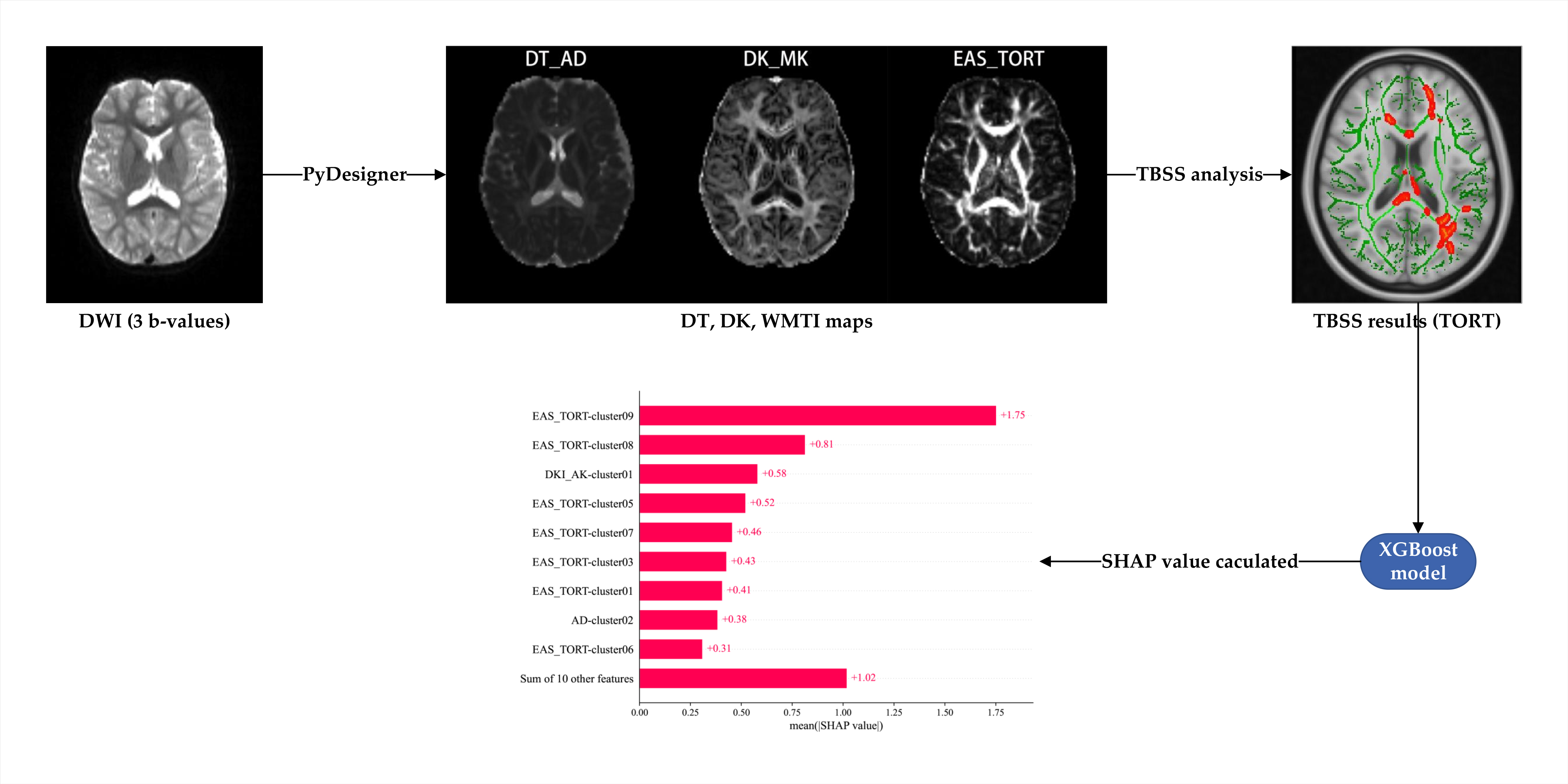
Moreover, we established an XGBoost model to classify children with ASD and their healthy counterparts, utilizing metrics that demonstrated significant differences between the two groups. Then, we calculated AUC, which was verified by the Bootstrap method. Additionally, we developed a logistic model, employing a single diffusion metric to assess the diagnostic performance of each individual indicator. Finally, we calculated the Shapley Additive explanations (SHAP) values for each feature to gauge their respective importance in the model's predictive capabilities. Pipeline of this study can be seen in Figure 1.
Results
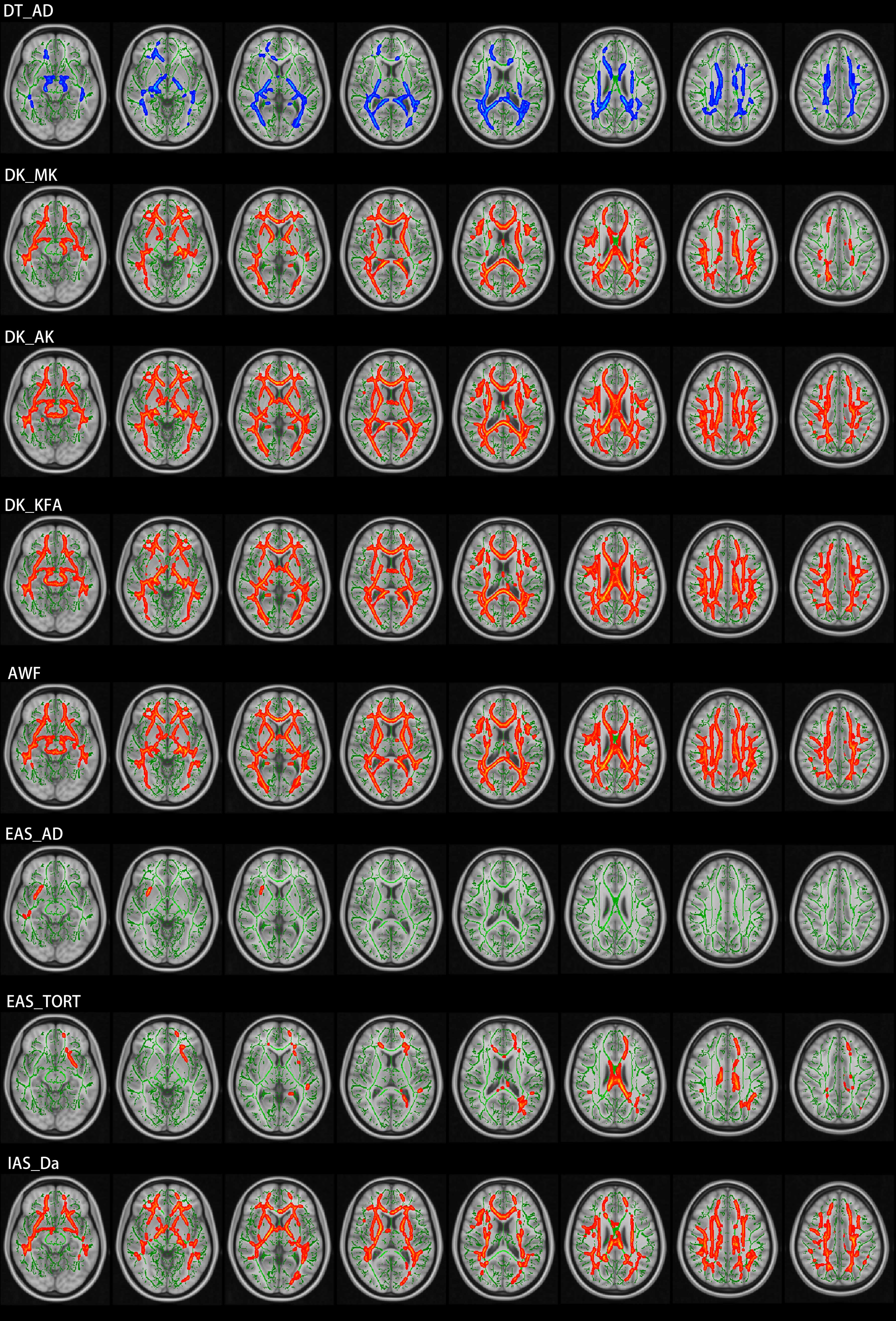
In our TBSS analysis, we observed a noteworthy decrease in axonal diffusivity (AD) within the left posterior corona radiata and the right superior corona radiata. Among the DK indicators, children with ASD displayed substantial increases in MK, AK, and FAK (Figure 2), while there was no significant difference in RK.Regarding WMTI metrics, we observed significant increases in AWF, EAS_AD, EAS TORT, and IAS_Da (Figure 2). These significant differences primarily clustered in the corpus callosum and fornix. Conversely, there was no significant difference in EAS_RD.
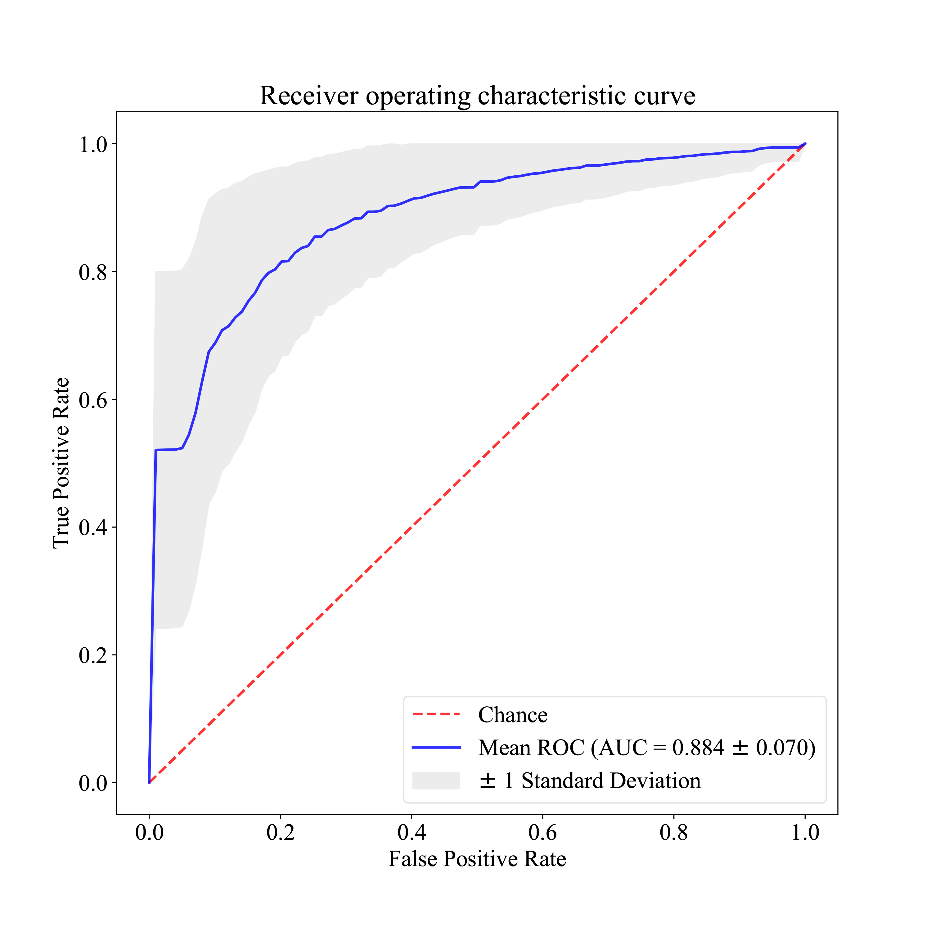
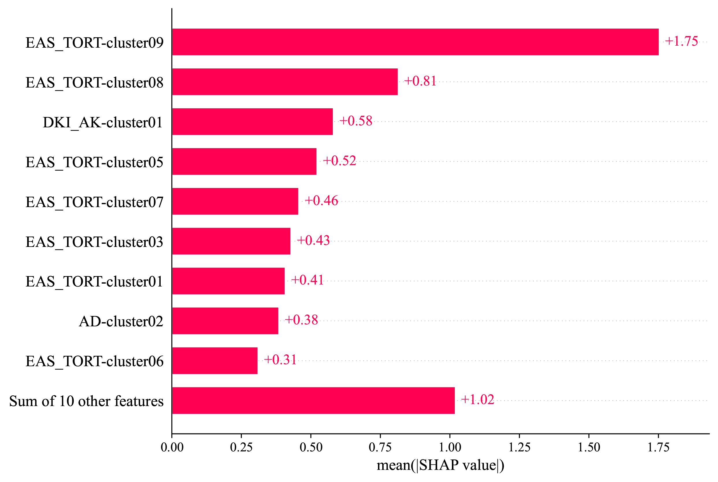
The XGBoost model we constructed demonstrated outstanding performance in classifying individuals with ASD and healthy controls (Figure 3). Furthermore, when assessing the importance of features within the XGBoost model using SHAP values, EAS_TORT emerged as the most influential, indicating its substantial contribution to the model's predictive performance (Figure 4).
Discussion and Conclusion
We conducted TBSS analyses utilizing DT, DK, and WMTI parameters extracted from DKI data. Our primary objective was to assess white matter abnormalities in children with ASD. The results revealed a noteworthy increase in DK and WMTI parameters, shedding light on potential mechanisms underlying early hyper-development in children with ASD. Furthermore, the machine learning model's performance was notably superior when utilizing the multi-shell diffusion model to classify individuals with ASD and healthy controls.In the context of SHAP values based on the XGBoost model, the EAS_TORT and AK indicators emerged as the most influential contributors to the model's predictions. These findings provide valuable insights into the nature of white matter abnormalities in young children with ASD and offer potential avenues for identifying biomarkers of ASD through machine learning techniques.
Acknowledgements
No acknowledgmentReferences
1. Lauritsen, M.B., Autism spectrum disorders. Eur Child Adolesc Psychiatry, 2013. 22 Suppl 1: p. S37-42.
2. Baio, J., et al., Prevalence of Autism Spectrum Disorder Among Children Aged 8 Years - Autism and Developmental Disabilities Monitoring Network, 11 Sites, United States, 2014. MMWR Surveill Summ, 2018. 67(6): p. 1-23.
3. Zeidan, J., et al., Global prevalence of autism: A systematic review update. Autism Res, 2022. 15(5): p. 778-790.
4. Ameis, S.H. and M. Catani, Altered white matter connectivity as a neural substrate for social impairment in Autism Spectrum Disorder. Cortex, 2015. 62: p. 158-81.
5. Travers, B.G., et al., Diffusion tensor imaging in autism spectrum disorder: a review. Autism Res, 2012. 5(5): p. 289-313.
6. Jensen, J.H., et al., Diffusional kurtosis imaging: the quantification of non-gaussian water diffusion by means of magnetic resonance imaging. Magn Reson Med, 2005. 53(6): p. 1432-40.
7. Umesh Rudrapatna, S., et al., Can diffusion kurtosis imaging improve the sensitivity and specificity of detecting microstructural alterations in brain tissue chronically after experimental stroke? Comparisons with diffusion tensor imaging and histology. Neuroimage, 2014. 97: p. 363-73.
8. Fieremans, E., J.H. Jensen, and J.A. Helpern, White matter characterization with diffusional kurtosis imaging. Neuroimage, 2011. 58(1): p. 177-88.
9. Fieremans, E., et al., Diffusion distinguishes between axonal loss and demyelination in brain white matter. Proc. Int. Soc. Magn. Reson. Med., 2012. 20.
10. Jelescu, I.O., et al., One diffusion acquisition and different white matter models: how does microstructure change in human early development based on WMTI and NODDI? Neuroimage, 2015. 107: p. 242-256.
11. Smith, S.M., et al., Tract-based spatial statistics: voxelwise analysis of multi-subject diffusion data. Neuroimage, 2006. 31(4): p. 1487-505.
12. Chen, T. and C. Guestrin, XGBoost: A Scalable Tree Boosting System, in Proceedings of the 22nd ACM SIGKDD International Conference on Knowledge Discovery and Data Mining. 2016, Association for Computing Machinery: San Francisco, California, USA. p. 785–794.
13. Siddhartha, D., et al., PyDesigner: A Pythonic Implementation of the DESIGNER Pipeline for Diffusion Tensor and Diffusional Kurtosis Imaging. bioRxiv, 2021: p. 2021.10.20.465189.
Figures