3075
Imaging Tumor Metabolism in Patients with Head & Neck Squamous Cell Carcinoma1Department of Imaging Physics, The University of Texas MD Anderson Cancer Center, Houston, TX, United States, 2The University of Texas MD Anderson Cancer Center UTHealth Graduate School of Biomedical Sciences, Houston, TX, United States, 3Department of Neuroradiology, The University of Texas MD Anderson Cancer Center, Houston, TX, United States, 4Department of Head & Neck Surgery, The University of Texas MD Anderson Cancer Center, Houston, TX, United States, 5Clinical Research Imaging, The University of Texas MD Anderson Cancer Center, Houston, TX, United States, 6Imaging Physics Residency Program, The University of Texas MD Anderson Cancer Center, Houston, TX, United States, 7Department of Radiation Oncology, The University of Texas MD Anderson Cancer Center, Houston, TX, United States, 8Department of Thoracic-Head & Neck Medical Oncology, The University of Texas MD Anderson Cancer Center, Houston, TX, United States, 9Center for Advanced Biomedical Imaging, The University of Texas MD Anderson Cancer Center, Houston, TX, United States, 10Bobby R. Alford Department of Otolaryngology - Head and Neck Surgery, Baylor College of Medicine, Houston, TX, United States
Synopsis
Keywords: Hyperpolarized MR (Non-Gas), Hyperpolarized MR (Gas), pyruvate, metabolism, head and neck, cancer
Motivation: Changes in metabolic imaging biomarkers offer tremendous potential for assessing response and optimizing treatment for patients with head and neck squamous cell carcinoma (HNSCC).
Goal(s): To assess the translational viability of metabolic MRI with hyperpolarized (HP) pyruvate in patients with HNSCC.
Approach: Patients with HNSCC are recruited into a prospective imaging study and scanned with HP [1-13C]-pyruvate in a test-retest paradigm before start of therapy.
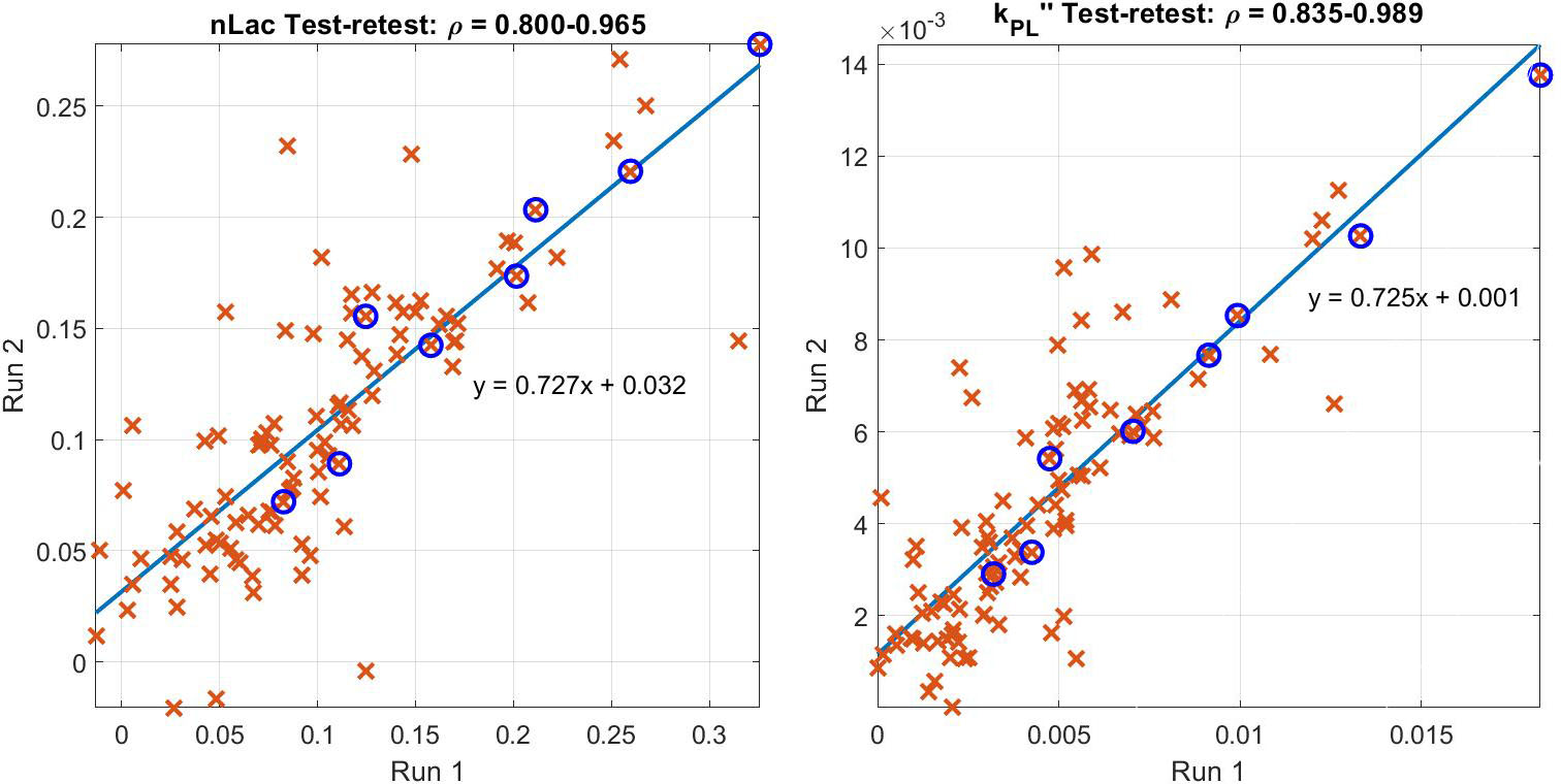
Results: Preliminary data indicates that reproducibility of semi-quantitative measures of tumor metabolism are high, with correlation between test-retest measurements >80% in all voxels that exceed a minimum SNR threshold and correlation >96% in regions identified as tumor.
Impact: These results demonstrate that metabolic MRI with hyperpolarized [1-13C]-pyruvate generates sufficient SNR and spatiotemporal resolution to permit reproducible measurement of tumor metabolism in patients with HNSCC.
Introduction
Altered metabolism is a hallmark of cancer, and metabolic shifts can provide important insight into tumor aggressiveness and response to therapy1-3. Metabolic MRI with hyperpolarized (HP) pyruvate provides a new mechanism for assessing tumor metabolism, and the clinical utility of this technology is currently being studied in the context of many different tumor sites and subtypes4. In this work, we seek to assess the clinical feasibility of HP MRI in patients with head and neck squamous cell carcinoma (HNSCC).Methods
All data that has been acquired in this study has been reviewed and approved by the FDA and our IRB. Patients are positioned head first and supine in the scanner inside the 13C “clamshell” transmit coil and with two four-channel 13C “paddle” array coils placed on either side of the patients’ oral cavity. Anatomic localizers were acquired through the 1H body coil. 250mM HP [1-13C]-pyruvate (0.43 mL/kg) was prepared using a SpinLab dDNP system (GE Healthcare). At the start of bolus injection, dynamic HP MRI data was acquired using a broad-band EPI sequence with spectral/spatial excitation pulses5. In this study, patients are administered two doses of HP pyruvate over a ~30-min interval during a single imaging session. SNR-thresholded dynamic data was analyzed on a voxel-by-voxel basis and the consistency of the two acquisitions was tested by assessing the correlation between measurements.Results
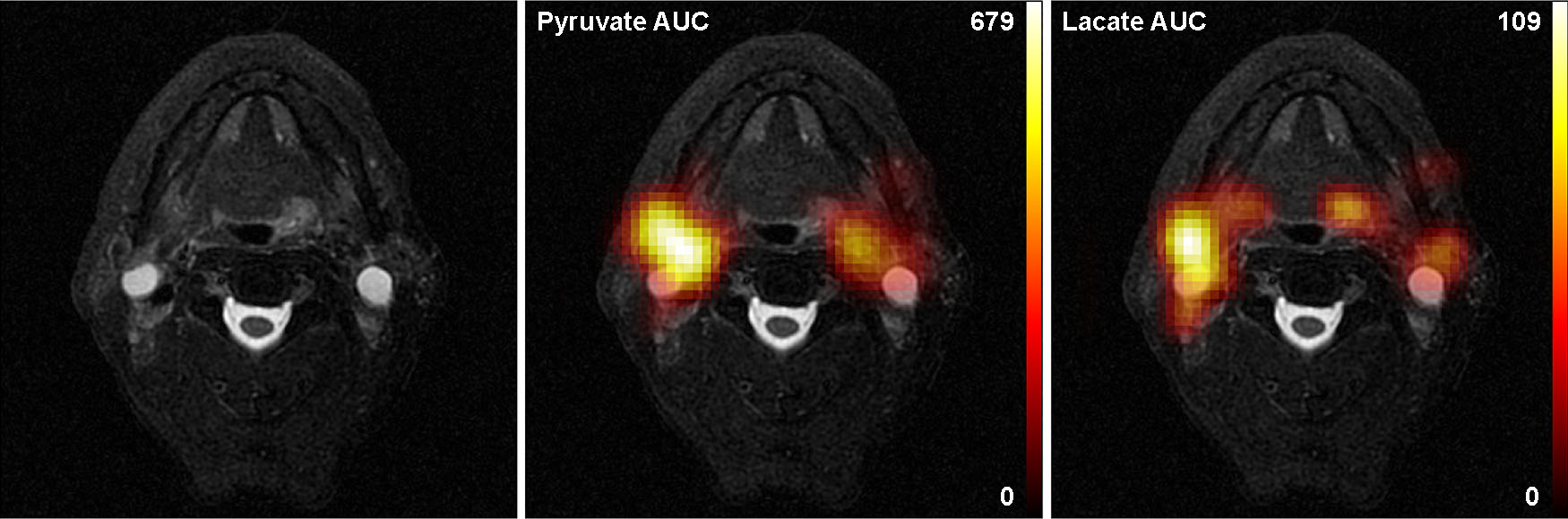
The test-retest paradigm was well tolerated without complications. Preliminary results demonstrate that dynamic HP MRI data is observed with sufficient SNR and image resolution to assess metabolism in primary tumor and in metastatic lymph nodes (Figure 1). Voxels that exceeded a threshold for total carbon SNR >10 showed a strong correlation between measurements (ρ>0.8) while voxels that are identified as being comprised of at least 30% tumor and tcSNR>10 showed a correlation of ρ>0.96 (Figure 2).Discussion
Metabolic MRI with HP pyruvate provides a minimally invasive mechanism for longitudinal assessment of tumor metabolism, enabling calculation of metabolic imaging biomarkers that can inform on disease aggressiveness and response to therapy. In patients with HNSCC, the resolution of these scans (1.5cm x 1.5cm in-plane) is sufficient to assess metabolism in the primary tumor and in nearby metastatic lymph nodes. General-purpose 13C coils that were used in this work are not optimized for this anatomic region; we anticipate that coils optimized for scanning the oral cavity and oropharynx would significantly improve the sensitivity and reproducibility of these measurements in future studies which will test the deployment of this technology at scale with measurements before and during treatment.Conclusions
These results support the clinical feasibility of HP MRI to assess tumor metabolism in patients with HNSCC.Acknowledgements
This work was supported in part by funding from the National Cancer Institute of the National Institutes of Health (R01CA211150, R01CA280980, U54CA274321) and GE Healthcare. This content is solely the responsibility of the authors and does not necessarily represent the views of its sponsors.References
- Yu W, Chen Y, Putluri N, Osman A, Coarfa C, Putluri V, Kamal AHM, Asmussen JK, Katsonis P, Myers JN, Lai SY, Lu W, Stephan CC, Powell RT, Johnson FM, Skinner HD, Kazi J, Ahmed KM, Hu L, Threet A, Meyer MD, Bankson JA, Wang T, Davis J, Parker KR, Harris MA, Baek ML, Echeverria GV, Qi X, Wang J, Frederick AI, Walsh AJ, Lichtarge O, Frederick MJ, Sandulache VC. Evolution of cisplatin resistance through coordinated metabolic reprogramming of the cellular reductive state. Br J Cancer 128(11):2013-24, 2023.
- Sandulache VC, Chen Y, Lee J, Rubinstein A, Ramirez MS, Skinner HD, Walker CM, Williams MD, Tailor R, Court LE, Bankson JA, Lai SY. Evaluation of hyperpolarized [1-¹³C]-pyruvate by magnetic resonance to detect ionizing radiation effects in real time. PLoS One 9(1):e87031, 2014.
- Chen Y, Maniakas A, Tan L, Cui M, Le X, Niedzielski JS, Michel KA, Harlan CJ, Lu W, Henderson YC, Mohamed ASR, Lorenzi PL, Putluri N, Bankson JA, Sandulache VC, Lai SY. Development of a rational strategy for integration of lactate dehydrogenase A suppression into therapeutic algorithms for head and neck cancer. Br J Cancer 124(10):1670-9, 2021.
- Larson PE, Bernard JM, Bankson JA, Bogh N, Bok RA, Chen AP, Cunningham CH, Gordon J, Hovener JB, Laustsen C, Mayer D, McLean MA, Schilling F, Slater J, Vanderheyden JL, von Morze C, Vigneron DB, Xu D, Group HCMC. Current Methods for Hyperpolarized [1-(13)C]pyruvate MRI Human Studies. Magn Reson Med, DOI: 10.1002/mrm.29875, forthcoming.
- Gordon JW, Chen HY, Autry A, Park I, Van Criekinge M, Mammoli D, Milshteyn E, Bok R, Xu D, Li Y, Aggarwal R, Chang S, Slater JB, Ferrone M, Nelson S, Kurhanewicz J, Larson PEZ, Vigneron DB. Translation of Carbon-13 EPI for hyperpolarized MR molecular imaging of prostate and brain cancer patients. Magn Reson Med 81(4):2702-9, 2019.
Figures