3073
Hyperpolarized 13C Metabolic Imaging in Human Subjects with Advanced Coronary Artery Disease: Initial Experience and Thoughts1Cardiovascular and Thoracic Surgery, UT Southwestern Medical Center, Dallas, TX, United States, 2Advanced Imaging Research Center, UT Southwestern Medical Center, Dallas, TX, United States, 3Radiology, UT Southwestern Medical Center, Dallas, TX, United States, 4Internal Medicine, UT Southwestern Medical Center, Dallas, TX, United States
Synopsis
Keywords: Hyperpolarized MR (Non-Gas), Hyperpolarized MR (Non-Gas), Coronary Artery Disease, FDG PET-CT, Ischemic Heart Disease
Motivation: Coronary Artery Disease (CAD) continues to be a significant health issue worldwide, necessitating enhanced diagnostic methods capable of imaging cardiac metabolism. Hyperpolarized Carbon-13 Magnetic Resonance Imaging (HP-13C MRI) offers a potential solution for the non-invasive evaluation of metabolism in the human heart.
Goal(s): Our goal is to demonstrate the feasibility of HP-13C MRI in two human subjects with advanced CAD.
Approach: By examining these subjects, we have successfully imaged metabolic abnormalities within the myocardium.
Results: Our results indicate promising potential for using this technology to visualize dynamic changes in cardiac energetics associated with CAD. These findings can potentially influence the management of CAD.
Impact: This study establishes HP 13C-MRI as a safe, non-invasive tool for visualizing metabolic abnormalities in CAD patients, paving the way for more precise management of ischemic heart disease and prompting further comparative research with traditional imaging methods.
Introduction
Coronary Artery Disease (CAD) remains a major global health challenge, demanding improved diagnostic methods capable of visualizing cardiac energetics1. Traditional imaging methods, such as PET scans, dobutamine stress echocardiography, etc., fall short of directly assessing mitochondrial metabolism. The hyperpolarized 13C-metabolic magnetic resonance imaging (HP-13C MRI), already proving its merit in imaging human hearts in clinical studies2,3,4, offers a promising avenue for the non-invasive investigation of mitochondrial function in CAD. In this study, we demonstrate the feasibility of using HP-13C MRI to image cardiac metabolism in human subjects with advanced CAD.Methods
We conducted an HP-13C MRI examination on two preoperative patients with advanced CAD. These individuals volunteered and provided informed consent according to a protocol approved by the Institutional Review Board and Protocol Review Committee. Subject 1, a 60-year-old male, exhibited obstructive coronary artery disease with significant stenosis in multiple arteries and occasional chest discomfort during physical activity. Subject 2, a 78-year-old male, had three-vessel coronary artery disease, accompanied by comorbid conditions such as hypertension and hyperlipidemia. Before the metabolic exam, baseline blood samples were collected and analyzed for pyruvate, triglycerides, free fatty acids, and insulin levels and were sent to the laboratory. After assessing vital signs and MRI screening, both subjects received 48 grams of glucose gel. The IND-approved metabolic probe, [1-13C] pyruvic acid, manufactured by Good Manufacturing Practice regulations, in clinical fluid paths (0.40 mL/kg body weight of 250-mM HP [1-13C] pyruvate solution). After 3-4 hours of polarization in a clinical polarizer (SPINlab™, GE Healthcare), we assessed pyruvate concentration, pH, temperature, volume, and radical concentration using a dedicated quality control (QC) device. The HP solution was administered intravenously to the subjects, followed by a saline flush. The 1H-SSFP-CINE images for anatomical reference and multi-echo images of [13C] bicarbonate, [1-13C] lactate, and [1-13C] pyruvate were obtained on a GE MR750 MR with transmit/receive Helmholtz loop-pair 13C coil (PulseTeq Limited, UK) as previously reported5. These images were acquired for the short axis during the first injection and for the long axis in the second HP injection. The HP 13C data were reconstructed and analyzed with MATLAB. Regions of interest (ROIs) with prominent 13C signals were delineated, and the average 13C signal within each ROI was subsequently modeled to calculate the ratio of bicarbonate and lactate.Results and Discussion
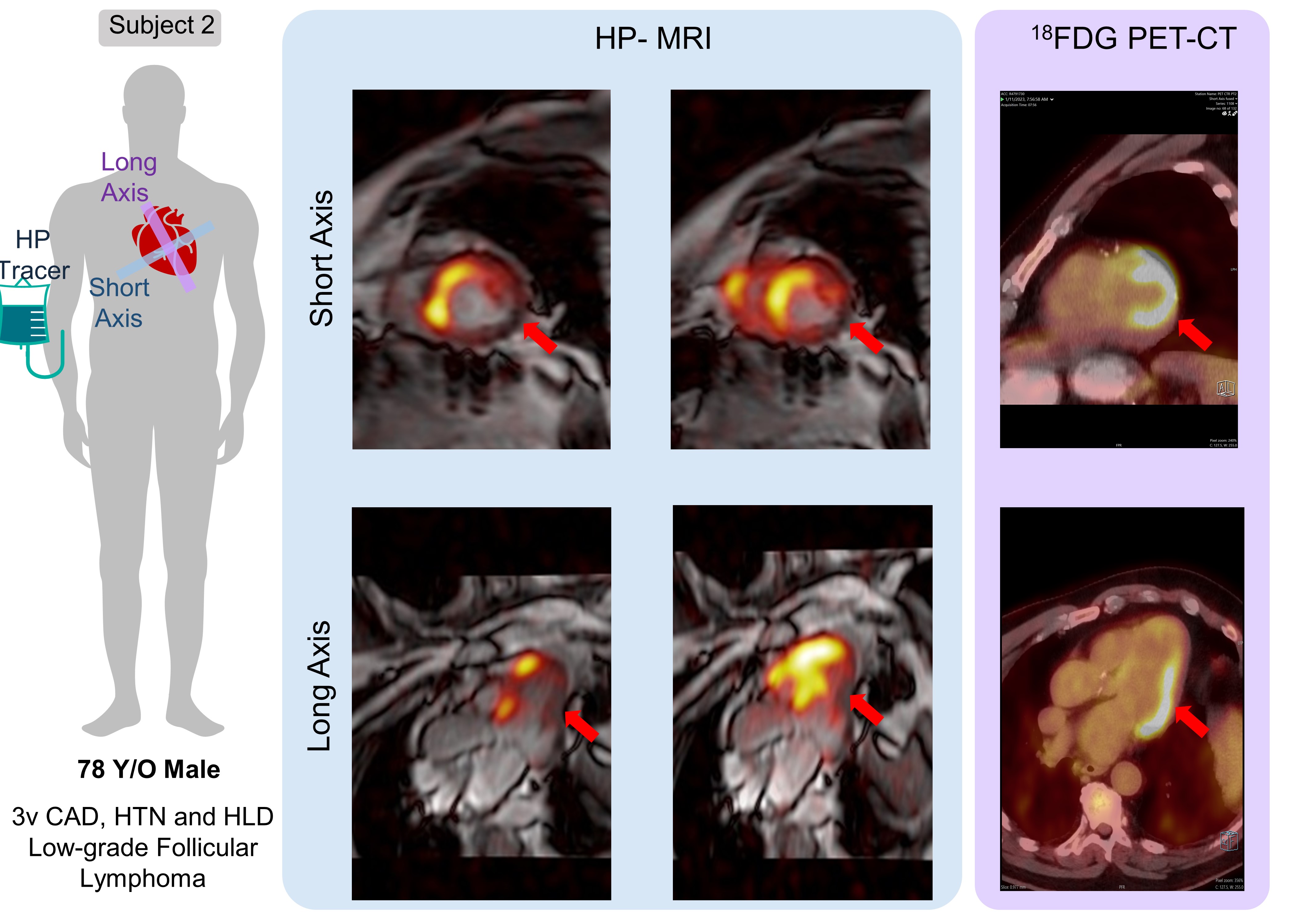
In this study, we used HP 13C-MRI to image hearts from two human subjects with advanced CAD. For Subject 1, a 60-year-old male with three-vessel diseases, dynamic 13C images were obtained in short (Fig. 2A) and long (Fig. 2B) axes. Subject 1 exhibited metabolic defect with reduced levels of 13C-bicarbonate and 13C-lactate in the mid-anterolateral regions. Figure 3 demonstrates the data from subject 2, a 78-year-old male with multiple conditions who displayed reduced bicarbonate and lactate in the mid-inferolateral myocardium. Comprehensive metabolic results, including bicarb/lactate ratios, glucose levels, and lab measurements, are depicted in Table 1. The bicarbonate/lactate ratios (Fig 4A) were similar between the short and long axes, indicating the reproducibility of results in different planes from two injections. The pre-and post-glucose load blood glucose measurements (Table 1.1) were within the normal range for both subjects. The blood measurements (Table 1.2) for pyruvate, triglycerides, free fatty acids, and insulin were also within the normal range for both subjects, except for insulin, which was elevated due to poor glucose control and significant carbohydrate load from breakfast in these subjects. Incidentally, we encountered 18F-FDG PET-CT data from subject 2 as this subject has low-grade follicular lymphoma. Figure 3 presents intriguing discordant results between PET-CT and HP 13C MRI data in this same subject. In regions where the 18F-FDG PET-CT scan reveals significant myocardial 18F-FDG uptake, the HP 13C MRI exam of the same subject displays contrasting data with reduced levels of bicarbonate and lactate. These contrasting results suggest that the two technologies (HP-MRI and PET-CT) may provide complementary information on myocardial energetics.Conclusion
Our HP 13C-MRI examination demonstrates the feasibility of imaging metabolic abnormalities in patients with ischemic heart disease, with both subjects tolerating this imaging well. This imaging method shows promise as a tool to visualize changes in cardiac energetics in hearts affected by advanced CAD. It may have a role in assessing areas of the myocardium with active mitochondrial function, which might influence management decisions in patients with this disease. Further research is warranted to evaluate the value of HP-MRI in ischemic heart disease and to compare the metabolic imaging capabilities of HP 13C-MRI with PET-CT and other currently available methods.Acknowledgements
We thank the American Heart Association (AHA Award Number: 23SCEFIA1154964 to Gaurav Sharma) for financial support. Additionally, we are thankful to Jennine Leary, RN, and Jeannie Baxter, RN, for their nursing support, as well as to Corey Mozingo and Daniel Tetrick for their MR technical support.References
1. Vaduganathan M, Mensah G, Turco J, et al. The Global Burden of Cardiovascular Diseases and Risk. J Am Coll Cardiol. 2022 Dec, 80 (25) 2361–2371.
2. Apps A, Lau JYC, Miller JJJJ, Tyler A, Young LAJ, Lewis AJM, Barnes G, Trumper C, Neubauer S, Rider OJ, Tyler DJ. Proof-of-Principle Demonstration of Direct Metabolic Imaging Following Myocardial Infarction Using Hyperpolarized 13C CMR. JACC Cardiovasc Imaging. 2021 Jun;14(6):1285-1288.
3. Rider OJ, Apps A, Miller JJJJ, Lau JYC, Lewis AJM, Peterzan MA, Dodd MS, Lau AZ, Trumper C, Gallagher FA, Grist JT, Brindle KM, Neubauer S, Tyler DJ. Noninvasive In Vivo Assessment of Cardiac Metabolism in the Healthy and Diabetic Human Heart Using Hyperpolarized 13C MRI. Circ Res. 2020 Mar 13;126(6):725-736.
4. Cunningham CH, Lau JY, Chen AP, Geraghty BJ, Perks WJ, Roifman I, Wright GA, Connelly KA. Hyperpolarized 13C Metabolic MRI of the Human Heart: Initial Experience. Circ Res. 2016 Nov 11;119(11):1177-1182.
5. Ma J, et al. Cardiac T2∗ measurement of hyperpolarized 13C metabolites using metabolite-selective multi-echo spiral imaging. Magn Reson Med. 2021 Sep;86(3):1494-1504.
Figures
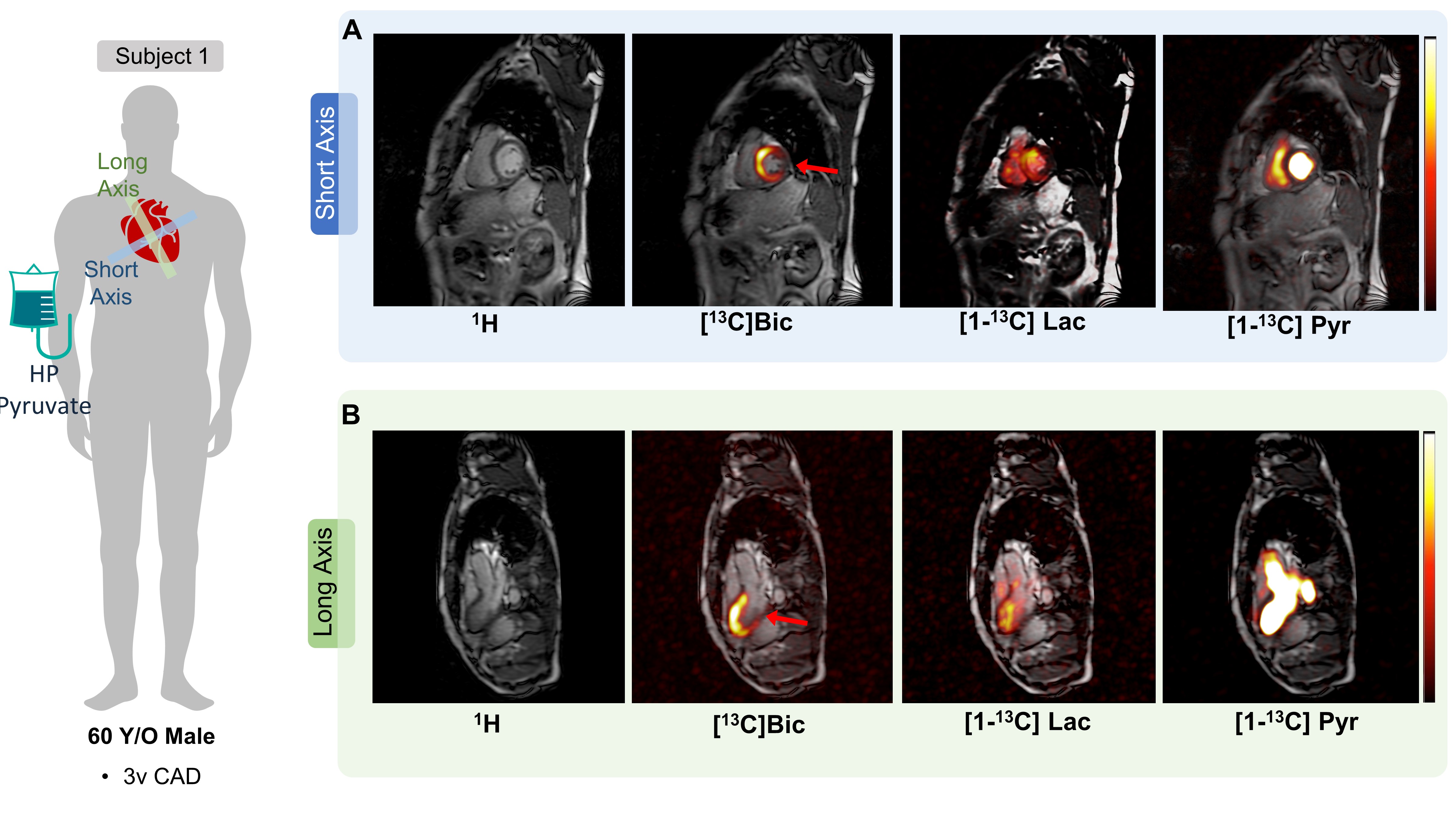
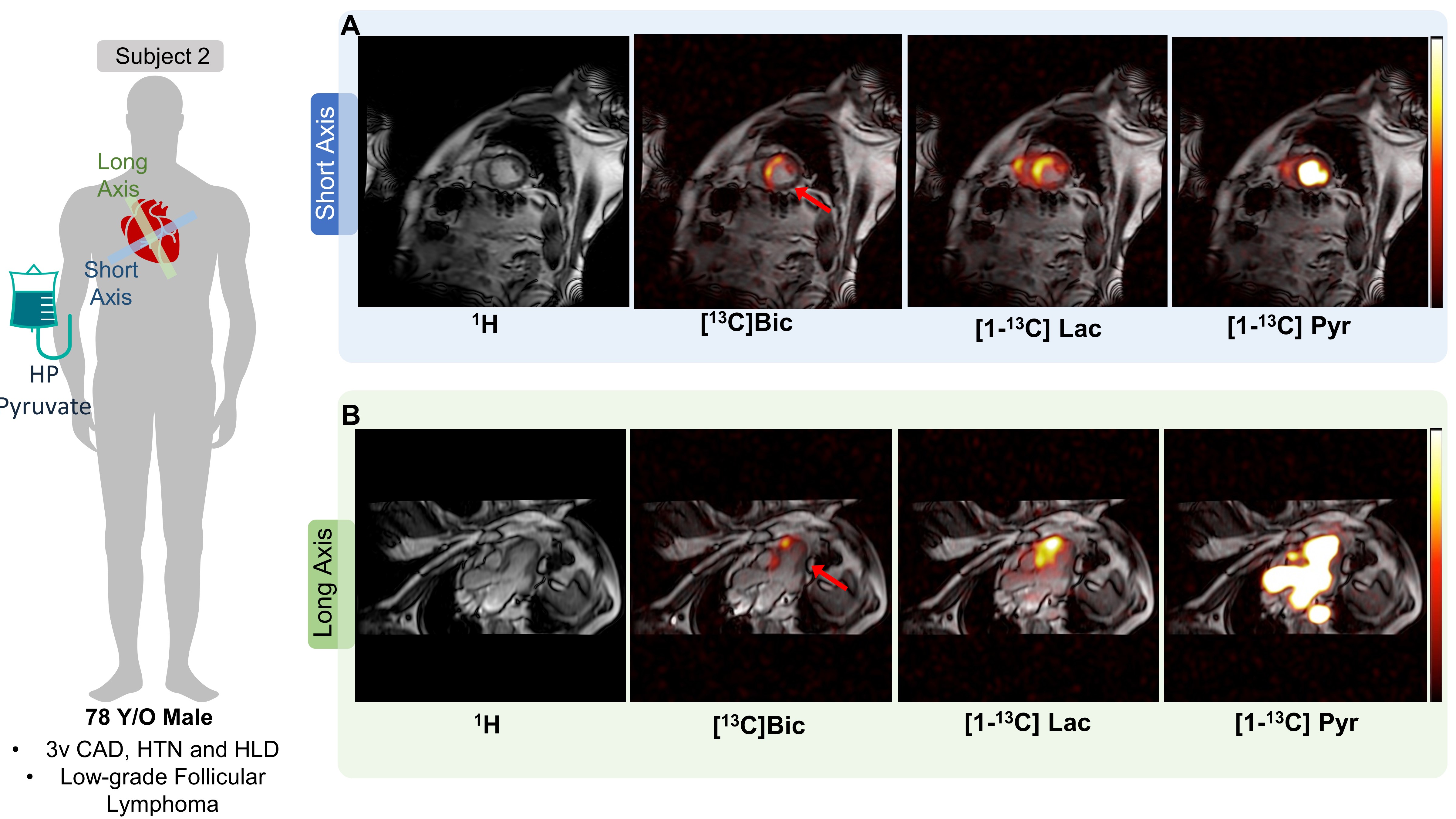
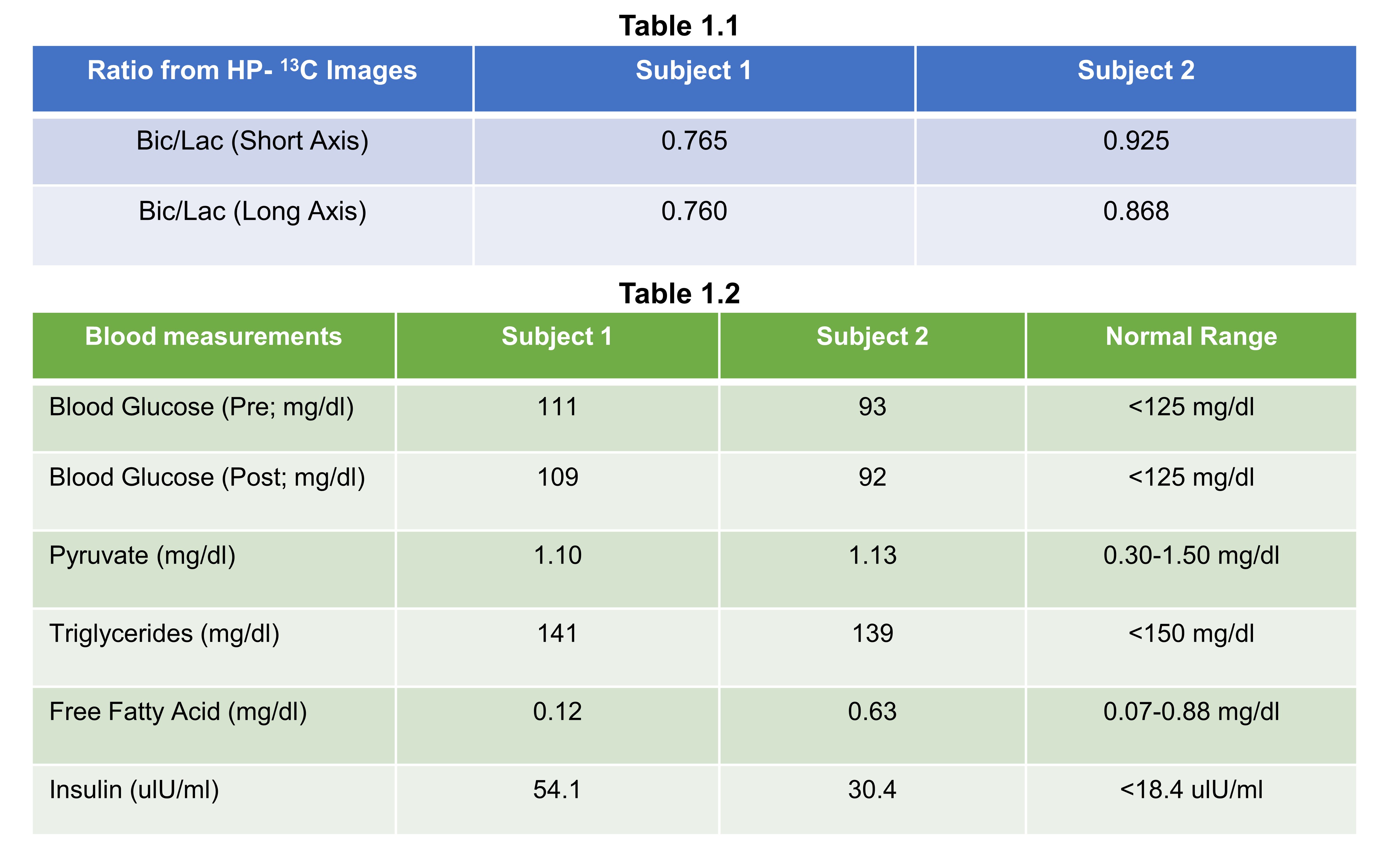
Table 1: Metabolic Examination Results. 1.1. Bicarb/lactate ratio calculated from short and long axes. 1.2. Blood glucose levels before (row 1) and after (row 2) a 45-minute glucose load preceding the metabolic examination; and laboratory measurements (rows 3-6) obtained before the metabolic examination for subjects 1 and 2, facilitating a metabolic correlation of findings. These measurements include pyruvate, triglycerides, free fatty acids, and insulin levels.