3069
Assessing Pre-Treatment Metabolic Profiles in High-Risk DLBCL via Hyperpolarized 13C-Pyruvate MR Spectroscopy1Clinical Metabolomics Core Laboratory, Chang Gung Memorial Hospital, Linkou, Taoyuan, Taiwan, 2Department of Medical Imaging and Intervention, Chang Gung Memorial Hospital, Linkou, Taoyuan, Taiwan, 3Research Center for Radiation Medicine, Chang Gung University, Taoyuan, Taiwan
Synopsis
Keywords: Hyperpolarized MR (Non-Gas), Metabolism
Motivation: The study addresses the need for early detection of treatment response in high-risk diffuse large B cell lymphoma, where traditional methods based on tumor size are inadequate.
Goal(s): The study aims to validate 13C-Pyruvate DNP MR spectroscopy as a sensitive technique for early evaluation of treatment efficacy in lymphoma patients.
Approach: Utilizing 13C MRS, the research monitors the metabolic conversion of 13C-labeled pyruvate to lactate, reflecting the treatment's impact on the tumor's metabolic activity.
Results: Preliminary data from three patients show different metabolic rates, indicating the potential of this method to discern early treatment responses, leading to more timely and effective clinical decisions.
Impact: These results could revolutionize protocols for lymphoma by enabling earlier assessment of therapy efficacy, thus informing more timely adjustments to treatment strategies. This advancement may spur further metabolic research and ultimately improving survival rates and quality of life for patients.
Introduction
Diffuse Large B Cell Lymphoma (DLBCL) is a prevalent malignancy with current treatment response assessments relying on tumor size changes post-therapy [1]. However, this method suffers from insensitivity and a lag in visible results, necessitating more advanced techniques like metabolic imaging [2]. Metabolic imaging, exploiting the Warburg effect, can potentially offer an early insight into treatment efficacy [3]. This study aims to utilize 13C-Pyruvate DNP MR Spectroscopy, a novel metabolic imaging approach that could improve early treatment response detection in high-risk DLBCL patients. Early detection is crucial as it can influence treatment decisions, which in turn affects morbidity and mortality. The aberrant metabolic pathways of tumor cells, particularly the heightened conversion of pyruvate to lactate, serve as the focal point for this study. This is the first known clinical investigation of its kind in lymphoma, aiming to establish a metabolic baseline pre-treatment and potentially aid in predicting response to therapy.Methods
This clinical study involved the recruitment of three male patients, IRB number 202002397A3C601A0, and TFDA:1110813745 for the drug safety, with newly diagnosed high-risk DLBCL. The production of hyperpolarized (HP) [1-13C]-pyruvate was carried out using sterile fluid path pharmacy kits under stringent conditions to ensure safety and maintain the integrity of the hyperpolarization process. The subsequent MR imaging was performed with a 3.0-T MRI scanner (Discovery MR750w; GE Healthcare), employing a flex RF coil optimized for the spleen's anatomy. Initially, a pulse-and-acquire sequence was employed to obtain the 13C whole spectrum, capturing a comprehensive range of metabolite resonances. Subsequently, an IDEAL spiral chemical shift imaging (CSI) sequence was applied, enhancing the detection and spatial mapping of 13C-labeled metabolites within the spleen. This two-step acquisition process allowed for detailed metabolic profiling, vital for understanding the treatment response. A kinetic model was used to calculate the apparent exchange rates from pyruvate to lactate in the spleen [4]. Additionally, the area under the curve (AUC) ratios of pyruvate, lactate, alanine, and bicarbonate were determined relative to the total carbon signal, providing a comprehensive metabolic profile.Results
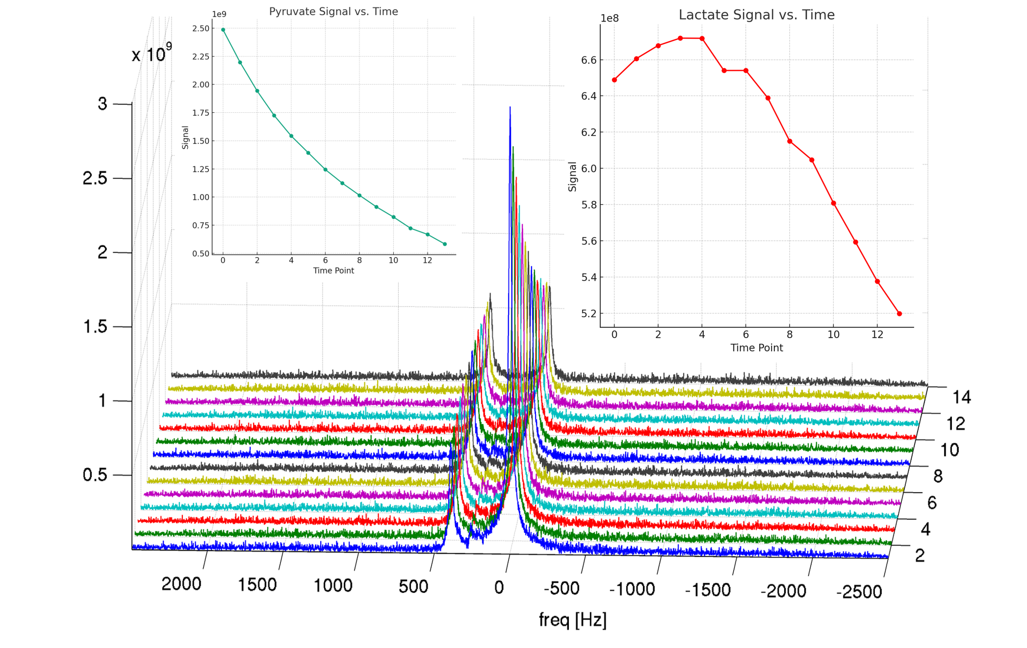
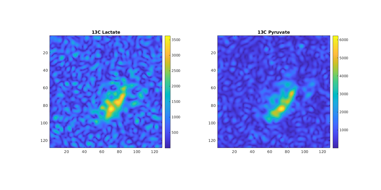
The apparent exchange rates from pyruvate to lactate for the patients were 0.012/s, 0.0081/s, and 0.019/s, respectively. These rates suggest variability in metabolic activity, which may correlate with differential treatment responses. The AUC ratios also varied among patients, indicating distinct metabolic profiles that could potentially be linked to the underlying tumor biology and responsiveness to treatment. The results showcase the ability of 13C-Pyruvate DNP MR Spectroscopy to detect nuanced metabolic changes in high-risk DLBCL patients, a crucial step towards personalized treatment strategies. Figure 1 presents the complete 13C spectrum and the specific plots for pyruvate and lactate across 14 acquisitions. Figure 2 provides the IDEAL Spiral CSI sequence images, illustrating the distribution of pyruvate and lactate, which may reflect the metabolic response to disease staus.Discussion
The variability in exchange rates and AUC ratios observed suggests that 13C-Pyruvate DNP MR Spectroscopy could be a sensitive indicator of metabolic alterations in DLBCL. These findings may allow for the early prediction of treatment outcomes, potentially guiding therapeutic decisions. The study opens new avenues for investigating metabolic imaging as a complementary tool alongside traditional imaging methods, potentially enhancing the understanding of lymphoma metabolism. The results also provide a foundation for future studies, including those on different lymphoma types, and underscore the potential of combining metabolic imaging with PET/CT to develop integrated diagnostic protocols. Moreover, this study positions Taiwan at the forefront of hyperpolarized DNP MRS research in Asia.Conclusion
This pioneering study demonstrates the feasibility and potential of 13C-Pyruvate DNP MR Spectroscopy in capturing the pre-treatment metabolic profile of high-risk DLBCL patients. The metabolic insights gained could play a significant role in tailoring patient-specific treatment plans and in the early prediction of treatment response, which is paramount in improving clinical outcomes. The study's findings advocate for the integration of metabolic imaging into the clinical workflow and lay the groundwork for expansive research that could redefine the management of DLBCL.Acknowledgements
The authors express their thanks to the great support from GE Healthcare, Taiwan, and Germany, and GE Research Circle Technology.References
1. Baris D, Zahm SH. Epidemiology of lymphomas. Curr Opin Oncol 2000;12(5):383-394. doi: 10.1097/00001622-200009000-00002
2. Day SE, Kettunen MI, Gallagher FA, Hu DE, Lerche M, Wolber J, Golman K, Ardenkjaer-Larsen JH, Brindle KM.Detecting tumor response to treatment using hyperpolarized 13C magnetic resonance imaging and spectroscopy. NatMed 2007;13(11):1382-1387. doi: 10.1038/nm1650
3. Dong Y, Eskandari R, Ray C, Granlund KL, Santos-Cunha LD, Miloushev VZ, Tee SS, Jeong S, Aras O, Chen YB,Cheng EH, Hsieh JJ, Keshari KR. Hyperpolarized MRI Visualizes Warburg Effects and Predicts Treatment Response tomTOR Inhibitors in Patient-Derived ccRCC Xenograft Models. Cancer Res 2019;79(1):242-250. doi: 10.1158/0008-5472.CAN-18-2231
4. Daniels, C.J.; McLean, M.A.; Schulte, R.F.; Robb, F.J.; Gill, A.B.; McGlashan, N.; Graves, M.J.; Schwaiger, M.; Lomas, D.J.;Brindle, K.M.; et al. A comparison of quantitative methods for clinical imaging with hyperpolarized 13C-pyruvate. NMR Biomed.2016, 29, 387–399.
Figures