3065
Fast repeated administrations of PHIP hyperpolarized [1-13C]pyruvate enables averaging of in vivo metabolism in a single MRI exam1NVision Imaging Technologies GmbH, Ulm, Germany, 2Department of Nuclear Medicine, TUM School of Medicine, Klinikum Rechts der Isar, Technical University of Munich, Munich, Germany
Synopsis
Keywords: Hyperpolarized MR (Non-Gas), Hyperpolarized MR (Non-Gas)
Motivation: Parahydrogen Induced Polarization (PHIP) is a potential alternative to d-DNP for the polarization of [1-13C]pyruvate, but has so far been limited to low polarizations, low concentrations, and high impurities.
Goal(s): We present, for the first time at a conference, recent advances in the polarization of [1-13C]pyruvate using PHIP via Side-Arm-Hydrogenation.
Approach: Demonstration of an automated production of hyperpolarized [1-13C]pyruvate using PHIP-SAH. The PHIP system was benchmarked against a d-DNP system and hyperpolarized [1-13C]pyruvate from both systems was injected in animals for comparative in vivo metabolic MRI.
Results: PHIP and d-DNP based hyperpolarization achieve comparable results, but PHIP is much faster.
Impact: PHIP is demonstrated as a fast, effective, and cost-efficient polarization method for [1-13C]pyruvate. With very short dose production times, researchers are enabled to administer multiple (4) injections into the same animal within reasonable anesthesia time (<1h), allowing averaging of metabolism.
Introduction
Metabolic MRI using hyperpolarized 13C-labelled probes has become a popular tool for the study of metabolic processes and has been used for a variety of applications, including assessment of tumor aggressiveness, tumor treatment response, or the study of heart and neurological diseases. Dissolution Dynamic Nuclear Polarization (d-DNP) is effectively the gold standard for hyperpolarization of [1-13C]pyruvate. However, the d-DNP method is complex and especially slow. Parahydrogen (pH2) Induced Polarization (PHIP) is a very fast, effective, and cost-efficient alternative to d-DNP for the polarization of [1-13C]pyruvate [1,2]. The use of PHIP [1-13C]pyruvate has so far been limited due to: (1) insufficient polarization and concentration for the required signal-to-noise ratio; (2) too high impurity profile, including rhodium from the catalyst and residual solvents inducing toxicity (e.g. chloroform). In this study, we present the culmination of several recent advances in the polarization of [1-13C]pyruvate via PHIP via Side-Arm Hydrogenation (PHIP-SAH) [3]. With the help of a fully automated system, purified and hyperpolarized solutions of pyruvate at physiological pH and preclinically relevant volumes were produced and injected sequentially into rats with delays as short as 15 min.Methods
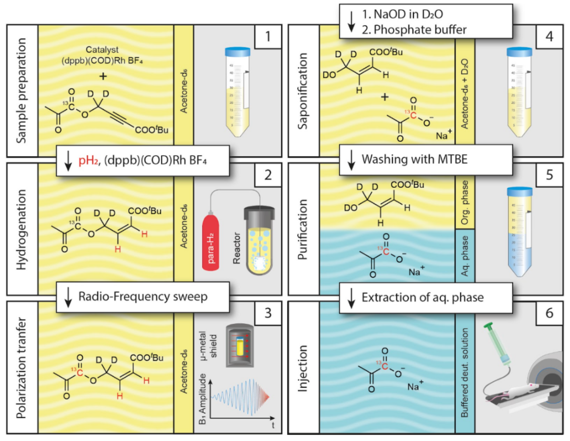
Three female Wistar rats (Charles River), one healthy and two tumor-bearing, were examined. The tumor-bearing animals were subcutaneously injected with MAT B III adenocarcinoma cells 13 - 17 days before imaging (tumor diameter Ø: 28mm). During the MRI exams, animals were monitored via breathing rate, O2 saturation, and rectal temperature. [1-13C]pyruvate was polarized using either a HyperSense (Oxford Instruments) d-DNP polarizer: microwave radiation at 94.133 GHz at 1.2 K for >45min, and rapid dissolution in phosphate buffer solution (D2O); or using a prototype PHIP polarizer (NVision Imaging Technologies). Inside the PHIP polarizer, a pyruvate ester solution was hydrogenated with pH2, the 13C-nuclei polarized using a radio-frequency sweep, and cleaved into sodium pyruvate and a side-arm. The aqueous phase was separated from the organic phase, washed with MTBE, and bubbled with nitrogen to reduce impurities. The final PHIP-SAH product is a 2 ml D2O solution containing up to 160 mM polarized sodium pyruvate (Figure 1). One tumor-bearing animal received two injections of 1.2 ml, 80 mM [1-13C]pyruvate sequentially from d-DNP and PHIP preparation with a delay of 30 min. The second and third animals received four repeated injections of 0.45 ml each, containing 160 mM [1-13C]pyruvate sequentially from PHIP with shortened time intervals of 15 min between injections. 13C spectroscopy was conducted after each injection using a preclinical 7T MRI (Bruker/Agilent) and a dynamic slab-selective single pulse sequence: flip angle 8°, slice thickness 16 mm (DNP vs PHIP) or 25.4 mm (4x PHIP), 100 dynamic scans with TR 2 s, bandwidth 2200 Hz, Ns 2048 spectral points. All signals were normalized by the respective peak pyruvate signal and timed t = 0 s at the start of injection.Results and Dicsussion
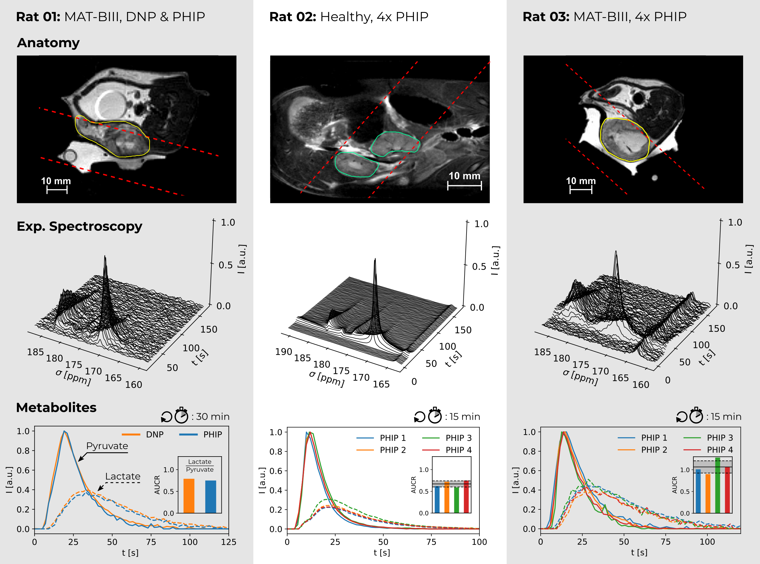
We first benchmarked the PHIP system against a d-DNP system and found that both methods provide samples of comparable volume (0.8-2 ml), concentration (70-160 mM), and polarization level (PHIP: 17.8±1.2%; d-DNP: 22.8±5.4%) at the time of injection, yielding quantitatively similar metabolic information in the in vivo experiment (Figure 2, Rat 01). Quantification of the pyruvate-to-lactate area under curve ratios (AUCR) resulted in Rat 01 in 0.79 (d-DNP) and 0.75 (PHIP) in the tumor region. The achieved dose-to-dose intervals of the polarization systems were approximately 45 min and 13 min for d-DNP and PHIP, respectively. The two exams with four quickly-repeated injections and spectroscopy experiments successfully demonstrated high throughput and reproducibility (Figure 2, Rat 02 & 03). Quantification of AUCR resulted in Rat 02: 0.67±0.07 (n = 4) in the healthy kidneys and in Rat 03: 1.06±0.14 (n = 4) in the tumor, demonstrating an elevated metabolism in the tumor.Conclusion
PHIP-based hyperpolarization of [1-13C]pyruvate can achieve comparable results to d-DNP regarding volume, concentration, and polarization levels at the time of injection. The dose-to-dose interval for the PHIP prototype is much shorter than that for the d-DNP system and thus allows researchers to repeat multiple injections in single MRI examinations within reasonable anesthesia time (<1h) for the animals. This may turn out to be advantageous for the detection of metabolites at low concentrations, e.g. alanine and bicarbonate, to increase the signal-to-noise ratio, to conduct averaging of the metabolic processes, or to create different contrasts by using various sequences or treatments with multiple injections.Acknowledgements
Research reported in this abstract was supported by the German Federal Ministry of Education and Research (BMBF) in the funding program “Quantum Technologies – from Basic Research to Market” under the project “QuE-MRT” (contract number: 13N16450). FS received support from the DFG (#391523415) and the Young Academy of the Bavarian Academy of Sciences and Humanities.References
1. Reineri, F., Boi, T. & Aime, S. ParaHydrogen Induced Polarization of 13C carboxylate resonance in acetate and pyruvate. Nat. Commun. 6, 5858 (2015).
2. Knecht, S. et al. Rapid hyperpolarization and purification of the metabolite fumarate in aqueous solution. Proc. Natl. Acad. Sci. 118, e2025383118 (2021).
3. Nagel, L. et al. Parahydrogen-Polarized [1-13C]Pyruvate for Reliable and Fast Preclinical Metabolic Magnetic Resonance Imaging. Adv. Sci. 10, 2303441 (2023).
Figures