3064
Implementing fully endogenous hyperpolarized molecular probes for imaging cerebral metabolism1LIFMET, EPFL, Lausanne, Switzerland, 2Health technology department, DTU, Kgs Lyngby, Denmark
Synopsis
Keywords: Hyperpolarized MR (Non-Gas), Brain
Motivation: Photo-induced non-persistent radicals for dissolution Dynamic Nuclear Polarization (dDNP) reduce the delay between the preparation and injection of hyperpolarized (HP) substrates by avoiding the need for filtering potentially toxic radicals.
Goal(s): Our goal was the in vivo implementation of probes hyperpolarized with endogenous non-persistent polarizing agents.
Approach: Brain metabolism of HP glucose was monitored in a group of 12h-fasted male mice.
Results: The lactate-to-glucose ratio (LGR) was found similar to previously reported values, with a trend of higher LGR when glucose was hyperpolarized with alpha-ketoglutarate (aKG) than Trityl radicals.
Impact: Our investigation demonstrates the successful in-vivo application of radical-free HP glucose, revealing metabolic responses comparable to those achieved with stable persistent radicals and indicating the potential benefits of hyperpolarizing glucose with aKG radicals.
Introduction
Recent developments in photo-induced non-persistent radicals for dissolution dynamic nuclear polarization (dDNP) enable reducing the delay between the preparation and injection of hyperpolarized (HP) substrates by eliminating the need for filtering potentially toxic radicals1–4. Additionally, the non-persistent nature of the DNP polarizing agent, combined with sample exposure to precise magnetic field and temperature control, currently provides the most efficient method for transporting hyperpolarized (HP) contrast agents5–7. These steps are critical for translating HP probes from animals to humans, particularly for compounds with short hyperpolarization lifetimes, such as 13C-glucose8–10. This abstract focuses on in vivo implementation of hyperpolarized probes polarized with endogenous non-persistent polarizing agents.
Methods
Hyperpolarization of Glucose with UV-Induced Radicals:
A stock solution containing 64 mg of [2H7, U-13C6] glucose, 16.5 mg of alpha-ketoglutarate (aKG), and 128 mg of a 1:1 water-glycerol (w/w) solution was prepared. A 6 µL droplet of the solution was rapidly frozen in liquid nitrogen and then exposed to UV light (using a deuterium lamp operating at 40 W/cm2). The radical concentration as a function of the UV irradiation time was measured using an EPR spectrometer. A 300s irradiation time was found to yield the targeted radical concentration (55 mM) and used for the remaining experiments. The irradiated samples were then loaded into a custom-built DNP polarizer operation at 7T and 1K11,12. Microwave frequency sweeps, both with and without frequency modulation, were carried out to determine the optimal DNP conditions. The polarization levels were calculated by hyperpolarizing and dissolving 12 beads (approximately 72 µL) in 4.5 mL superheated D2O and comparing the carbon signal of the hyperpolarized (HP) solution to the thermal equilibrium signal, following complete relaxation, using a 9.4T MR scanner.
In Vivo MRS of Glucose Cerebral Metabolism in the Mouse Brain:
In vivo spectra were acquired in a 9.4T MRI system (Varian/Magnex) using a home-built quadrature 1H-coil/13C surface coil positioned on top of the mouse head. Brain metabolism of HP glucose was monitored in a group of 12h-fasted male C57BL6/J mice (n=3). Mice were initially anesthetized with 1.3-1.6% isoflurane during a surgical procedure to place a femoral catheter for bolus injection. Subsequently, anesthesia was transitioned to a combination of medetomidine (0.3 mg/kg bolus followed by 0.03 mg/kg/h i.v.) and isoflurane (0.25-0.50%) one hour before injecting HP [2H7, U-13C6]D-glucose. To monitor real-time de novo synthesis of [1-13C]lactate, a 540 µL bolus of HP solution, generated as previously described using 25 beads (approx. 150 µL), was injected via the femoral vein. A pulse-acquire sequence was triggered 5.5s post-injection with 25° frequency selective Gaussian pulse (250 μs) centred at 182 ppm, repeated every 1s for 70s. Lactate-to-glucose ratio (LGR) was calculated from the summed spectra and compared to previously studies using persistent radicals10 (Figure 2).
Results
UV irradiation of 300s generated a radical concentration of 55±5 mM (Figure 1A). The optimal microwave parameters were a central frequency of 196.7 GHz with a microwave frequency modulation amplitude of 50 MHz (Figure 1B). Under these experimental conditions, the liquid state polarization reached 21.3±0.7% by integrating the signal around C2-C5 carbons (Figure 1C). Lactate production from HP glucose was detectable in all spectra acquired in mouse brain. The calculated lactate-to-glucose ratios (LGR) were consistent with previously reported values10, showing a notable trend of higher LGR when hyperpolarizing glucose with aKG radicals compared to Trityl (0.23±0.11 and 0.14±0.06, mean±SD, respectively) (Figure 2).
Discussion
In this work, we report the first in vivo MRS measurements of cerebral metabolism in mice following the administration of radical-free HP glucose. This preparation yielded a metabolic response comparable to what has been observed with HP glucose polarized using stable persistent radicals, with a trend of higher LGR values when using aKG radicals for glucose hyperpolarization compared to Trityl radicals. Further investigations are required to confirm the significance and reasons of this metabolic difference. Given the possibility of transporting hyperpolarized glucose samples when prepared with UV-induced radicals5, our results emphasize the applicability of the technique for interrogating cerebral metabolism. This will eventually lead to an accessible route for real-time molecular imaging.
Conclusion
We demonstrated that measuring cerebral metabolism of HP glucose is feasible using a fully endogenous hyperpolarized substrate preparation.
Acknowledgements
This work was generously supported by the Swiss National Science Foundation (193276 assigned to Andrea Capozzi, 214069 assigned to Mor Mishkovsky). The authors gratefully thank Prof. Rolf Gruetter for the fruitful discussion, Drs. Estelle Gerossier and Stefan Mitrea for their assistance in the animal preparation, and the CIBM Center for Biomedical Imaging, co-founded and supported by the Lausanne University Hospital, University of Lausanne, École Polytechnique Fédérale de Lausanne, University of Geneva and Geneva University Hospitals.
References
1. Capozzi, A.; Patel, S.; Gunnarsson, C. P.; Marco-Rius, I.; Comment, A.; Karlsson, M.; Lerche, M. H.; Ouari, O.; Ardenkjær-Larsen, J. H. Efficient Hyperpolarization of U-13C-Glucose Using Narrow-Line UV-Generated Labile Free Radicals. Angewandte Chemie International Edition 2019, 58 (5), 1334–1339. https://doi.org/10.1002/anie.201810522.
2. Eichhorn, T. R.; Takado, Y.; Salameh, N.; Capozzi, A.; Cheng, T.; Hyacinthe, J.-N.; Mishkovsky, M.; Roussel, C.; Comment, A. Hyperpolarization without Persistent Radicals for in Vivo Real-Time Metabolic Imaging. Proc Natl Acad Sci U S A 2013, 110 (45), 18064–18069. https://doi.org/10.1073/pnas.1314928110.
3. Gaunt, A. P.; Lewis, J. S.; Hesse, F.; Cheng, T.; Marco-Rius, I.; Brindle, K. M.; Comment, A. Labile Photo-Induced Free Radical in α-Ketoglutaric Acid: A Universal Endogenous Polarizing Agent for In Vivo Hyperpolarized 13C Magnetic Resonance. Angewandte Chemie 2022, 134 (2), e202112982. https://doi.org/10.1002/ange.202112982.
4. Zanella CC, Capozzi A, Yoshihara HAI, Radaelli A, Mackowiak ALC, Arn LP, Gruetter R, Bastiaansen JAM. Radical-free hyperpolarized MRI using endogenously occurring pyruvate analogues and UV-induced nonpersistent radicals. NMR Biomed. 2021 Nov;34(11):e4584. doi: 10.1002/nbm.4584. Epub 2021 Jul 10. PMID: 34245482; PMCID: PMC8518970.
5. Capozzi, A.; Cheng, T.; Boero, G.; Roussel, C.; Comment, A. Thermal Annihilation of Photo-Induced Radicals Following Dynamic Nuclear Polarization to Produce Transportable Frozen Hyperpolarized 13C-Substrates. Nat Commun 2017, 8 (1), 15757. https://doi.org/10.1038/ncomms15757.
6. Capozzi, A.; Kilund, J.; Karlsson, M.; Patel, S.; Pinon, A. C.; Vibert, F.; Ouari, O.; Lerche, M. H.; Ardenkjær-Larsen, J. H. Metabolic Contrast Agents Produced from Transported Solid 13C-Glucose Hyperpolarized via Dynamic Nuclear Polarization. Commun Chem 2021, 4 (1), 1–11. https://doi.org/10.1038/s42004-021-00536-9.
7. Capozzi, A. Design and Performance of a Small Bath Cryostat with NMR Capability for Transport of Hyperpolarized Samples. Sci Rep 2022, 12 (1), 19260. https://doi.org/10.1038/s41598-022-23890-7.
8. Mishkovsky, M.; Anderson, B.; Karlsson, M.; Lerche, M. H.; Sherry, A. D.; Gruetter, R.; Kovacs, Z.; Comment, A. Measuring Glucose Cerebral Metabolism in the Healthy Mouse Using Hyperpolarized 13C Magnetic Resonance. Sci Rep 2017, 7, 11719. https://doi.org/10.1038/s41598-017-12086-z.
9. Flatt, E.; Lanz, B.; Pilloud, Y.; Capozzi, A.; Lerche, M. H.; Gruetter, R.; Mishkovsky, M. Measuring Glycolytic Activity with Hyperpolarized [2H7, U-13C6] D-Glucose in the Naive Mouse Brain under Different Anesthetic Conditions. Metabolites 2021, 11 (7), 413.https://doi.org/10.3390/metabo11070413.
10. Mishkovsky, M.; Gusyatiner, O.; Lanz, B.; Cudalbu, C.; Vassallo, I.; Hamou, M.-F.; Bloch, J.; Comment, A.; Gruetter, R.; Hegi, M. E. Hyperpolarized 13C-Glucose Magnetic Resonance Highlights Reduced Aerobic Glycolysis in Vivo in Infiltrative Glioblastoma. Sci Rep 2021, 11, 5771. https://doi.org/10.1038/s41598-021-85339-7.
11. Cheng, T.; Capozzi, A.; Takado, Y.; Balzan, R.; Comment, A. Over 35% Liquid-State 13 C Polarization Obtained via Dissolution Dynamic Nuclear Polarization at 7 T and 1 K Using Ubiquitous Nitroxyl Radicals. Physical Chemistry Chemical Physics 2013, 15 (48), 20819–20822. https://doi.org/10.1039/C3CP53022A.
12. Comment, A.; van den Brandt, B.; Uffmann, K.; Kurdzesau, F.; Jannin, S.; Konter, J. a.; Hautle, P.; Wenckebach, W. Th.; Gruetter, R.; van der Klink, J. j. Design and Performance of a DNP Prepolarizer Coupled to a Rodent MRI Scanner. Concepts in Magnetic Resonance Part B: Magnetic Resonance Engineering 2007, 31B (4), 255–269. https://doi.org/10.1002/cmr.b.20099.
Figures
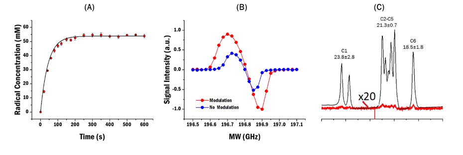
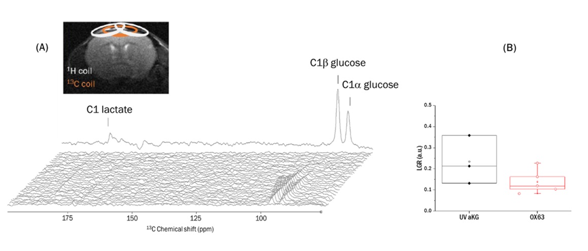
Figure 2: Cerebral metabolism of HP glucose polarized with UV-induced polarizing agents: (A) Typical time course and summed spectra (top). (B) Lactate-to-glucose ratio (LGR) measured in mice receiving glucose hyperpolarized using aKG radicals (black full diamonds) and previously reported LGR measured using persistent OX63 radicals10 (red circles) (p = 0.16).