3058
Fluorinated gelatin-based scaffolds traceable by 19F MRI1Center for Advanced Preclinical Imaging (CAPI), First Faculty of Medicine, Charles University, Prague, Czech Republic, 2Department of Organic and Macromolecular Chemistry, Ghent University, Ghent, Belgium
Synopsis
Keywords: Non-Proton, Preclinical, Gelatin scaffolds, 19F MRI/MRS
Motivation: Gelatin-based scaffolds may be seeded by cells or may serve as depots for drugs and ensure their slow release.
Goal(s): The goal of the study was to prepare scaffolds with tunable properties and monitor their stability in time in a mouse model in vivo.
Approach: The scaffolds implanted to mice were monitored by 1H/19F MRI, fluorine content was quantified by 19F MR spectroscopy.
Results: The scaffolds can be easily monitored by 19F MRI/MRS. The study confirmed that the design of the scaffolds can be fine-tuned for future applications, biodegradation rates may be set from several weeks up to one year.
Impact: Gelatin-based scaffolds pave the road towards qualitatively different drug applications and regenerative medicine. The study evaluated their applicability, traceability by 1H/19FMRI, and stability in vivo. Future work should investigate release speed of different compounds (drugs) both hydrophilic and hydrophobic.
Introduction
Biomaterials with tunable mechanical properties that can be processed in various sizes and shapes are potentially very interesting for various clinical applications. Porous biocompatible scaffolds may carry drugs and ensure their slow release, or they can be seeded with stem cells and enable regeneration of the desired tissue. The scaffolds need to mimic the mechanical properties of the target tissue and be degradable in a chosen time frame. In this study, we tested 3D-printed gelatin-based scaffolds with an implemented 19F MRI tracer for their monitoring in vivo using 19F magnetic resonance spectroscopy (MRS) and magnetic resonance imaging (MRI). While 19F MRI provides specific hotspot imaging, which can be easily colocalized with standard anatomical 1H MRI, MRS enables quantification for estimation of the scaffold dissolution speed. Combination of 1H and 19F MRI supplemented by 19F MRS represents a tool applicable even in future clinical practice.Materials and Methods
Gelatin-based scaffolds with implemented 19F MRI tracer were prepared. Amine moieties of the starting material (gelatin-based hydrogel) were converted into methacrylamide moieties (methacrylated portion was ≈ 100% - G1, ≈ 70% - G2, ≈ 30% - G3)1. Subsequently, the gels were crosslinked with N-(2,2‑difluoroethyl)acrylamide (DFEA) monomers, which enabled scaffold detection by 19F MRI/MRS and controlled modification of mechanical properties and biodegradability. Scaffolds G1 – G3 were 3D printed, samples with different sizes (50 – 400 mm3) were scanned in vitro by 1H/19F MRI to estimate the detection limit. In vivo animal experiments included implantation of the scaffolds subcutaneously through an incision in the abdominal area and MR scanning of mice immediately after the implantation, 1, 2, 7, 14, 28 days after and then monthly up to nine months. Each scaffold type was tested on 7 mice. Both in vitro and in vivo MR experiments were performed using an animal 7 T scanner with a 1H/19F rat whole-body volume coil. 1H MR images were obtained using a gradient echo sequence (echo/repetition time TE/TR = 3.1/200 ms, flip angle FA = 50°, number of acquisitions NA = 4, matrix 256×256, field of view FOV = 30×30 mm2 in axial, and 512×256, FOV = 60×30 mm2 in coronal directions, slice thickness 1 mm). 19F MR images were measured using both turbospin echo (TE/TR = 8/800 ms, turbofactor 8, NA = 128, matrix 64×64, FOV = 30×30 mm2 in axial, and matrix 128×64, FOV = 60×30 mm2 in coronal directions, slice thickness 10 mm) and gradient echo (TE/TR = 2.5/120 ms, same geometry) sequences. 1H and 19F images were interpolated to the same image matrix, color coded (1H – grayscale, 19F – red scale) and merged using ImageJ software2. Unlocalized 19F MR spectra for signal quantification were acquired using an FID sequence (FA = 90°, TE/TR = 3.1/1000 ms, NA = 128) and processed using a home-made script written in Matlab3.Results and Discussion
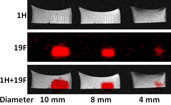
The incorporation of DFEA monomer into the reaction mixture increased the fluorine content of the cross-linked hydrogels, which enabled reliable detection by 19F MRI/MRS and decreased hydrophilicity.Suitable fluorine relaxation times (T1 ≈ 400 ms, T2 ≈ 120 ms) enabled scaffold visualization by both gradient and turbospin echo sequences within acceptable scanning times (15 minutes). In vitro MRI (Fig. 1) confirmed visibility of scaffolds down to 50 mm3 of all tested samples (G1, G2, G3), nevertheless, we consider this volume as a detection of limit for the sample with the lowest 19F content (G3).
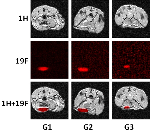
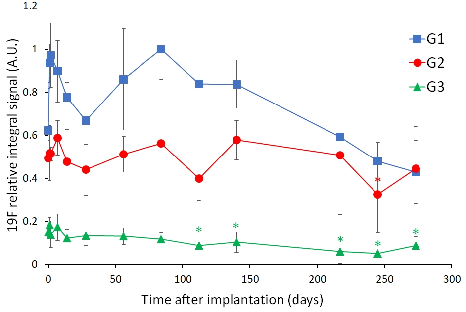
In vivo imaging (Fig. 2) confirmed that the implanted scaffolds can be reliably visualized and monitored in time by 19F MRI. The scaffolds degraded very slowly. Dissolution depended on the level of crosslinking, as was proved by 19F MRS (Fig. 3). Signal decrease corresponding to dissolution of the scaffolds was observed 4 months after implantation in the case of G3, while G1 and G2 implants remained stable for up to 9 months. The implants caused no adverse reaction (inflammation) in experimental animals.
Conclusion
Fluorinated hydrogel scaffolds are biocompatible and suitable for regenerative medicine either as drug carriers or as matrices for cell therapies. Mechanical properties and biodegradability can be fine-tuned by controlled crosslinking. Their fate after implantation can be easily monitored in vivo by 19F MRI/MRS, which enables their visualization and quantification.Acknowledgements
The facility infrastructure was supported by European Regional Development Fund No. CZ.02.1.01/0.0/0.0/18_046/0016045 (OPVVV project) and by Ministry of Education, Youth and Sports of the Czech Republic (Large RI Project LM2023050 Czech-BioImaging), financial support was provided by Research Foundation – Flanders (FWO, Fonds Wetenschappelijk Onderzoek – Vlaanderen, project no. 1229422N), and by European Union’s Horizon 2020 research and innovation program under grant agreement no. 828835.References
1. Van Hoorick J, Gruber P, Markovic M, et al. Cross-linkable gelatins with superior mechanical properties through carboxylic acid modification: increasing the two-photon polymerization potential. Biomacromolecules 2017;18(10):3260–3272.
2. Schneider, CA, Rasband WS, Eliceiri KW. NIH Image to ImageJ: 25 Years of Image Analysis. Nat Methods 2012;9(7):671–675.
3. The MathWorks Inc. (2018). MATLAB version: 9.5.0.944444 (R2018b), Natick, Massachusetts: The MathWorks Inc. https://www.mathworks.com
Figures