3057
Investigation of the methionine uptake of glioma cells through deuterium magnetic resonance spectroscopy1Paul C. Lauterbur Research Center for Biomedical Imaging, Shenzhen Institute of Advanced Technology, Chinese Academy of Sciences, Shenzhen, China, 2College of Engineering, Southern University of Science and Technology, Shenzhen, China
Synopsis
Keywords: Deuterium, Deuterium
Motivation: Glioma tumor cells have abnormally higher uptake of methionine, compared to normal brain tissues.
Goal(s): To characterize the methionine preference of glioma cells through deuterium magnetic resonance spectroscopy.
Approach: Deuterium magnetic resonance spectroscopy in glioma C6 and CTX-TNA2 glial cells incubated by [1-2H3]-methionine
Results: Abnormal uptake of deuterated methionine in glioma cells was revealed by using magnetic resonance deuterium spectroscopy, compared to normal cells
Impact: Deuterium magnetic resonance imaging with deuterium labeled methionine may have the potential in accurate diagnosis and treatment assessment of glioma.
Introduction
Glioma is a malignant brain tumor with high mortality. Accurate diagnosis and dynamic monitoring the progress of glioma are of the vital importance for the disease management. Recent studies have shown that glioma tumor cells have abnormally higher uptake of methionine, compared to normal brain tissues. In this study, the abnormal uptake of methionine in glioma cells was revealed by using magnetic resonance deuterium spectroscopy. Our findings may indicate a new way to characterize glioma[1] from the aspect of metabolic anomaly.Methods
C6 tumor cell and CTX-TNA2 glial cell were used for comparison our study. When the C6 and CTX-TNA2 cells reached 80% growth density in the T75 cell culture flasks, they were transferred to five sets of 6-well cell culture dishes (n=6). Cell numbers were counted before inoculation to ensure the presence of 10^6 cells in each well. Once the cells completely adhered to the 6-well culture dishes, they were washed twice with phosphate buffered saline (PBS). The cells were then incubated in the mixture of 1900 μL of methionine-free DMEM and 100 μL of 10 mM [1-2H3]-methionine (Dingbang Biotechnology Co., Ltd. in Shenzhen, China) solution (dissolved in PBS), . Samples was extracted at 0, 6, 24, and 48 hours[2]. Meanwhile, all of the cells were digested and counted. The solution contained 640 μL of extract and 160 μL of 1 mM d4-pyrazine (pyrazine-1,4-dioxide-d4, Sigma Aldrich ). Later, 500 μL of the combination were drawn from 800 μL of the mixed solution and transferred to a 5 mm NMR tube (Wilmad WG-1000-7), then scanned using deuterium NMR spectroscopy. All cell cultures were maintained in a humidified environment at 37 °C and 5% CO2. Methionine-free DMEM mentioned above was prepared using DMEM (Gibco 21013024, with glutamine, cysteine, and methionine deprived). The DMEM was then supplemented with L-Cystine dihydrochloride (Solarbio YS148316), 200 mM liquid L-glutamine (Solarbio G0200), and pyruvate sodium (Solarbio P8380) to create methionine-free DMEM (DMEM Met-free). Magnetic resonance deuterium spectrum was scanned on a Bruker 600MHz NMR . The scanning parameters were as follows: TR=3s, average =1024, scanning spectrum width SW=1013.51Hz sampling points = 1024. The deuterium spectra were processed in Mestrenova (version 14.3.3, Mestrelab Research, Spain) with line broadening = 0.3 Hz and phase correction. Spectra were then fitted to a mixed Lorentzian model. Signal intensities of HDO and methionine were normalized according to the pyrazine to measure the quantity of HDO and methionine (in μmol) in each sample. The methionine consumption rate were then evaluated by the methionine reduction per unit time (in hour) and per cell number (in 10^6).Results
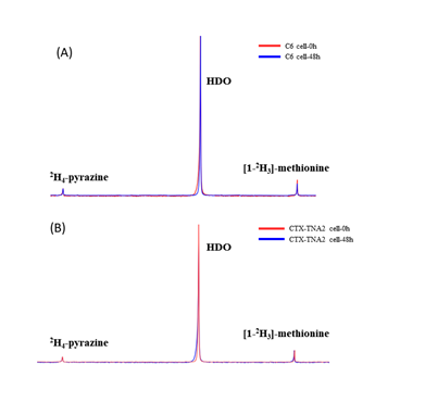
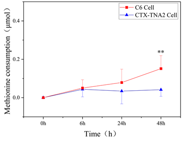
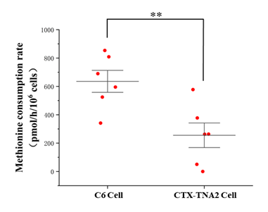
Figure 1 shows the 2H spectra acquired at 48 hours after the addition of methionine of two cells. Significant reduction in methionine peak can be observed in C6 cell indicating that the tumor cells have higher uptake of methionine than normal glial cells. Figure 2 compares the methionine consumption over time in C6 cells and CTX-TNA2 cells. It can be observed that the consumption of [1-2H3]-methionine by tumor cells increased over time. At 48 hours after the addition of methionine, there existed a significant difference(P=0.005) in the consumption between tumor and normal cells. Comparatively, normal neuroglial cells had little uptake throughout the whole experiment. After normalized to cell number, the methionine consumption rate in C6 tumor cells is also higher than the glial cells, as shown in Fig.3.Discussion
The application of 11C-methionine for the diagnosis of glioma has been demonstrated in several other studies[3]. Likewise, based on the results of our in vitro cell experiments, it was demonstrated that glioma cells have a higher uptake of methionine compared to normal glial cells using stable isotope labeled instead of radioactive labeled methionine. Our next efforts will be made to evaluate the applicability of using deuterated methionine in the diagnosis of glioma through animal model and human studies.Acknowledgements
This work was supported in part by the Project on Global Common Challenges of Chinese Academy of Sciences (No. 321GJHZ2022081GC), the NSFC grant (81627901), the Key Laboratory for Magnetic Resonance and Multimodality Imaging of Guangdong Province (2023B1212060052), the Funding Program of Shenzhen, China (RCYX20200714114735123), the Chinese Academy of Sciences Youth Innovation Promotion Association funded project (Y2021098), the Funding Program of Shenzhen and Guangdong Province, China (2022B1515120068).References
[1] Deuschl C, Goericke S, Grueneisen J, et al. Simultaneous 11C-methionine positron emission tomography/magnetic resonance imaging of suspected primary brain tumors[J]. PloS one, 2016, 11(12): e0167596.
[2] Spratt S J, Oguchi K, Miura K, et al. Probing methionine uptake in live cells by deuterium labeling and stimulated Raman scattering[J]. The Journal of Physical Chemistry B, 2022, 126(8): 1633-1639.
[3] Schaff L R, Mellinghoff I K. Glioblastoma and other primary brain malignancies in adults: a review[J]. Jama, 2023, 329(7): 574-587.
Figures