3054
Reproducibility of estimating total sodium concentration in the putamen and substantia nigra using sodium MRI at 3T1Department of Radiology, Ruijin Hospital, Shanghai Jiao Tong University School of Medicine, Shanghai, China, Shanghai, China, 2Philips Healthcare, Shanghai, China, shanghai, China, 3Faculty of Medical Imaging Technology, College of Health Science and Technology, Shanghai Jiao Tong University School of Medicine, shanghai, China
Synopsis
Keywords: Non-Proton, Metabolism
Motivation: Brain sodium (23Na) MRI can provide sodium concentration information in vivo, but there are still some challenges, such as low SNR and relatively low resolution.
Goal(s): To assess the reproducibility of total sodium concentration (TSC) evaluation in the putamen and substantia nigra of healthy individuals using 3D 23Na MRI.
Approach: Ten healthy subjects were scanned twice consecutively using the 3D TFE UTE and T1W sequence to quantify the TCS in the putamen and substantia nigra. The reproducibility was evaluated.
Results: Concentration of sodium in phantoms can be accurately quantified and TSC in putamen, substantia nigra had good consistency for 23Na MRI.
Impact: 23Na-MRI shows potential in becoming a stable and useful biomarker for the diagnosis of patients with neurodegenerative diseases.
Introduction
MRI has been widely used in clinical practice for the diagnosis of various diseases. However, conventional 1H MRI does not provide direct biochemical and metabolic information about tissues, such as cell integrity, tissue viability, or predictions about possible treatment outcomes1. 23Na MRI, on the other hand, offers a quantitative and non-invasive method to provide some of this complementary information. Despite sodium ions having low magnetic resonance sensitivity, which can limit image acquisition time, advancements in magnetic field strength have led to improved image quality and resolution. This makes 23Na MRI a potential new biomarker for neurodegenerative disease. This study represents a preliminary investigation into the reproducibility of 23Na MRI quantification.Methods
Ten volunteers were scanned twice with a 3T MR Scanner (Elition X, Philips Healthcare). A double-tuned 1H/ 23Na birdcage head coil (Rapid Biomedical GmbH, Rimpar, Germany) was used for scanning. For sodium MRI, a 3D TFE UTE sequence was scanned in the sagittal orientation in 14:28 min. The sequence parameters were: TR/TE = 17/0.1 ms, FOV = 272×272×180 mm3, acquired resolution = 4.75×4.75×4.75 mm3, reconstructed resolution = 2.43×2.43×2.38 mm3, radial acquisition, matrix = 56×56 with 76 slices, NSA = 1, acquisition bandwidth = 40 Hz, and flip angle = 90. Five phantoms with known sodium concentrations (20, 40 ,60 ,80, and 100 mM) were placed within the FOV to serve as external references for quantification. A 32-channel 1H head coil was used for the high-spatial-resolution STAGE (strategically acquired gradient echo) sequence 2,3 with a scan time of 3:11 min Other parameters were: FOV = 256×192×150 mm3, SENSE factor 2, matrix = 384×192 with 112 slices, acquired resolution = 0.67×1×1.34 mm3, reconstructed resolution = 0.67×0.67×1.34 mm3, TR = 27 ms, five echoes, TE first = 2.9 ms, and echo spacing = 4.7 ms.For the quantification of the total sodium concentration, 5 ROIs were drawn within each of the 5 phantoms , with one ROI per slice. Subsequently, linear regression was performed between the mean signal intensities within the 5 phantoms and their concentrations. Subsequently, the signal intensities of the sodium MRI were converted to TSC. Following that, the TSC maps were co-registered to the STAGE images which serve as a reference of the anatomical structures. For each of the two measurements, 5 circular ROIs were manually drawn on 5 different slices of the fused images of STAGE and TSC, for each of the brain regions including the Substantia Nigra (SN) and Putamen, which are important regions for neurodegenerative diseases such as Parkinson’s disease. Subsequently, the mean sodium concentrations across the 5 ROIs were extracted for each brain region.Statistical analyses were performed using R (version 4.3.2). The intraclass correlation coefficient (ICC) analyses were conducted to assess the degree of agreement between the two measurements. Correlation analysis was also performed, and the difference between two different scans was plotted against their means with their 95% limits of agreement.
Results
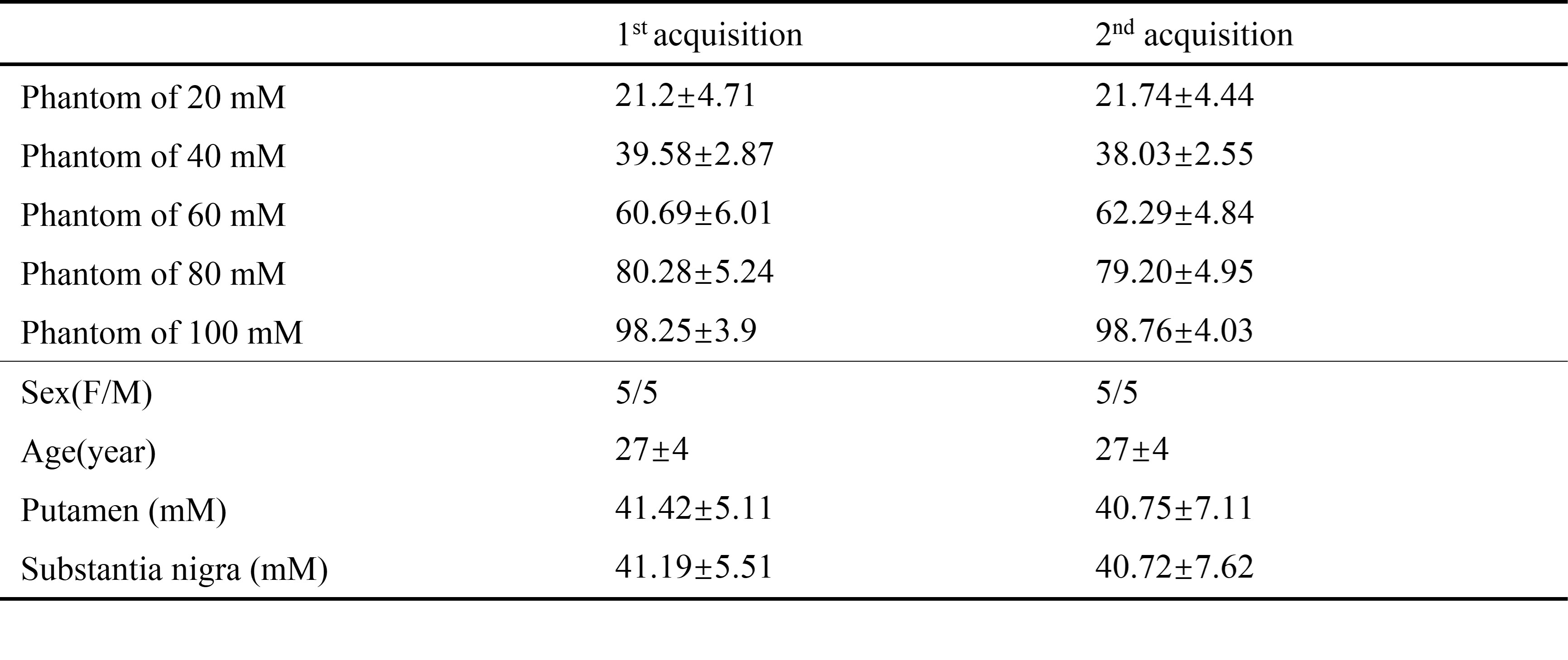
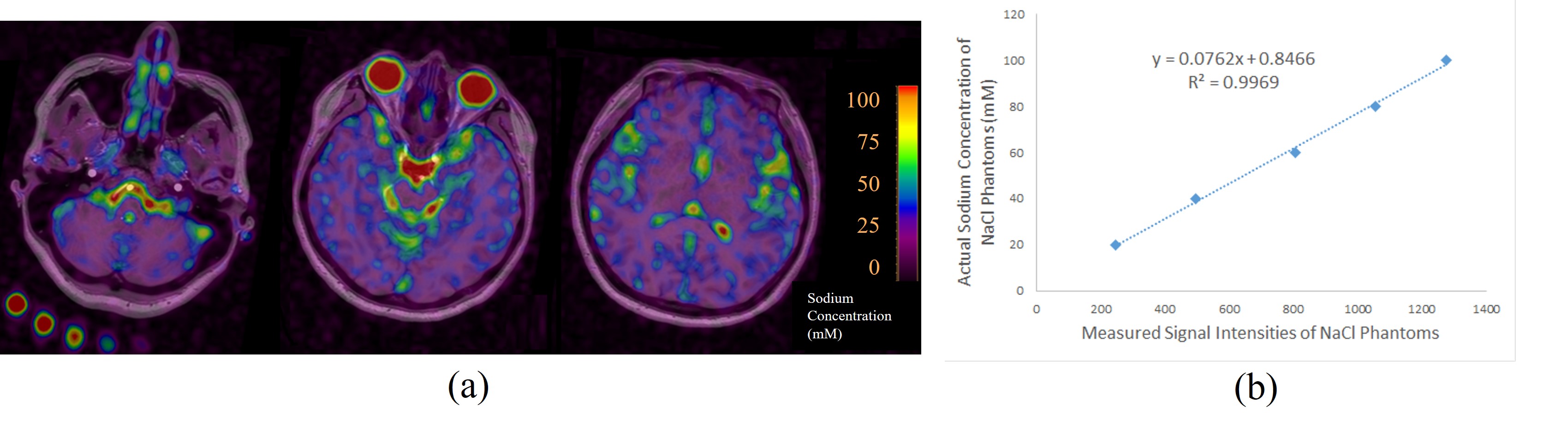
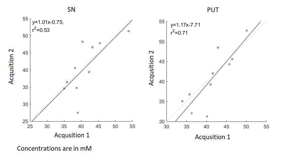
The demographics of the participants are shown in Table 1. The concentration of sodium in five phantoms had good consistency. Figure 1 shows in a representative case, the fused images of STAGE and TSC, and the excellent linear correlation (R2 = 0.997) between the signal intensities and sodium concentrations in the five phantoms . The calculated sodium concentrations of the five phantoms and two regions of brain (substantia nigra and putamen) for the two measurements were calculated and analyzed (shown in Table 1), showing good consistency and reproducibility. The ICCs were 0.89, 0.71, 0.64, 0.8 and 0.9 for the phantoms with 20, 40, 60, 80 and 100mM sodium concentrations, respectively. And for substantia nigra and putamen, the ICCs were 0.72 and 0.81 respectively. The correlation plot of the two sodium concentration measurements are shown in Figure2.Discussion and Conclusion
Our study investigated the reproducibility of tissue sodium intensity measured with 23Na MRI. Our results showed good reproducibility between the two measurements of 23Na MRI and TSCs in healthy volunteers in both the Substantia Nigra and Putamen regions, which are of particular importance to study neurodegenerative diseases. This indicates 23Na MRI can be an effective method with a good reproducibility and consistency for evaluating tissue sodium content. To the best of our knowledge, currently there is no reproducibility study published for 23Na MRI at 3T. Compared to ultra-high field, 3T 23Na MRI can be readily added to current clinical scans in patients and can provide valuable information about cell function and brain tissue viability in the neurodegenerative diseases4.Acknowledgements
This work was supported, in part, by the National Natural Science Foundation of China (grant number: 82271954, 81971576); Chinese National Science & Technology Pillar Program (grant number: 2022YFC2009900/2022YFC2009905) and the Innovative Research Team of High-level Local Universities in Shanghai.References
1. Madelin Guillaume. Sodium Magnetic Resonance Imaging: Biomedical Applications. https://doi.org/10.48550/arXiv.1212.4400
2. Chen Y, Liu S, Wang Y, Kang Y, Haacke EM. STrategically Acquired Gradient Echo (STAGE) imaging, part I: Creating enhanced T1 contrast and standardized susceptibility weighted imaging and quantitative susceptibility mapping. Magn Reson Imaging 2018, 46,130-139.
3. Haacke EM, Chen Y, Utriainen D, et al. Strategically acquired gradient echo (STAGE) imaging, part III: Technical advances and clinical applications of a rapid multi-contrast multi-parametric brain imaging method. Magnetic Resonance Imaging 2020, 65, 15-26.
4. Haeger A, Boumezbeur F, Bottlaender M, Rabrait-Lerman C, Lagarde J, Mirzazade S, Krahe J, Hohenfeld C, Sarazin M, Schulz JB, Romanzetti S, Reetz K. 3T sodium MR imaging in Alzheimer's disease shows stage-dependent sodium increase influenced by age and local brain volume. Neuroimage Clin. 2022; 36:103274.
Figures