3051
Deuterium metabolic imaging of early response to targeted therapy in pediatric brain tumors.1Radiology, University of California San Francisco, San Francisco, CA, United States, 2Neurology, University of California San Francisco, San Francisco, CA, United States
Synopsis
Keywords: Deuterium, Cancer, Preclinical
Motivation: Diffuse midline gliomas (DMGs) are devastating pediatric brain tumors. Although MRI is the mainstay for DMG imaging, it does not reliably report on response to therapy.
Goal(s): The goal of this study was to assess the utility of deuterium metabolic imaging for DMGs.
Approach: To this end, we interrogated [6,6’-2H]-glucose metabolism in patient-derived and syngeneic models.
Results: [6,6’-2H]-glucose enables non-invasive visualization of the metabolically active tumor lesion in mice bearing intracranial DMGs. Importantly, [6,6’-2H]-glucose provides an early readout of response to targeted therapy in vivo. Collectively, our studies highlight the potential of [6,6’-2H]-glucose for imaging tumor burden and response to therapy in DMGs.
Impact: Diffuse midline gliomas are deadly childhood brain tumors. Using clinically relevant patient-derived and murine tumor models, we show that deuterium metabolic imaging using [6,6’-2H]-glucose enables non-invasive assessment of tumor burden and early response to therapy in diffuse midline gliomas.
Introduction
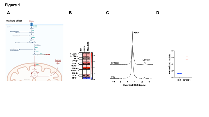
Diffuse midline gliomas (DMGs) harboring histone H3K27 alterations are devastating pediatric brain tumors with overall survival of ~9 months1-3. ONC201 is the first drug in decades to extend survival to ~27 months in clinical trials4. Magnetic resonance imaging (MRI) is the gold standard for DMG imaging5. However, there is no consensus on what constitutes a biologically meaningful reduction in tumor volume following treatment and multiple MRI scans separated by ≥8 weeks are required to assess response5,6.The Warburg effect is a metabolic hallmark of cancer that is characterized by elevated glucose uptake and preferential conversion to lactate in tumor tissues7. In contrast, normal tissues oxidize glucose via the tricarboxylic acid cycle to produce glutamate (denoted by convention as glx; see Fig. 1A)7. Deuterium metabolic imaging (DMI) is a novel, clinically translatable method of imaging [6,6’-2H]-glucose metabolism in vivo8-10. The goal of this study was to determine whether [6,6’-2H]-glucose provides a readout of tumor burden and response to therapy in DMGs.
Methods
Cell culture: Patient-derived BT245 cells were cultured in serum-free medium. SF7761, SF8628 and immortalized human astrocytes were cultured in DMEM with 10% fetal calf serum. The murine SB28-DMG cells were generated by transfecting sleeping-beauty transposon-based SB28 cells with H3.3 K27M-expressing lentivirus and grown in RPMI-1640 with 10% fetal calf serum.DMI in cells: SF7761 or IHA cells were incubated in media containing 12.5mM [6,6’-2H]-glucose for 72h. SB28-DMG or BT245 cells were treated with vehicle (DMSO) or 1mM ONC201 for 72h and incubated in media containing 11mM [6,6’-2H]-glucose. 2H-MR spectra were acquired from live cell suspensions on a Varian 14.1T (SF7761, IHA) or Bruker 3T (SB28-DMG, BT245) scanner using a 16mm 2H surface coil and a pulse-acquire sequence11.
DMI in vivo: SB28-DMG cells were intracranially implanted in C57BL/6J mice and SF8628 cells in SCID mice. Tumor volume was determined by T2-weighted MRI on a Varian 14.1T (SF8628) or Bruker 3T scanner as described10,11. Once tumors were ~30mm3, mice were treated with vehicle (3% DMSO in saline) or ONC201 (100 mg/kg every 4 days). Following intravenous injection of a bolus of [6,6’-2H]-glucose (1 g/kg), non-localized spectra were acquired over 48min with a pulse-acquire sequence on a Varian 14.1T or Bruker 3T scanner10,11. For spatial localization, we used a 2D chemical shift imaging (CSI) sequence (FOV = 30x30 mm2, 8x8, 128 points, 512.8 Hz spectral width, slice thickness=8mm, TE/TR = 1.04/265.9 ms, NA = 30 over 8min 30s) on a Bruker 3T scanner.
Results
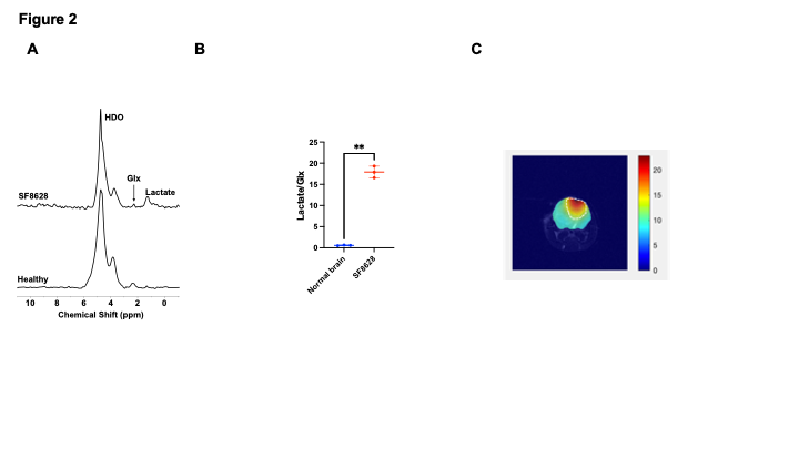
[6,6-2H]-glucose provides a readout of the Warburg effect in DMG cells: We confirmed that SF7761 and SB28-DMG cells upregulate key enzymes and transporters involved in the Warburg effect relative to immortalized human astrocytes (IHAs; Fig. 1A-1B). Importantly, examination of [6,6’-2H]-glucose metabolism showed elevated lactate production in SF7761 cells relative to IHAs (Fig. 1C-1D), indicating that the Warburg effect is a metabolic phenotype of DMGs.[6,6'-2H]-glucose monitors tumor burden in DMGs in vivo: Next, we examined [6,6’-2H]-glucose metabolism in mice bearing intracranial SF8628 tumors and compared to tumor-free mice. As shown in Fig. 2A-2B, we observed elevated lactate and reduced glx in SF8628 tumor-bearing mice while tumor-free mice produced only glx. To validate these results and assess the spatial distribution of [6,6’-2H]-glucose metabolism, we performed 2D CSI at the clinical field strength of 3T in mice bearing intracranial SB28-DMG tumors. Our results indicate that lactate production from [6,6’-2H]-glucose demarcates tumor from normal brain (Fig. 2C). These results suggest that [6,6’-2H]-glucose allows visualization of the metabolically active tumor lesion in DMG-bearing mice.
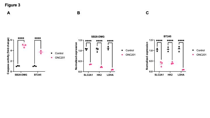
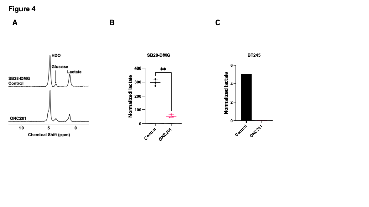
Reduced lactate production from [6,6'-2H]-glucose is a biomarker of response to therapy: ONC201 is a novel drug that disrupts metabolic and epigenetic pathways and induces apoptosis in DMGs4. We confirmed that ONC201 induced apoptosis and downregulated expression of the glucose transporter SLC2A1, hexokinase 2, and lactate dehydrogenase A in SB28-DMG and BT245 cells (Fig. 3A-3B). Importantly, lactate production from [6,6’-2H]-glucose was reduced in ONC201-treated cells relative to controls (Fig. 3C-3E).
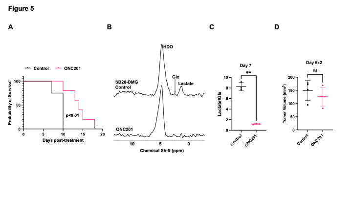
[6,6'-2H]-glucose provides an early readout of response to therapy in vivo: We examined whether [6,6’-2H]-glucose provides a readout of response to ONC201 in vivo at 3T. ONC201 significantly extended survival of mice bearing intracranial SB28-DMG tumors (Fig. 4A). Importantly, lactate production from [6,6’-2H]-glucose was significantly reduced in ONC201-treated mice at D7, when no change could be observed on T2-weighted MRI (Fig. 4B-4D).
Conclusions
DMGs are deadly brain tumors in children. The inability to reliably assess response to therapy is a significant challenge in clinical translation of novel therapies. Our studies indicate that of [6,6’-2H]-glucose provides a readout of tumor burden and response to therapy in clinically relevant DMG models in vivo.Acknowledgements
This study was supported by the Chad Tough Defeat DIPG Foundation and the Violet Foundation for Pediatric Brain Cancer.References
1- Ostrom, Q. T. et al. CBTRUS Statistical Report: Primary Brain and Other Central Nervous System Tumors Diagnosed in the United States in 2016-2020. Neuro-oncology 25, iv1-iv99, doi:10.1093/neuonc/noad149 (2023).
2- Price, M. et al. Childhood, adolescent, and adult primary brain and central nervous system tumor statistics for practicing healthcare providers in neuro-oncology, CBTRUS 2015–2019. Neuro-Oncology Practice, doi:10.1093/nop/npad061 (2023).
3- Jackson, E. R. et al. A review of the anti-tumor potential of current therapeutics targeting the mitochondrial protease ClpP in H3K27-altered, diffuse midline glioma. Neuro-oncology, doi:10.1093/neuonc/noad144 (2023).
4- Venneti, S. et al. Clinical Efficacy of ONC201 in H3K27M-Mutant Diffuse Midline Gliomas Is Driven by Disruption of Integrated Metabolic and Epigenetic Pathways. Cancer discovery, OF1-OF24, doi:10.1158/2159-8290.Cd-23-0131 (2023).
5- Cooney, T. M. et al. Response assessment in diffuse intrinsic pontine glioma: recommendations from the Response Assessment in Pediatric Neuro-Oncology (RAPNO) working group. Lancet Oncol 21, e330-e336, doi:10.1016/s1470-2045(20)30166-2 (2020).
6- Hargrave, D., Chuang, N. & Bouffet, E. Conventional MRI cannot predict survival in childhood diffuse intrinsic pontine glioma. J Neurooncol 86, 313-319, doi:10.1007/s11060-007-9473-5 (2008).
7- Venneti, S. & Thompson, C. B. Metabolic Reprogramming in Brain Tumors. Annu Rev Pathol 12, 515-545, doi:10.1146/annurev-pathol-012615-044329 (2017).
8- Lu, M., Zhu, X. H., Zhang, Y., Mateescu, G. & Chen, W. Quantitative assessment of brain glucose metabolic rates using in vivo deuterium magnetic resonance spectroscopy. Journal of cerebral blood flow and metabolism : official journal of the International Society of Cerebral Blood Flow and Metabolism 37, 3518-3530, doi:10.1177/0271678x17706444 (2017).
9- De Feyter, H. M. et al. Deuterium metabolic imaging (DMI) for MRI-based 3D mapping of metabolism in vivo. Sci Adv 4, eaat7314, doi:10.1126/sciadv.aat7314 (2018).
10- Taglang, C. et al. Deuterium magnetic resonance spectroscopy enables noninvasive metabolic imaging of tumor burden and response to therapy in low-grade gliomas. Neuro-oncology 24, 1101-1112, doi:10.1093/neuonc/noac022 (2022).
11- Batsios, G. et al. Deuterium Metabolic Imaging Reports on TERT Expression and Early Response to Therapy in Cancer. Clinical cancer research : an official journal of the American Association for Cancer Research 28, 3526-3536, doi:10.1158/1078-0432.Ccr-21-4418 (2022).
Figures