3049
Advancing Brain Tumour Sodium Imaging: Prospectively Accelerated eSISTINA and Compressed Sensing at 7T1Institute of Neurscience and Medicine - 4, Forschungszentrum Juelich GmbH, Juelich, Germany, 2Department of Neurology, RWTH Aachen University, Aachen, Germany, 3Department of Nuclear Medicine, RWTH Aachen University, Aachen, Germany, 4Institute of Neurscience and Medicine - 11, Forschungszentrum Juelich GmbH, Juelich, Germany
Synopsis
Keywords: Non-Proton, Non-Proton, Brain Tumours, Sodium Imaging, Multiple Quantum Filteres, Compressed Sensing
Motivation: Leverage cutting-edge enhanced simultaneous single and triple quantum filtered sodium imaging sodium imaging (eSISTINA) at 7T for unparalleled image precision, enhancing diagnostic efficacy and pushing the clinical frontier in brain tumour analysis.
Goal(s): Optimise imaging and relaxometry performance of the eSISTINA sequence without compromising clinically applicability due to exessive measurement times.
Approach: FLORET spiral trajectories coupled with pseudo-randomised undersampling and compressed sensing reconstruction yield significantly enhanced image quality and relaxometry performance of eSISTINA.
Results: Compressed sensing reconstructed images of a brain tumour patient highlight substantial improvements in quality and aquicistion efficiency of eSISTINA at 7T, surpassing prior work at lower field strengths.
Impact: This study demonstrates clearly that eSISTINA at ultra-high field strength is a promising diagnostic tool, offering invaluable insights for clinicians and researchers. Future patient cohort studies will undoubtedly reveal novel perspectives on brain tumours and metabolism.
Introduction
Sodium is an essential component of the human metabolism and is a proficient biomarker for various pathological conditions1, including brain tumours. Imaging of sodium has been shown to deliver valuable insights into tumour metabolism, complementing other imaging techniques such as conventional hydrogen MRI and amino acid PET. For example, studies at 4T have shown that sodium MRI can indicate the status of the IDH mutation, thereby giving valuable information as to patient prognosis non-invasively. This is achieved by analysing the images produced by Enhanced Simultaneous Single Quantum and Triple Quantum Filtered Imaging of 23Na (eSISTINA)2,3 either qualitatively by examining patterns in the tumour region in these image sets4 or via relaxometry and sodium quantification.5 Multiple quantum filtered sodium MRI at 7T can produce images with better quality, higher resolution or shorter acquisition time, thereby enhancing the overall performance of this method and making its clinical application more promising. In this study, a two-fold prospectively accelerated eSISTINA sequence with improved relaxometry performance is applied to a brain tumour patient for the first time, marking a significant step forward in the exploration and potential clinical utility of this technique.Methods
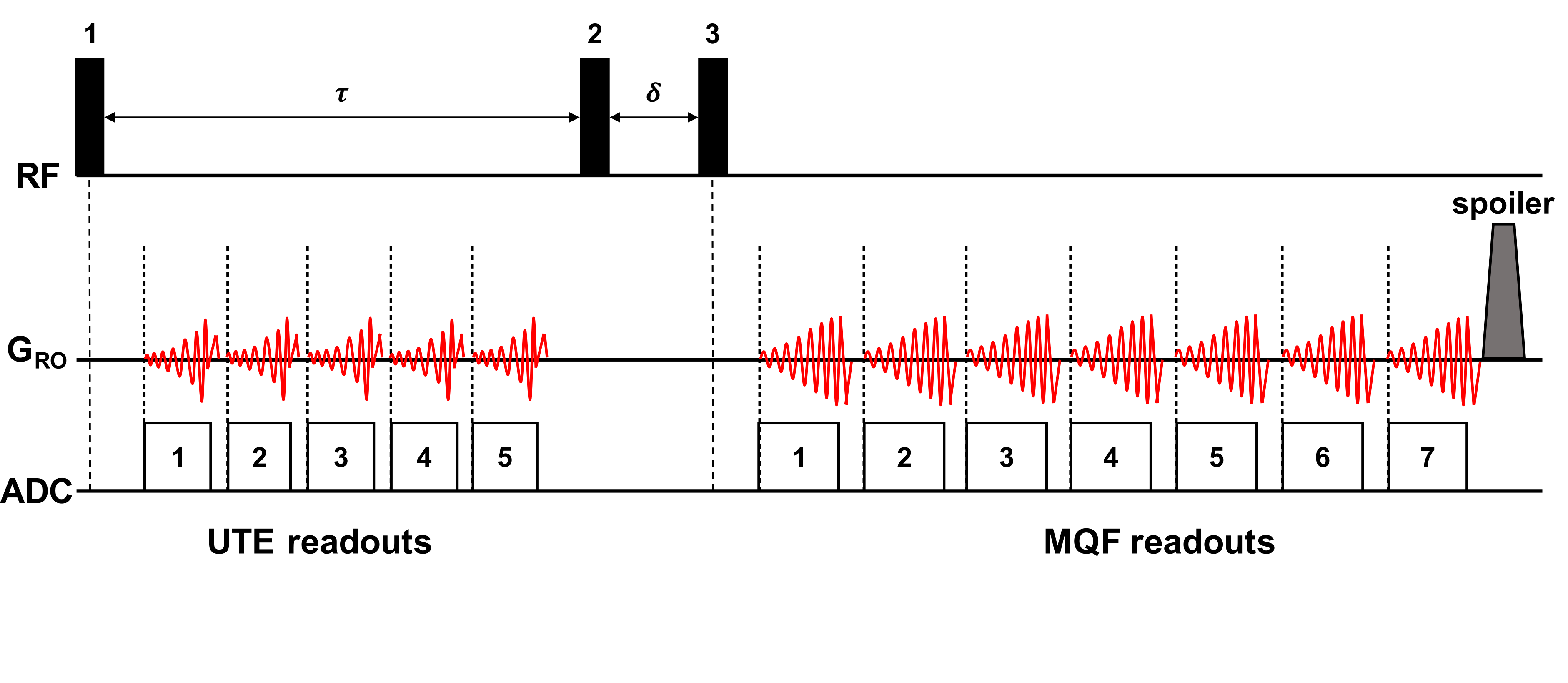
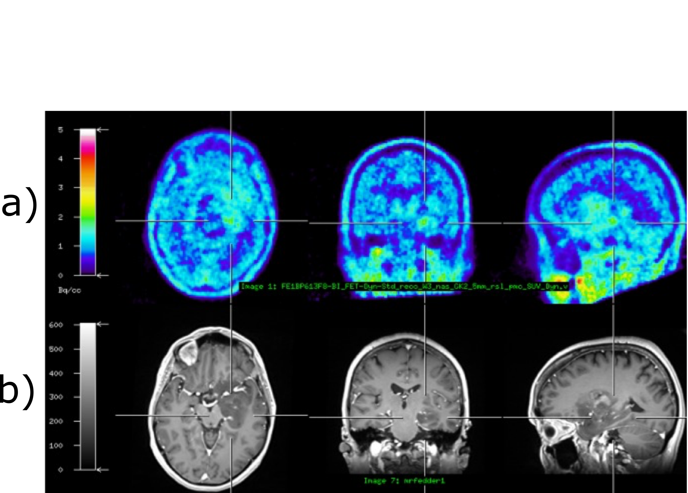
eSISTINA employs a three-pulse triple quantum filter with readout trains following the first and third radiofrequency (RF) pulses (Fig. 1). This configuration yields images weighted towards both restricted and non-restricted sodium, achieved through multiplex phase cycling. In this application, eSISTINA is coupled with a two-fold undersampled 3D spiral-based centre-out FLORET for k-space traversal6 , boasting excellent sampling efficiency and serving as a foundation for acceleration techniques such as compressed sensing (CS)7. The sequence has a repetition time of 150 ms, yielding full brain coverage (FOV: 320 mm) in 11 min. With a preparation time (τ) of 9.5 ms and an evolution time (δ) of 60 μs, the ultra-short echo time (UTE) readout train following the first RF pulse generates five images, starting at 0.56 ms with 1.85 ms increments. Meanwhile, the multiple quantum-filtered readout after the third RF pulse comprises seven images, starting at 10.40 ms with 9.13 ms increments—a configuration recommended for optimal relaxometry performance8. FWHM of the point spread function is 6.8mm and 13.7mm, respectively, for these readouts. The k-space is pseudo-randomly undersampled by a factor of 2.9 Images were acquired on a Siemens 7T Terra scanner (SIEMENS Healthineers, Erlangen, Germany) with an 80 mT/m gradient coil. A dual-tuned 1H/23Na birdcage coil (RAPID Biomedical, Germany) was used for both hydrogen and sodium imaging. The FLORET raw data were reconstructed using CS.10 The patient had undergone previous examination in a hybrid 3T MR/PET device (SIEMENS Healthineers, Erlangen, Germany) using an 18F-FET as a tracer (Fig. 2).Results
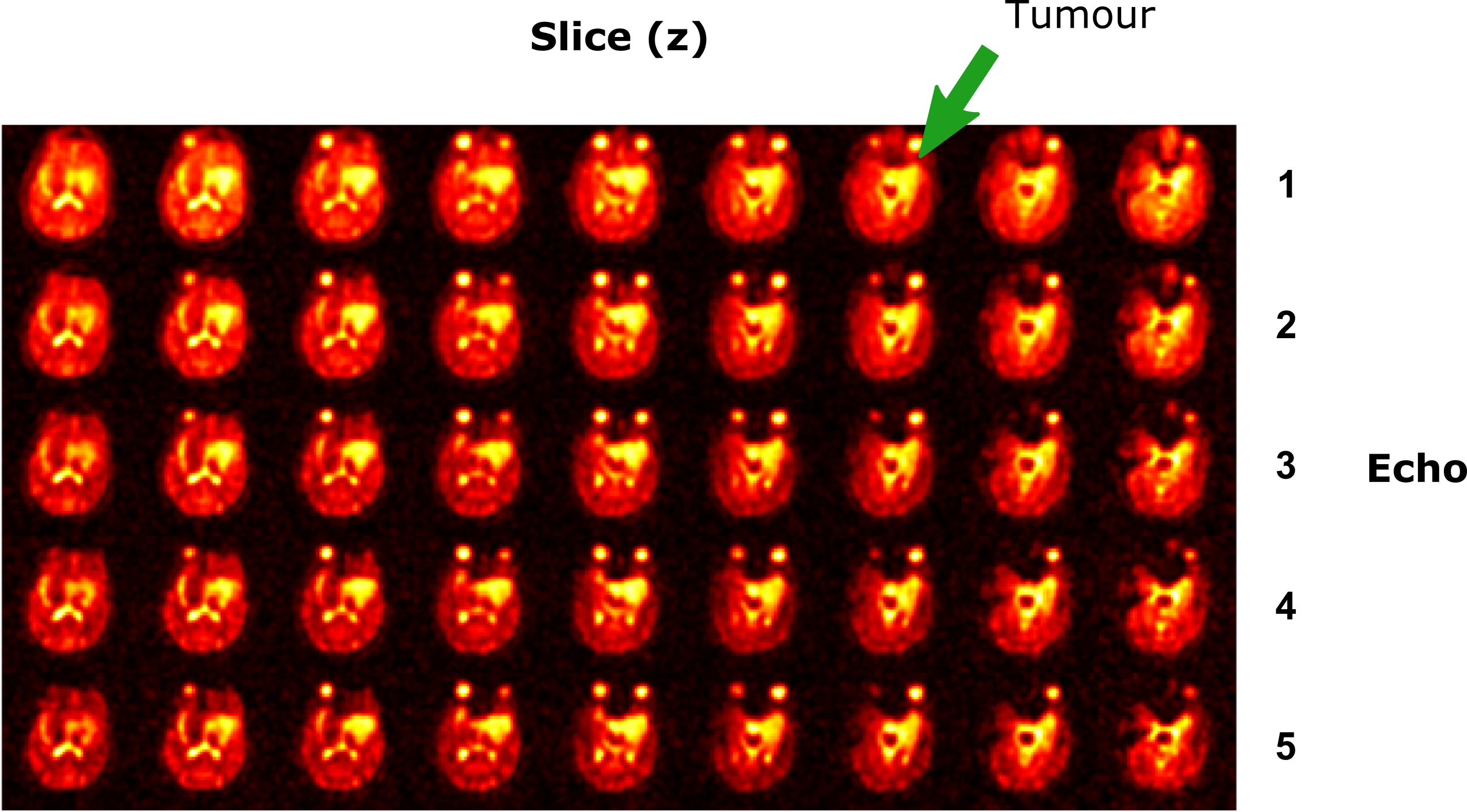
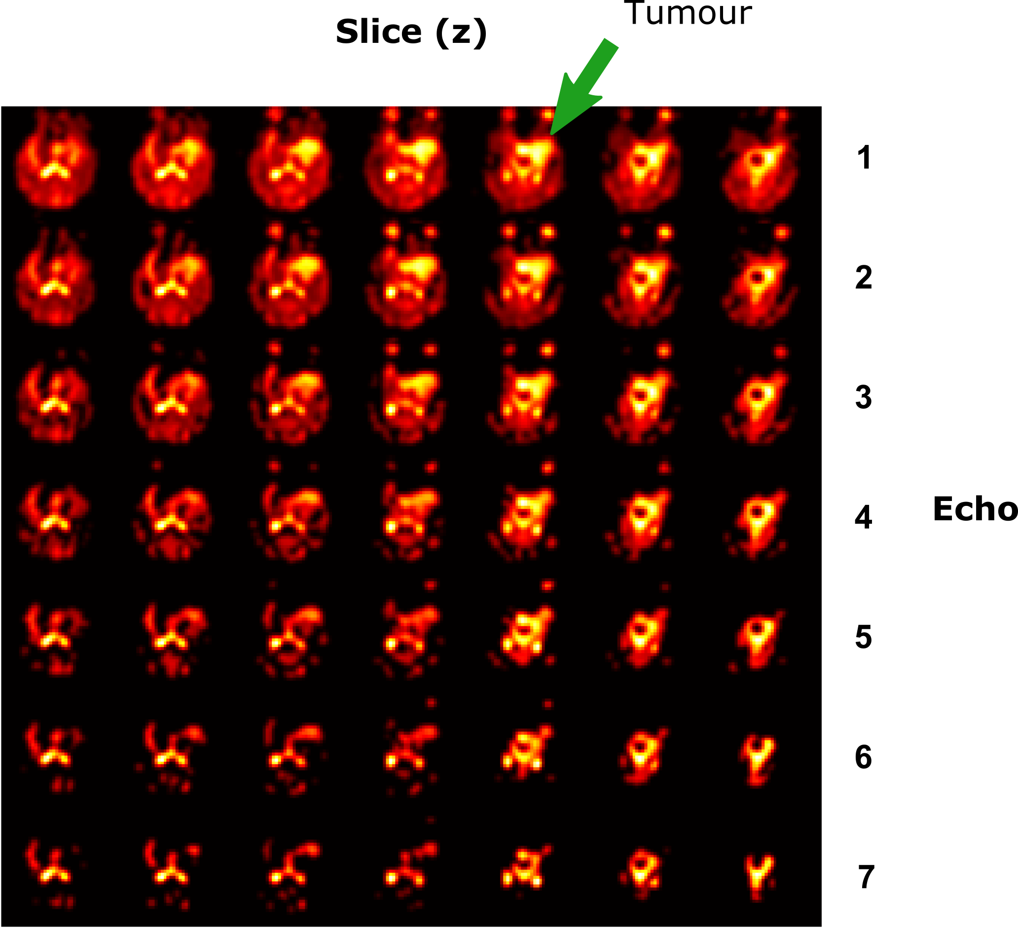
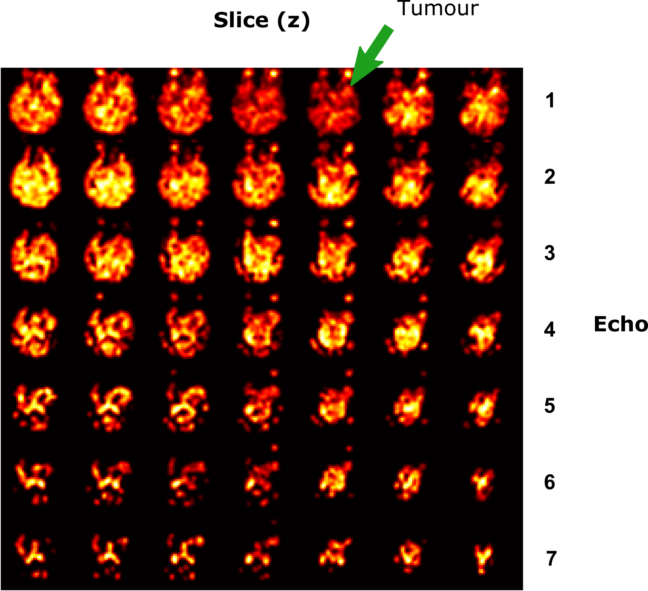
The patient was diagnosed with a meningioma located in the mesial temporal lobe (left). The tumour region exhibited a diffuse increase in FET uptake, resembling patterns often observed in low-grade gliomas. Despite the absence of metabolic tumour activity in the FET PET scan, eSISTINA images revealed a hyperintensity in both total sodium-weighted images (UTE) and non-restricted, mainly single-quantum-weighted (SQ-weighted) sodium images. In Figures 3-5, various brain slices (columns) and echoes (rows) are presented for total sodium (UTE), non-restricted (SQ), and restricted, mainly triple-quantum (TQ) weightings.Discussion
The images effectively portray the extent of the affected tissue. Notably, in comparison to prior studies, there is a marked improvement in image quality within a similar imaging duration when employing 7T. In the region of the tumour, restricted sodium-weighted images exhibit minimal contrast changes, while total sodium and non-restricted sodium showcase hyperintensity.Conclusion
The prospectively accelerated eSISTINA sequence excels in providing high-quality images for tumour analysis within a clinically relevant timeframe. Employing advanced reconstruction techniques, like CS has the potential of further elevating the image quality, thereby amplifying the overall performance and clinical significance of brain tumour imaging using eSISTINA.Acknowledgements
The authors express their sincere gratitude towards Elke Bechholz, Anita Koeth, Petra Engels and Claire Rick.References
1. Madelin G, Lee JS, Regatte RR, Jerschow A. Sodium MRI: methods and applications. Prog Nucl Magn Reson Spectrosc. 2014;79:14-47.
2. Fiege DP, Romanzetti S, Mirkes CC, Brenner D, Shah NJ. Simultaneous single-quantum and triple-quantum-filtered MRI of 23Na (SISTINA). Magn Reson Med. 2013;69(6):1691-1696.
3. Worthoff WA, Shymanskaya A, Shah NJ. Relaxometry and quantification in simultaneously acquired single and triple quantum filtered sodium MRI. Magn Reson Med. 2019;81:303-315.
4. Shymanskaya A, Worthoff WA, Stoffels G, Lindemeyer J, Neumaier B, Lohmann P, Galldiks N, Langen KJ, Shah NJ. Comparison of [(18)F]Fluoroethyltyrosine PET and Sodium MRI in Cerebral Gliomas: a Pilot Study. Mol Imaging Biol 2019.
5. Worthoff WA, Shymanskaya A, Lindemeyer J, Langen KJ, Shah NJ. Relaxometry and quantification in sodium MRI of cerebral gliomas: A FET-PET and MRI small-scale study. NMR Biomed. 2020;33(10):e4361.
6. Pipe JG, Zwart NR, Aboussouan EA, Robison RK, Devaraj A, Johnson KO. A new design and rationale for 3D orthogonally oversampled k-space trajectories. Magn Reson Med. 2011;66(5):1303-1311.
7. Chen Q, Shah NJ, Worthoff WA. Compressed Sensing in Sodium Magnetic Resonance Imaging: Techniques, Applications, and Future Prospects. J Magn Reson Imaging. 2022;55(5):1340-1356.
8. Worthoff WA, Chen Q, Shah NJ Optimisation of Simultaneously Acquired Single and Triple Quantum Filtered Sodium MRI for Relaxometry at 7T. ISMRM 2023 abstract 4625.
9. Chen Q, Shah NJ, Worthoff WA. Accelerated Enhanced Simultaneous Single-Quantum and Triple-Quantum-Filtered Sodium MRI Using Compressed Sensing at 7T. ISMRM 2023 abstract 4715.
10. Lustig M, Donoho D, Pauly JM. Sparse MRI: the application of compressed sensing for rapid MR imaging. Magn Reson Med. 2007;58(6):1182-1195.
Figures