3041
Compressed sensing reconstruction for high-SNR, rapid dissolved 129Xe gas exchange MRI1POLARIS, Division of Clinical Medicine, School of Medicine and Population Health, University of Sheffield, Sheffield, United Kingdom, 2Insigneo Institute for in silico Medicine, University of Sheffield, Sheffield, United Kingdom
Synopsis
Keywords: Hyperpolarized MR (Gas), Image Reconstruction
Motivation: 3D hyperpolarized 129Xe gas exchange imaging is limited by low SNR and long breath-holds, which is problematic for patients with dyspnea and/or low gas transfer. Compressed sensing (CS) reconstruction can accelerate 129Xe MRI whilst improving SNR.
Goal(s): To assess whether gas exchange ratio maps are quantitatively preserved with CS dissolved 129Xe imaging and investigate the feasibility of reduced-cost natural abundance (NA) CS dissolved 129Xe imaging.
Approach: CS-reconstructed gas exchange ratios were evaluated in healthy volunteers and COPD patients and prospectively with NA 129Xe.
Results: CS increased image SNR, allowed 3-fold acquisition acceleration and maintained ratio map fidelity, even with NA 129Xe.
Impact: Compressed sensing reconstruction of dissolved 129Xe spectroscopic imaging improved image quality even with decreased scan time, whilst preserving key gas exchange metrics. This will benefit patients with breathlessness and/or low gas transfer and enables natural abundance dissolved 129Xe imaging.
Introduction
Pulmonary gas exchange can be assessed with hyperpolarized 129Xe MRI via quantification of the signal from 129Xe dissolved in the alveolar membrane (M) and capillary red blood cells (RBCs)1,2. However, dissolved 129Xe MRI is SNR limited with signal ~2% that of the gaseous 129Xe3. SNR is often further reduced in patients with lung disease, who may have inherently lower RBC signal due to reduced gas transfer and find it difficult to complete the ~15s breath-hold. Most 129Xe imaging is performed with enriched Xe (>85% 129Xe isotopic abundance), due to its increased signal strength compared to NA (26% 129Xe). However, this has a significantly greater cost, and feasibility of NA 129Xe gas-phase ventilation imaging has been demonstrated4.The denoising properties of CS5 can improve image quality and enable reduced scan time for non-Cartesian 129Xe imaging6. The effects of CS reconstruction on the key clinical metrics of gas exchange imaging, the RBC:M, RBC:Gas and M:Gas signal ratios, have yet to be explored. Here, we compare the gas exchange ratios from both CS and conventional radial gridding reconstruction in a 4-echo radial EPSI implementation in healthy volunteers and patients with COPD. We also assess the feasibility of imaging dissolved NA 129Xe with CS.
Theory
CS: non-Cartesian k-space data are reconstructed by solving a nonlinear regularized optimization problem5:$$x =argmin_x\parallel Ax-y\parallel_2+\lambda_1\parallel\Psi x\parallel_1+\lambda_2\parallel Tx\parallel_1,\hspace{12cm}(1)$$
where $$$x$$$=reconstructed image, $$$y$$$=k-space data, $$$\lambda_{1,2}$$$=regularization parameters, $$$\Psi$$$=sparsity operator and $$$T$$$=finite difference transform. $$$A$$$ is the ‘forwards model’:
$$y=Ax+\nu,\hspace{20cm}(2)$$
$$A=PF,\hspace{20.7cm}(3)$$
where $$$\nu$$$=noise, $$$P$$$=sampling density, $$$F$$$=Fourier transform.
Conventional reconstruction: $$$x$$$ is calculated via matrix inversion ($$$A^{-1}$$$) following gridding to a Cartesian matrix7-9.
Methods
One dissolved 129Xe healthy dataset was retrospectively analyzed to optimize the CS reconstruction parameters. This was acquired with a four-echo 3D radial spectroscopic imaging sequence7 on a 1.5T GE HDx scanner, with a 129Xe transmit-receive vest coil and 1L hyperpolarized10 enriched 129Xe inhaled from functional residual capacity. CS reconstruction was performed using the Berkeley Advanced Reconstruction Toolbox (BART)11, and identity and total variation regularization in the image domain. The gas, RBC and M resonances were separated in k-space using matrix inversion and the chemical shifts and T2* obtained from calibration spectra7,12 before solving Equation 1. $$$\lambda_1$$$, $$$\lambda_2$$$ and the acceleration factor (AF) for random undersampling were chosen empirically to find the combination of parameters which minimized the mean absolute error (MAE) and maximized the structural similarity index measure (SSIM) of the dissolved 129Xe ratio maps. The mean ratio values from the optimized CS and conventional reconstructions were compared for ten healthy volunteers and five COPD patients. One healthy female was prospectively imaged with 800ml NA 129Xe on a 1.5T GE Artist scanner to assess the feasibility of NA dissolved 129Xe CS imaging.Results and Discussion
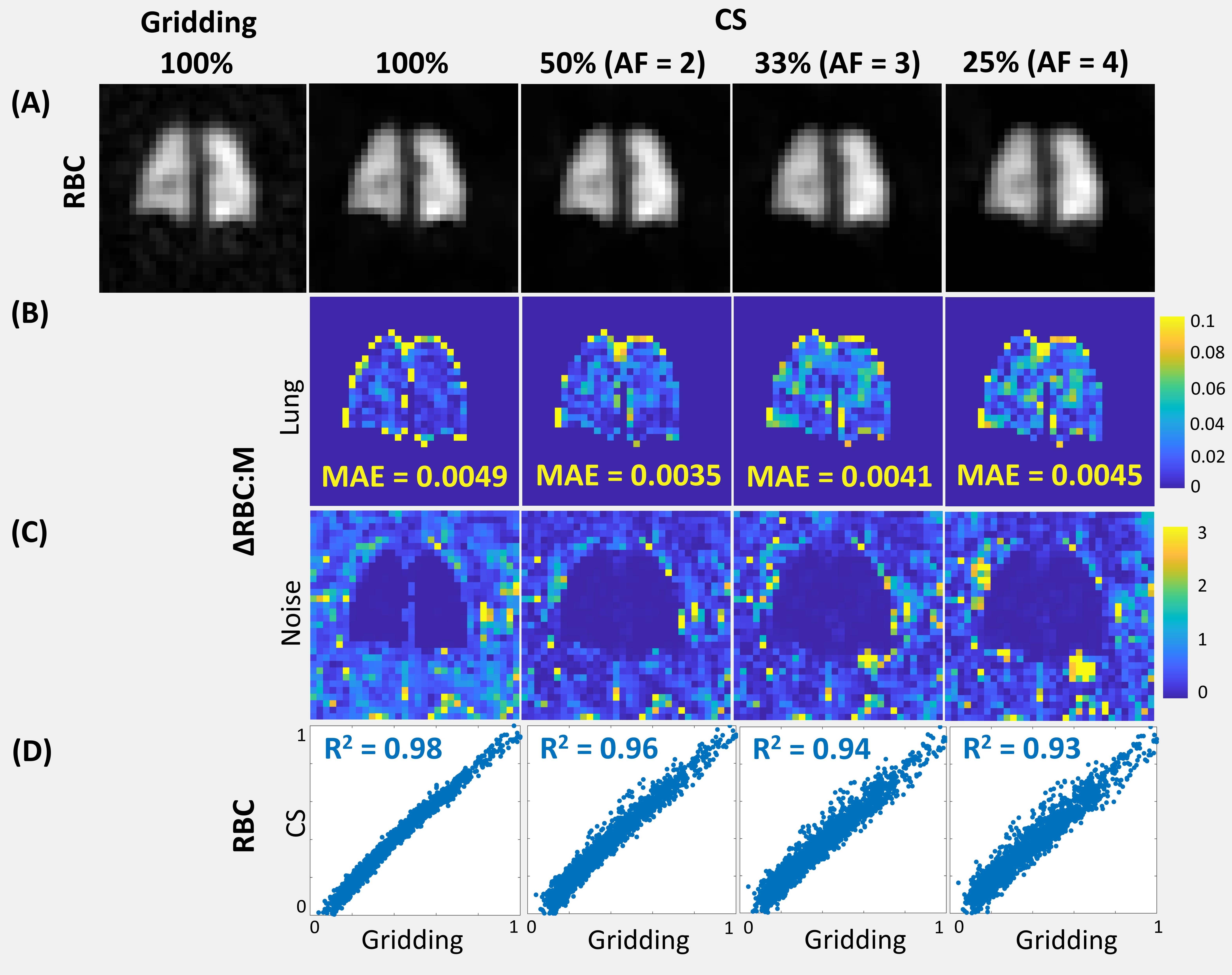
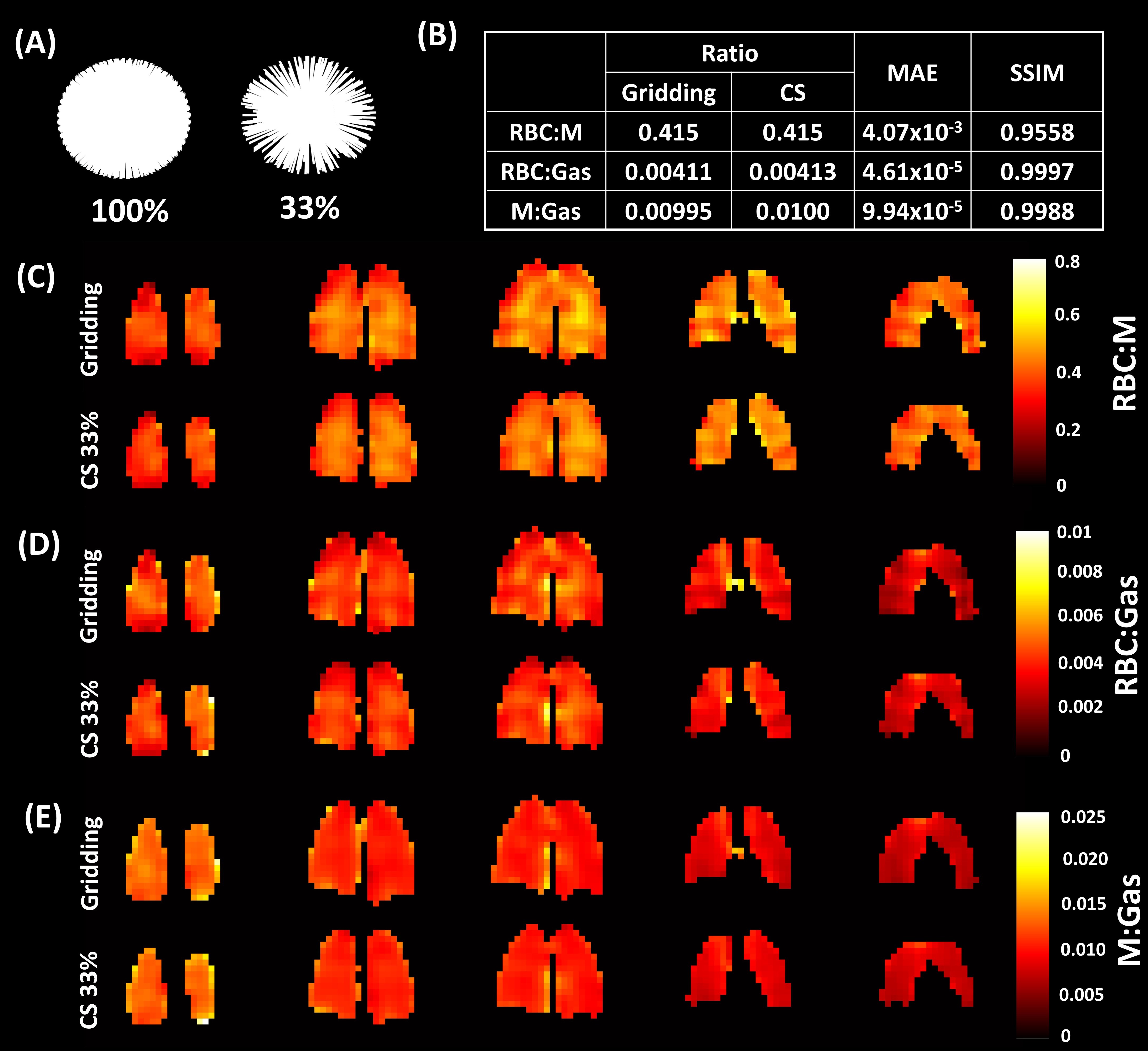
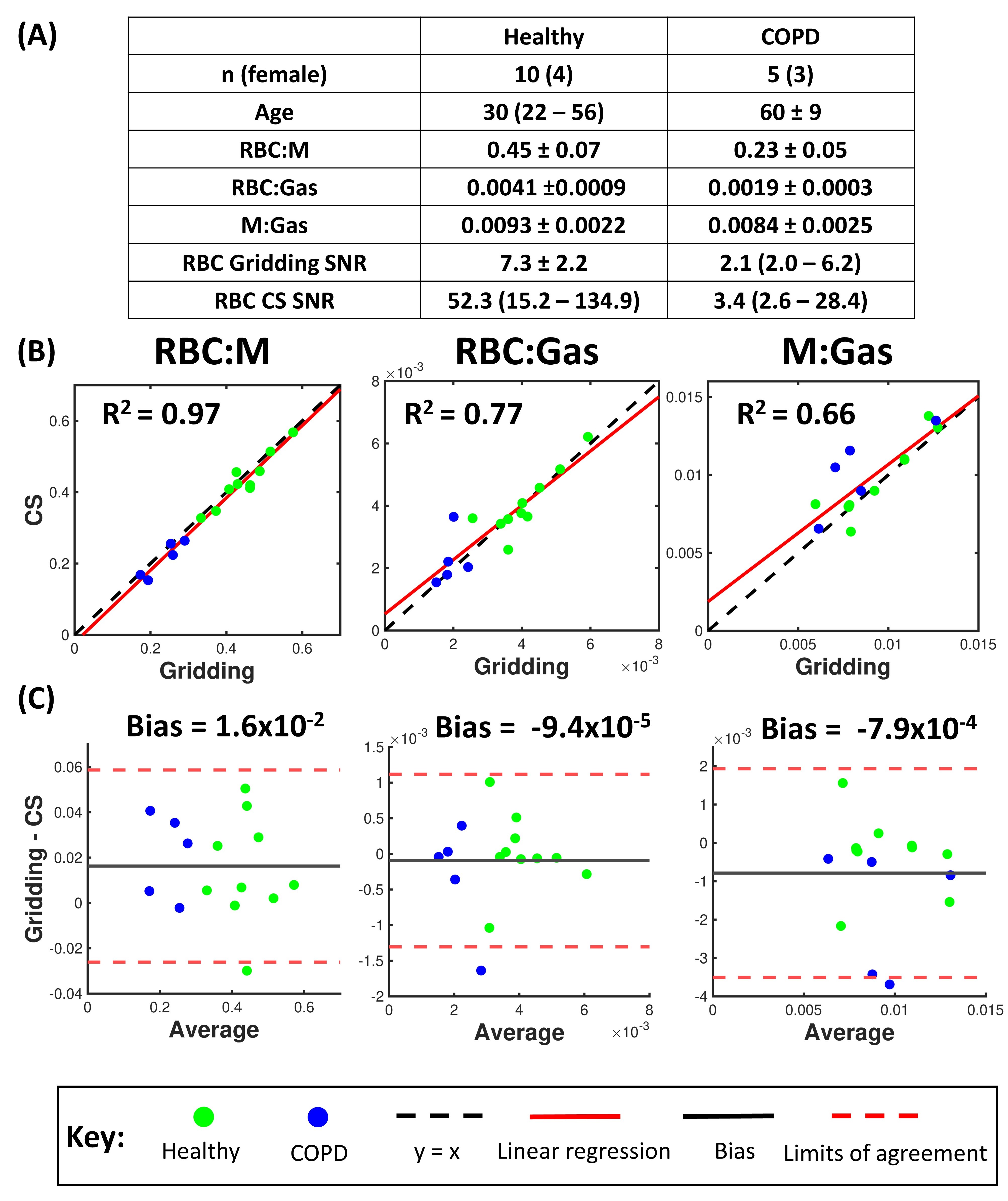
Figure 1 shows the conventional and CS reconstructed RBC images with different AFs, the corresponding RBC:M difference maps and pixel-wise linear regression of the RBC signal. To balance image fidelity with reduction of scan time, AF=3 was chosen for the final implementation, with $$$\lambda_1$$$=0.005 and $$$\lambda_2$$$=0.001. With this AF, only 311 radial spokes are required (Figure 2A) and the breath-hold is reduced to ~5s compared with the routine clinical implementation7. This should help improve success rates of 129Xe gas exchange imaging in patients with breathlessness.CS image fidelity metrics are shown in Figure 2B and ratio maps are shown in Figure 2C-E. Close agreement was found between the mean ratio values and maps from each reconstruction. In healthy volunteers and COPD patients, the mean ratio values were preserved with the CS reconstruction, even for low SNR images (Figure 3). The CS reconstruction was effective in suppressing noise, and RBC SNR increased by an average factor of 9 for healthy subjects and 1.7 for COPD subjects. This denoising effect is especially desirable for imaging patients with intrinsically weak RBC signal from reduced gas transfer or capillary perfusion.
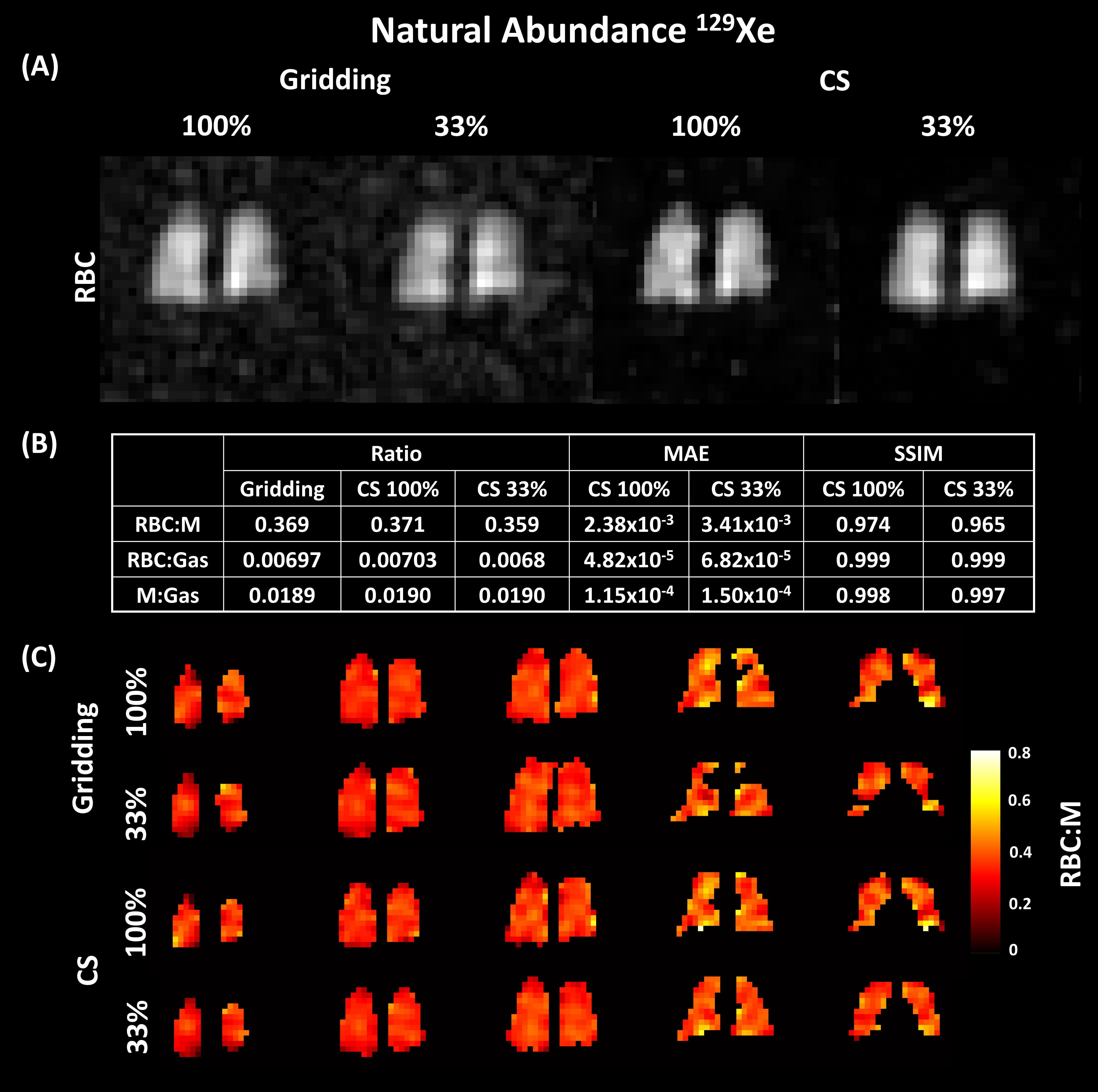
Using CS, high SNR RBC images were achieved with NA 129Xe for 100% and 33% sampling (Figure 4A). The MAE values (Figure 4B) for the quantitative ratio maps (Figure 4C) were comparable to those found for enriched 129Xe. Further prospective evaluation in increased subject numbers, including patients with lung disease, is required to validate this approach, but these initial results are promising for low-cost NA dissolved 129Xe imaging.
Conclusion
CS reconstruction of dissolved 129Xe spectroscopic imaging improves image SNR and enables reduced scan time, whilst maintaining ratio map fidelity. This benefits patients with breathlessness and/or low gas transfer and permits NA dissolved 129Xe imaging, allowing for cheaper and faster gas exchange imaging.Acknowledgements
We thank Dr Rod Lawson and the study teams and patients of the SUMMER study. The work was supported by GlaxoSmithKline and MRC grant MR/M008894/1 awarded to J.M.W.
J.H.P-M is funded by a PhD scholarship from the Discovery Medicine North (DiMeN) Doctoral Training Partnership.
References
1. Kaushik SS, Robertson SH, Freeman MS, et al., Single-Breath Clinical Imaging of Hyperpolarized Xe-129 in the Airspaces, Barrier, and Red Blood Cells Using an Interleaved 3D Radial 1-Point Dixon Acquisition. Magnetic Resonance in Medicine, 2016. 75(4): p. 1434-1443.
2. Wang JM, Robertson SH, Wang ZY, et al., Using hyperpolarized Xe-129 MRI to quantify regional gas transfer in idiopathic pulmonary fibrosis. Thorax, 2018. 73(1): p. 21-28.
3. Marshall H, Stewart NJ, Chan HF, et al., In vivo methods and applications of xenon-129 magnetic resonance. Progress in Nuclear Magnetic Resonance Spectroscopy, 2021. 122: p. 42-62.
4. Stewart NJ, Norquay G, Griffiths PD, and Wild JM, Feasibility of human lung ventilation imaging using highly polarized naturally abundant xenon and optimized three-dimensional steady-state free precession. Magn Reson Med, 2015. 74(2): p. 346-52.
5. Lustig M, Donoho D, and Pauly JM, Sparse MRI: The application of compressed sensing for rapid MR imaging. Magn Reson Med, 2007. 58(6): p. 1182-95.
6. Plummer JW, Bdaiwi AS, Soderlund SA, et al. Compressed sense reconstructions improve non-Cartesian hyperpolarized 129Xe MRI. in Proc. 31st Intl. Soc. Mag. Reson. Med. 0856. 2023.
7. Collier GJ, Eaden JA, Hughes PJC, et al., Dissolved Xe-129 lung MRI with four-echo 3D radial spectroscopic imaging: Quantification of regional gas transfer in idiopathic pulmonary fibrosis. Magnetic Resonance in Medicine, 2021. 85(5): p. 2622-2633.
8. Wiesinger F, Weidl E, Menzel MI, et al., IDEAL spiral CSI for dynamic metabolic MR imaging of hyperpolarized [1-13C]pyruvate. Magnetic Resonance in Medicine, 2012. 68(1): p. 8-16.
9. Beatty PJ, Nishimura DG, and Pauly JM, Rapid gridding reconstruction with a minimal oversampling ratio. IEEE Transactions on Medical Imaging, 2005. 24(6): p. 799-808.
10. Norquay G, Collier GJ, Rao M, Stewart NJ, and Wild JM, 129Xe-Rb Spin-Exchange Optical Pumping with High Photon Efficiency. Physical Review Letters, 2018. 121(15): p. 153201.
11. Blumenthal M, Holme C, Roeloffs V, et al., mrirecon/bart: version 0.8.00. 2022, Zenodo.
12. Kaushik S, Freeman M, Yoon S, et al., Measuring diffusion limitation with a perfusion-limited gas—Hyperpolarized 129Xe gas-transfer spectroscopy in patients with idiopathic pulmonary fibrosis. J Appl Physiol (1985), 2014. 117(6): p. 577-85.
Figures