3040
Enabling SENSE Accelerated 2D CSI For Hyperpolarized Carbon-13 Imaging1Oxford Centre for Clinical Magnetic Resonance Research, University of Oxford, Oxford, United Kingdom, 2Department of Physiology, Anatomy, and Genetics, University of Oxford, Oxford, United Kingdom, 3Department of Clinical Medicine, Aarhus University, Aarhus, Denmark, 4JD Coils, Hamubrg, Germany, 5GE HealthCare, Munich, Germany, 6Department of Radiology, Oxford University Hospitals, Oxford, United Kingdom
Synopsis
Keywords: Hyperpolarized MR (Non-Gas), Metabolism, carbon-13 imaging, x-nuclei MRI, metabolic imaging, flexible RF coil, CSI
Motivation: For hyperpolarized 13C metabolic imaging studies, a challenge is to achieve high temporal resolution without decreasing spatial and/or spectral resolution.
Goal(s): To accelerate hyperpolarized 13C MRI by combining a 2D Chemical Shift Imaging (CSI) sequence with SENSitivity Encoding (SENSE) reconstruction.
Approach: Due to the low natural abundance of 13C, the sensitivity maps needed for SENSE reconstruction cannot be pre-acquired. As such, in this work, the novel approach of using sodium sensitivity maps was demonstrated.
Results: SENSE reconstruction corrected aliased images, where in-vivo metabolic information was acquired with a 4-fold temporal acceleration.
Impact: As hyperpolarized 13C metabolic imaging is clinically translated, there is a need for easy-to-implement, fast, and robust imaging techniques. Therefore, this study implemented a novel 13C technique to accelerate Chemical Shift Imaging: a ubiquitous and robust sequence.
Introduction
Hyperpolarized (HP) MRI is a non-invasive clinical technique that detects low-signal metabolites through the injection of pre-polarized 13C-labelled substrates1,2. However, a challenge in HP-13C studies is to achieve high temporal resolution while preserving spatial and spectral resolution3. Fast imaging approaches have been developed but all have specific trade-offs: spectral-spatial imaging provides good spatial resolution but offers limited spectral information4,5; fast spectroscopic imaging offers high temporal resolution but limited spectral bandwidth and spatial resolution due to hardware limitations6; multi-echo approaches require well-defined spectral profiles7,8; and phase-encoded Chemical Shift Imaging (CSI) provides a robust solution but is RF-intensive and slow9. Parallel imaging methods, such as SENSitivity Encoding (SENSE), offer acceleration opportunities, where fast acquisition results in a reduced field of view (FOV) and folding artifacts10. For artifact correction, SENSE requires a pre-acquired sensitivity profile, which is challenging for 13C with low natural abundance11. Prior work with a dual-tuned sodium-carbon coil has successfully obtained sodium sensitivity maps representing the carbon profile12. As such, in this work, a clinically robust and easy-to-use sequence, CSI, was accelerated by leveraging the previously presented sodium-carbon coil while preserving spatial and spectral quality.Methods
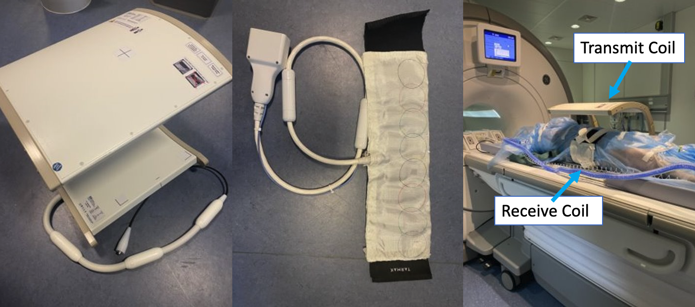
Compliant with Danish law on animal experiments, four female Danish domestic pigs were imaged (figure 1) using a 3T GE MR750 MRI scanner (GE Healthcare, WI). Proton imaging was acquired with the body coil using a T2 fast spin echo sequence. Sodium and carbon imaging were acquired using a clamshell transmit coil (Rapid Biomedical, Germany) and an eight-channel flexible 23Na/13C receive coil (JD Coils, Germany) (figure 1). Pigs received two injections of 30ml of 250mM hyperpolarized pyruvate, prepared in a DNP system (SpinAligner, Polarize) as previously described13, administered intravenously at ∼5ml/s and flushed with 20 ml saline. Imaging commenced 20 seconds after injection completion. Injections were separated by ~2 hours. A single slice sodium CSI was acquired (FOV=240mm, number of averages(NEX)=16, flip angle (FA)=90◦, Repetition Time (TR)=38.5ms, bandwidth=5kHz, matrix =24x24, slice thickness=10mm). Either a fully sampled or under sampled 13C CSI was acquired after each HP injection (FOV=240 or 120mm, NEX=1, FA=10◦, TR=64.1ms, bandwidth=5kHz, matrix =24x24 or 12x12, slice thickness=10mm, temporal resolution = 36 or 9s). Metabolite signals were extracted from the spectra using a deconvolution spectral fit14. Reconstructions used sodium sensitivity and carbon images with a 2D SENSE algorithm in MATLAB (R2023b, Mathworks).Results
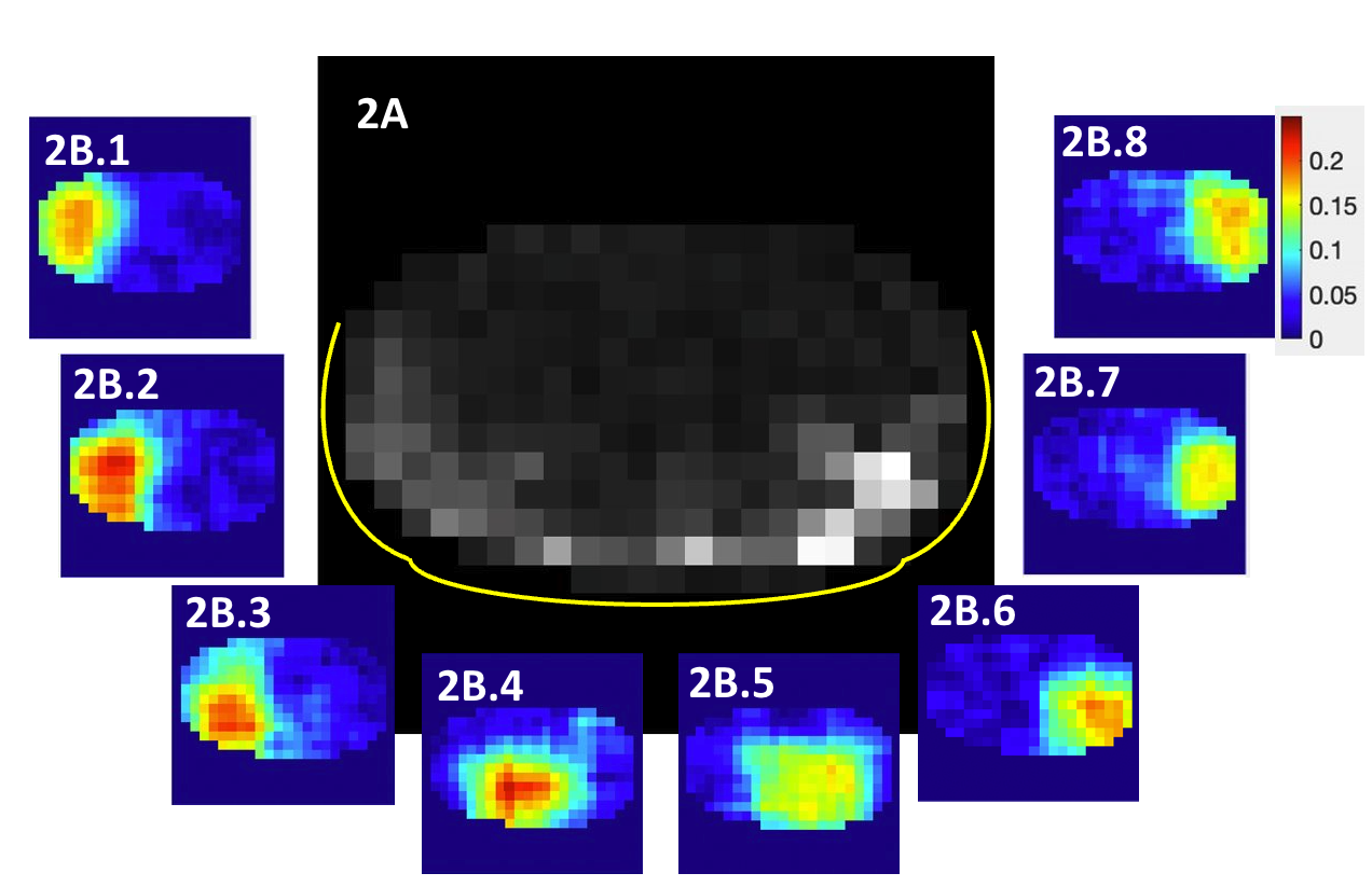
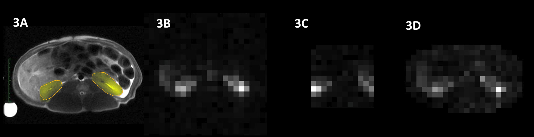
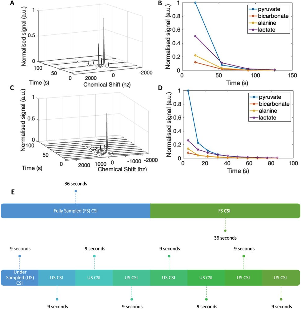
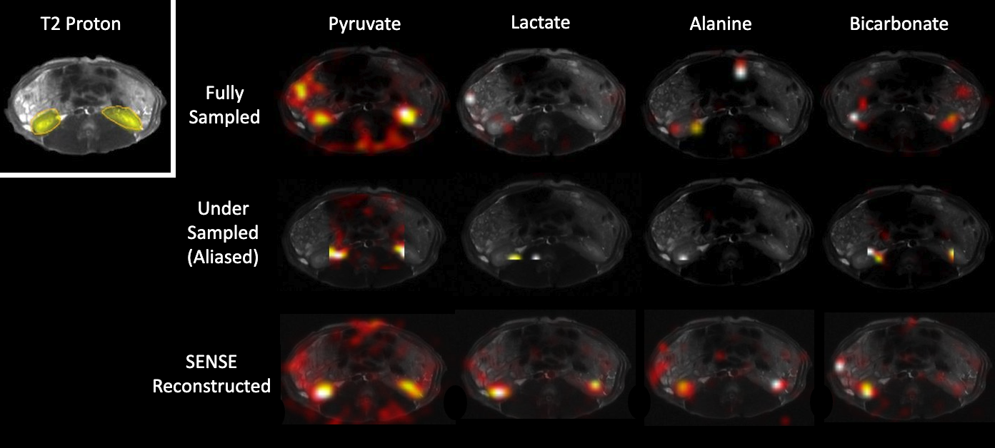
In figure 2A, the combination of eight channels aggregates the low signal per loop into a sensitivity image. Figures 2B.1-8 show the corresponding localised sensitivity on the porcine torso closest to each receiver element. Sensitivity drops rapidly away from the coil. Figure 3A shows the anatomy in the axial proton image. Pyruvate was expected to localise at the yellow kidney ROIs. Figure 3B is the pyruvate signal from the fully sampled CSI (SNR = 37 ± 19) showing two high intensity regions corresponding to the kidneys. Figure 3C demonstrates the folded aliasing due to reduced FOV in the under sampled CSI. Figure 3D is the SENSE reconstruction with two bright regions at the kidneys, illustrating the structural similarity between the fully sampled image and corrected aliasing (SNR = 23 ± 14). Figure 4 shows tracked metabolism dynamics (pyruvate, bicarbonate, lactate, and alanine) in the kidneys of a second porcine subject. Increased temporal resolution from the accelerated acquisition is seen in figures 4C/4D compared to that in figures 4A/4B. The imaging scheme in figure 4E illustrates the four-fold temporal acceleration, allowing four under sampled CSIs to be taken in a span of one fully sampled CSI. In figure 5, another subject’s fully sampled CSIs show bright spots in the pyruvate image. The under sampled images illustrate the aliasing due to the reduced FOV. SENSE reconstructed images for pyruvate, lactate, and alanine exhibited two clear bright spots at the kidneys.Discussion
In vivo accelerated CSI using SENSE reconstruction demonstrated successful aliasing corrections with good SNR. Sodium sensitivity maps eliminate the need for co-registration and additional HP injections. High temporal resolution produced a detailed decay curve, useful for further metabolite kinetic modelling or denoising. In cases with unknown metabolite spectra or limited HP MRI specialist knowledge, this robust acceleration method ensures comprehensive capture of metabolic signals, mitigating the risk of missing spectral data.Conclusion
The novel approach of using sodium sensitivity profiles with accelerated SENSE-reconstructed CSI demonstrated alias-corrected images, where metabolic information was acquired with a 4-fold temporal acceleration. Further work will look to optimise image post-processing, signal fitting, and k-space acquisitions for increased SNR (spiral, radial and rosette sampling). This robust acceleration technique offers a promising solution for metabolic imaging in diverse clinical scenarios.Acknowledgements
This work was supported by the Oxford-MRC Doctoral Training Partnership iCASE award, and the Oxford Clarendon Scholarship.References
1. Kurhanewicz, J. et al. Hyperpolarized 13C MRI: Path to Clinical Translation in Oncology. Neoplasia 21, 1–16 (2019).
2. Wolber, J. et al. Generating highly polarized nuclear spins in solution using dynamic nuclear polarization. Nucl Instrum Methods Phys Res A 526, 173–181 (2004).
3. Grist, J. T. et al. Quantifying normal human brain metabolism using hyperpolarized [1–13C]pyruvate and magnetic resonance imaging. Neuroimage 189, 171–179 (2019).
4. Meyer, C. H., Pauly, J. M., Macovskiand, A. & Nishimura, D. G. Simultaneous spatial and spectral selective excitation. Magn Reson Med 15, 287–304 (1990).
5. Gordon, J. W., Vigneron, D. B. & Larson, P. E. Z. Development of a symmetric echo planar imaging framework for clinical translation of rapid dynamic hyperpolarized 13 C imaging. Magn Reson Med 77, 826–832 (2017).
6. Gordon, J. W. et al. Fast Imaging for Hyperpolarized MR Metabolic Imaging HHS Public Access. J Magn Reson Imaging 53, 686–702 (2021).
7. Bieri, O. & Scheffler, K. Fundamentals of balanced steady state free precession MRI. Journal of Magnetic Resonance Imaging 38, 2–11 (2013).
8. Reeder, S. B. et al. Iterative decomposition of water and fat with echo asymmetry and least-squares estimation (IDEAL): application with fast spin-echo imaging. Magn Reson Med 54, 636–644 (2005).
9. Wang, J. X., Merritt, M. E., Sherry, A. D. & Malloy, C. R. Accelerated Chemical Shift Imaging of Hyperpolarized 13C Metabolites. Magn Reson Med 76, 1033 (2016).
10. Pruessmann, K. P., Weiger, M., Scheidegger, M. B. & Boesiger, P. SENSE: Sensitivity Encoding for Fast MRI. doi:10.1002/(SICI)1522-2594(199911)42:5.
11. Arunachalam, A. et al. Accelerated spectroscopic imaging of hyperpolarized C-13 pyruvate using SENSE parallel imaging. NMR Biomed 22, 867–873 (2009).
12. Sanchez-Heredia, J. D. et al. RF Coil Design for Accurate Parallel Imaging on 13 C MRSI using 23 Na Sensitivity Profiles.
13. Laustsen, C. et al. Acute porcine renal metabolic effect of endogastric soft drink administration assessed with hyperpolarized [1-13c]pyruvate. Magn Reson Med 74, (2015).
14. Schroeder, M. A. et al. Measuring intracellular pH in the heart using hyperpolarized carbon dioxide and bicarbonate: a 13C and 31P magnetic resonance spectroscopy study. Cardiovasc Res 86, 82–91 (2010).
Figures