3038
Advancement of a novel 31P MRS-based approach for the in vivo determination of pH and magnesium ion content1Division of Medical Physics in Radiology, German Cancer Research Center (DKFZ), Heidelberg, Germany, 2Faculty of Physics and Astronomy, Heidelberg University, Heidelberg, Germany, 3International Max Planck Research School for Quantum Dynamics in Physics, Chemistry and Biology (IMPRS-QD), Max Planck Institute for Nuclear Physics (MPIK), Heidelberg, Germany, 4Faculty of Medicine, Heidelberg University, Heidelberg, Germany
Synopsis
Keywords: Non-Proton, Spectroscopy, Phosphorus, 31P, pH, magnesium, brain
Motivation: The reliability of conventional 31P MRS-based methods for the determination of pH and magnesium ion content (Mg) is hampered when applied to pathologies due to their calibration for physiological conditions.
Goal(s): The aim of this study was the advancement of a novel approach for pH and Mg mapping to improve its reliability for application in vivo.
Approach: This advancement was performed by incorporating an additional input parameter into the approach and tested on in vivo 31P MRSI brain datasets.
Results: Compared to the original algorithm, the advanced version resulted in robust mapping of pH and Mg yielding homogeneous brain maps for healthy volunteers.
Impact: The advancement of a novel approach for the in vivo determination of pH and magnesium ion content under different chemical conditions improves its reliability and can now potentially be used for the investigation of pathologies such as cancer.
Introduction
Phosphorus magnetic resonance spectroscopic imaging (31P MRSI) enables the non-invasive imaging of pH values and magnesium ion content (Mg) in living tissues by using the chemical shift changes of inorganic phosphate (Pi) and adenosine-5’-triphosphate (ATP)1,2. However, the reliability of the conventional methods (e.g. the modified Henderson-Hasselbalch equation in case of pH3) for application in pathologies is hampered due to their calibration based on specific physiological conditions. To overcome this reliability issue, a novel approach for the in vivo determination of pH and Mg under different chemical conditions was recently proposed4,5.The purpose of this study was to further advance this novel approach in order to overcome the current challenges, such as an under-determined assignment of pH and Mg values, and therewith increase the reliability and robustness of the in vivo determination of pH and Mg under different chemical conditions.
Methods
The novel approach for the in vivo determination of pH and Mg under different chemical conditions is based on a multi-parametric look-up algorithm introduced in4,5. The basic idea of this algorithm is to use the chemical shifts of β- and γ-ATP to determine the output values pH, Mg and the parameter Ion (representing the ionic strength in general). To overcome the current under-determination (three output parameters from two input parameters), the look-up algorithm was advanced in this study by (i) additionally incorporating the chemical shift of Pi and (ii) changing the reference peak from Phosphocreatine (PCr) to δα, i.e. using δαβ, δγα and δPi-α. The reference peak was changed because the referencing to PCr was assumed to cause data mismatches between the look-up elements acquired in model solutions and the in vivo data6. An overview of the basic concept of the developed advanced look-up algorithm can be found in Figure 1. Note that the magnesium ion content is defined as R = [Mgtot]/[ATPtot]. To advance the algorithm, new model functions were developed in analogy to5, which are also based on the Hill equation7,8. In order to test the new algorithm, it was applied to in vivo 31P MRSI brain datasets of seven healthy volunteers acquired at B0 = 7T. The measurement and evaluation protocol can be found in9.Results
The development and implementation of new model functions into the algorithm could successfully be performed for the new input chemical shifts δαβ, δγα and δPi-α. Application of the advanced look-up algorithm to the in vivo 31P MRSI datasets resulted in over 90% solution triple (pH,R,Ion) being found for all tissue voxels. The volumetric maps for pH, R and Ion of five representative volunteers are shown in Figures 2 – 4. Compared to the original algorithm, which showed unexpected hot spots of altered values, the new maps show a homogeneous distribution of output values over the entire brain tissue. The mean values across all volunteers are (6.87 ± 0.04) for pH, (0.98 ± 0.04) for R and (0.15 ± 0.07) for the parameter Ion. The parameter Ion shows the largest variance of values. The mean values of each volunteer individually can be found in Table 1. The determined pH values show a systematic shift of about -0.1 pH compared to the pH values calculated with the conventional modified Henderson-Hasselbalch equation3.Discussion
The advanced approach performed in this study resulted in a more robust algorithm for the determination of pH and magnesium ion content. This improvement is presumably due to the incorporation of an additional input chemical shift to overcome the under-determination of the previous algorithm. As expected for healthy brain tissue, the resulting maps for pH, R and Ion of the advanced algorithm show homogeneous value distributions without any large hotspots of altered values. Furthermore, the maps reveal a white matter / gray matter difference that is also observed in pH maps calculated with the conventional method. Nevertheless, a systematic shift in pH values (about 0.1 lower compared to pH values calculated with the Henderson-Hasselbalch equation3) is still present. This is presumably due to differences between the model solutions used for the development of the algorithm, i.e. generation of the look-up table, and the chemical conditions of the brain tissue, which has to be investigated further in the future.Conclusion
The advancement of the recently proposed look-up algorithm for in vivo determination of pH and magnesium ion content resulted in more robust results for human brain tissue than the original version. In the future, this advanced algorithm could be used for the investigation of pH and Mg of diseases affecting the brain, where larger deviations from the baseline values identified in this study are expected.Acknowledgements
No acknowledgement found.References
1. Korzowski A, Weinfurtner N, Mueller S, et al. ”Volumetric mapping of intra- and extracellular pH in the human brain using 31P MRSI at 7T”. Magn Reson Med. (2020); 84: pp. 1707– 1723. https://doi.org/10.1002/mrm.28255
2. Taylor JS, Vigneron DB, Murphy-Boesch J, et al. “Free magnesium levels in normal human brain and brain tumors: 31P chemical-shift imaging measurements at 1.5 T”. Proc Natl Acad Sci U S A. (1991); 88(15): pp. 6810-6814. doi:10.1073/pnas.88.15.6810
3. de Graaf RA. “In Vivo NMR Spectroscopy: Principles and Techniques”: 2nd Edition.; 2007. doi: 10.1002/9780470512968.
4. Franke VL, Breitling J, Bangert R, et al. ”Estimation of pH values and magnesium ion content in vivo using a chemical shift dictionary for 31P MRSI at UHF”. Proceedings of the 31th Annual Meeting of the ISMRM, Toronto, CA. (2023); Program #0022.
5. Franke VL. “In vivo determination of pH and magnesium ion concentration by means of 31P MRSI: A multi parametric look-up approach”. Dissertation. University of Heidelberg, Germany. (2023).
6. Franke VL, Breitling J, Boyd PS, et al. “A versatile look-up algorithm for mapping pH values and magnesium ion content using 31P MRSI”. In: NMR in Biomedicine (2023). (Under revision)
7. JW Pettegrew, Withers G, Panchalingam K, et al. “Considerations for brain pH assessment by 31P NMR”. In: Magnetic resonance imaging 6.2. (1988), pp. 135–142
8. John L Markley. “Observation of histidine residues in proteins by nuclear magnetic resonance spectroscopy”. In: Accounts of Chemical Research 8.2. (1975), pp. 70– 80.
9. Korzowski A, Weckesser N, Franke VL, et al. “Mapping an Extended Metabolic Profile of Gliomas Using High-Resolution 31P MRSI at 7T”. In: Frontiers in neurology 12.735071. (2021)
Figures
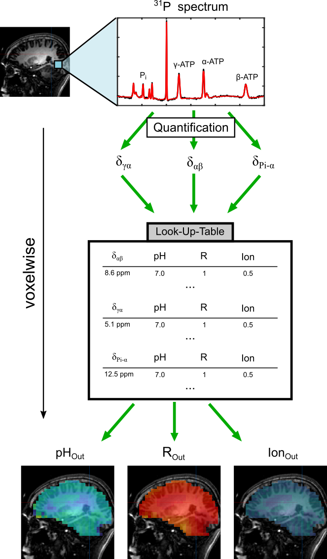
Figure 1: Schematic diagram explaining the basic principle of the advanced look-up algorithm. For each voxel of a 31P MRSI dataset, the corresponding 31P spectrum is processed and quantified. The quantified chemical shift differences of ATP (δαβ, δγα) and inorganic phosphate to α-ATP (δPi-α), are fed voxelwise into the look-up algorithm consisting of a database acquired from measurements in model solutions. For each voxel, a solution triple (pH,R,Ion) is determined to yield 3D maps for these three parameters.
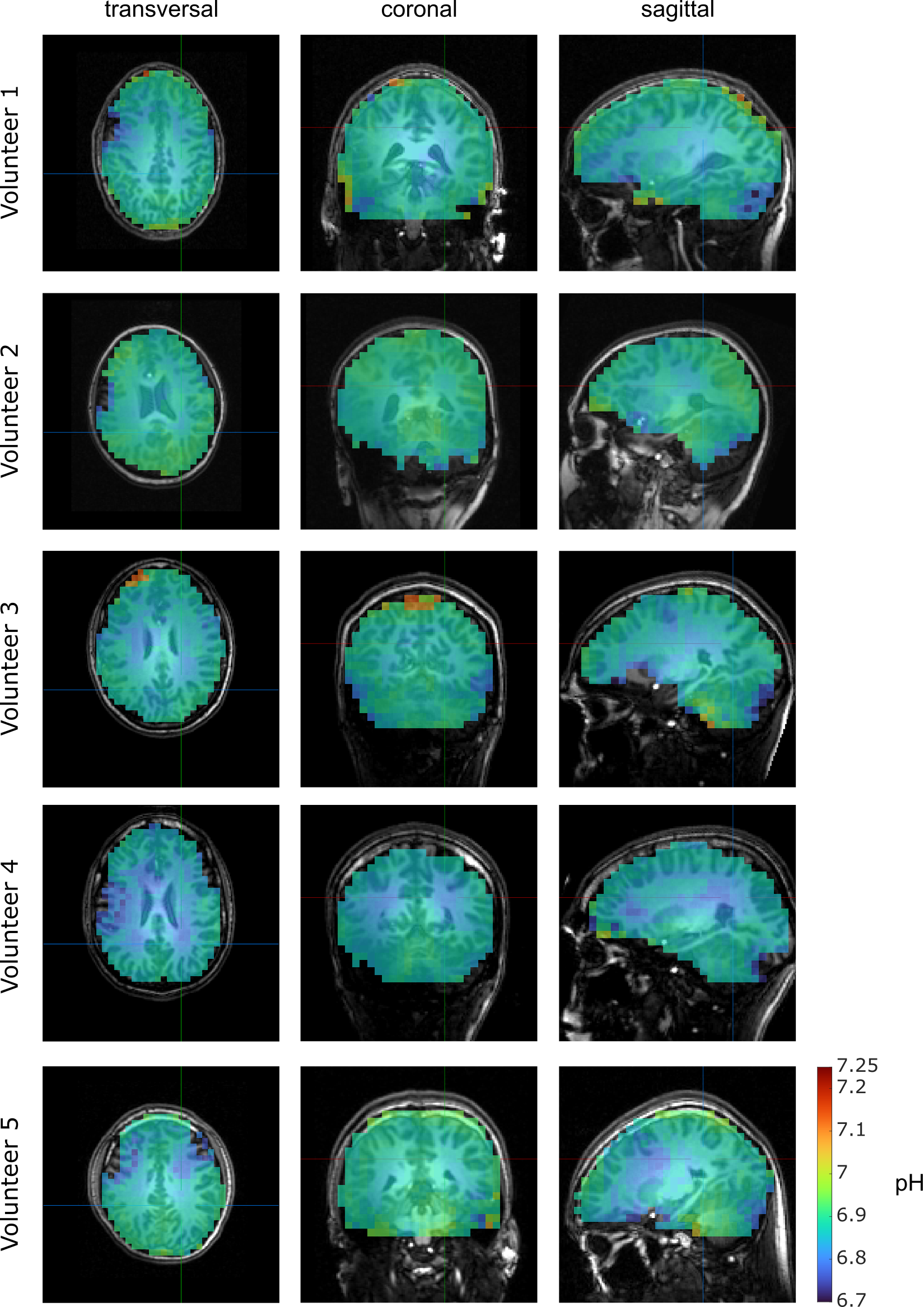
Figure 2: Volumetric maps of the determined pH values resulting from the application of the advanced look-up algorithm to 3D 31P MRSI datasets from the brains of healthy volunteers. Representative transversal, coronal and sagittal slices from five volunteers are shown.
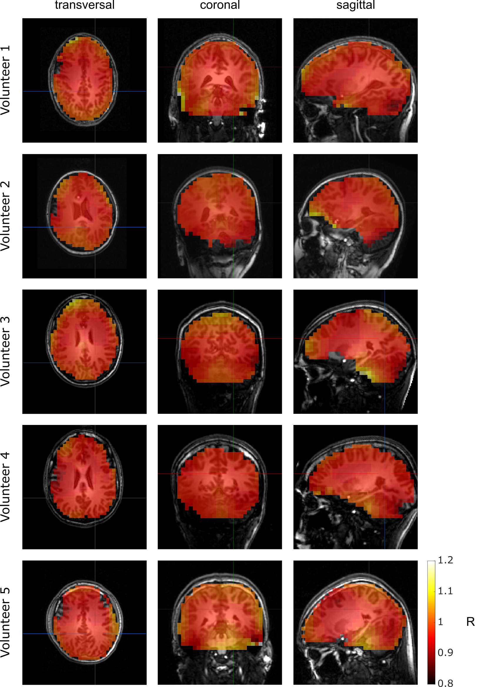
Figure 3: Volumetric maps of the determined magnesium ion content given as R = [Mgtot]/[ATPtot] resulting from the application of the advanced look-up algorithm to 3D 31P MRSI datasets from the brains of healthy volunteers. Representative transversal, coronal and sagittal slices from five volunteers are shown.
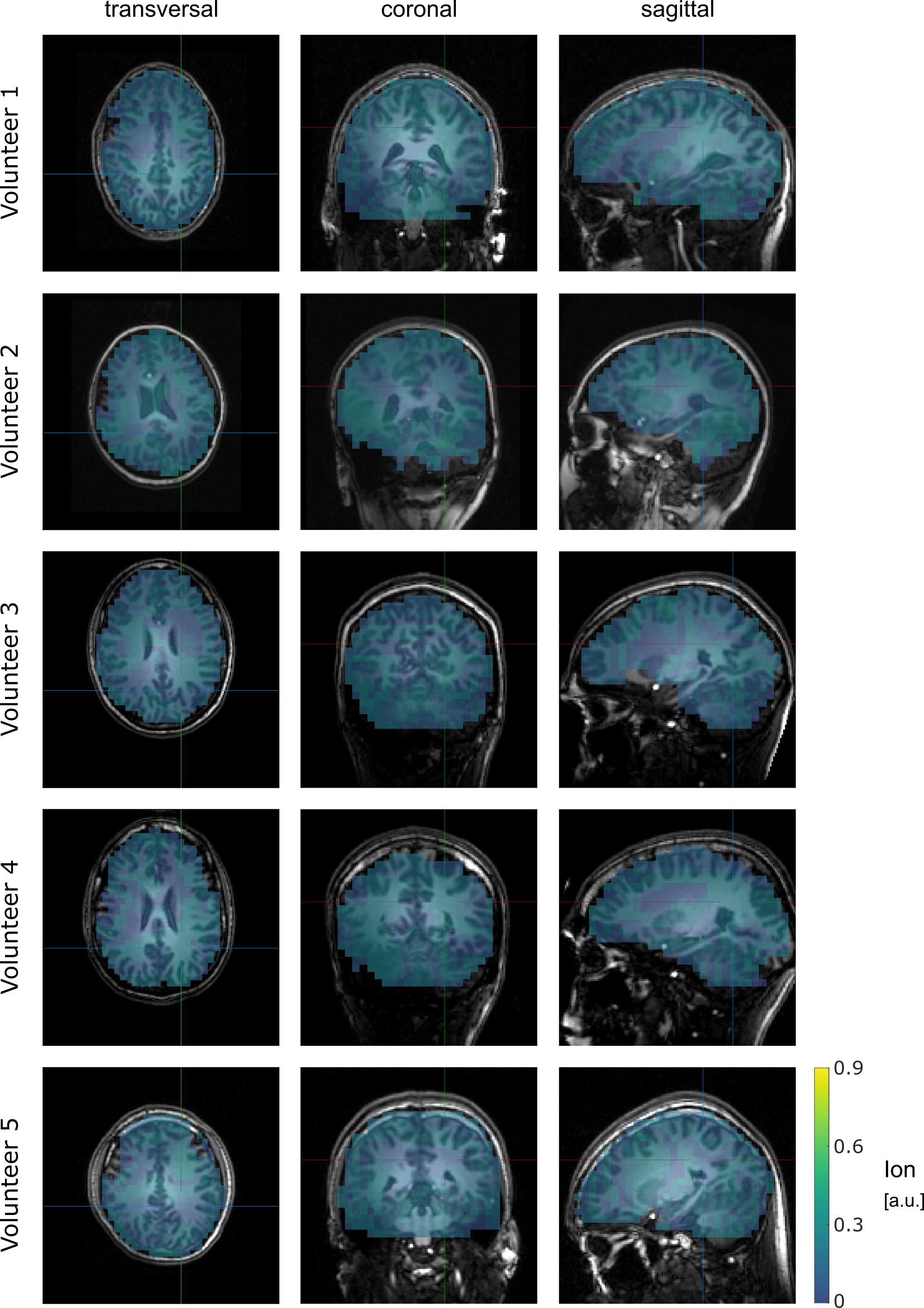
Figure 4: Volumetric maps of the determined Ion values resulting from the application of the advanced look-up algorithm to 3D 31P MRSI datasets from the brains of healthy volunteers. Representative transversal, coronal and sagittal slices from five volunteers are shown. The parameter Ion represents a measure for the ionic composition in arbitrary units.
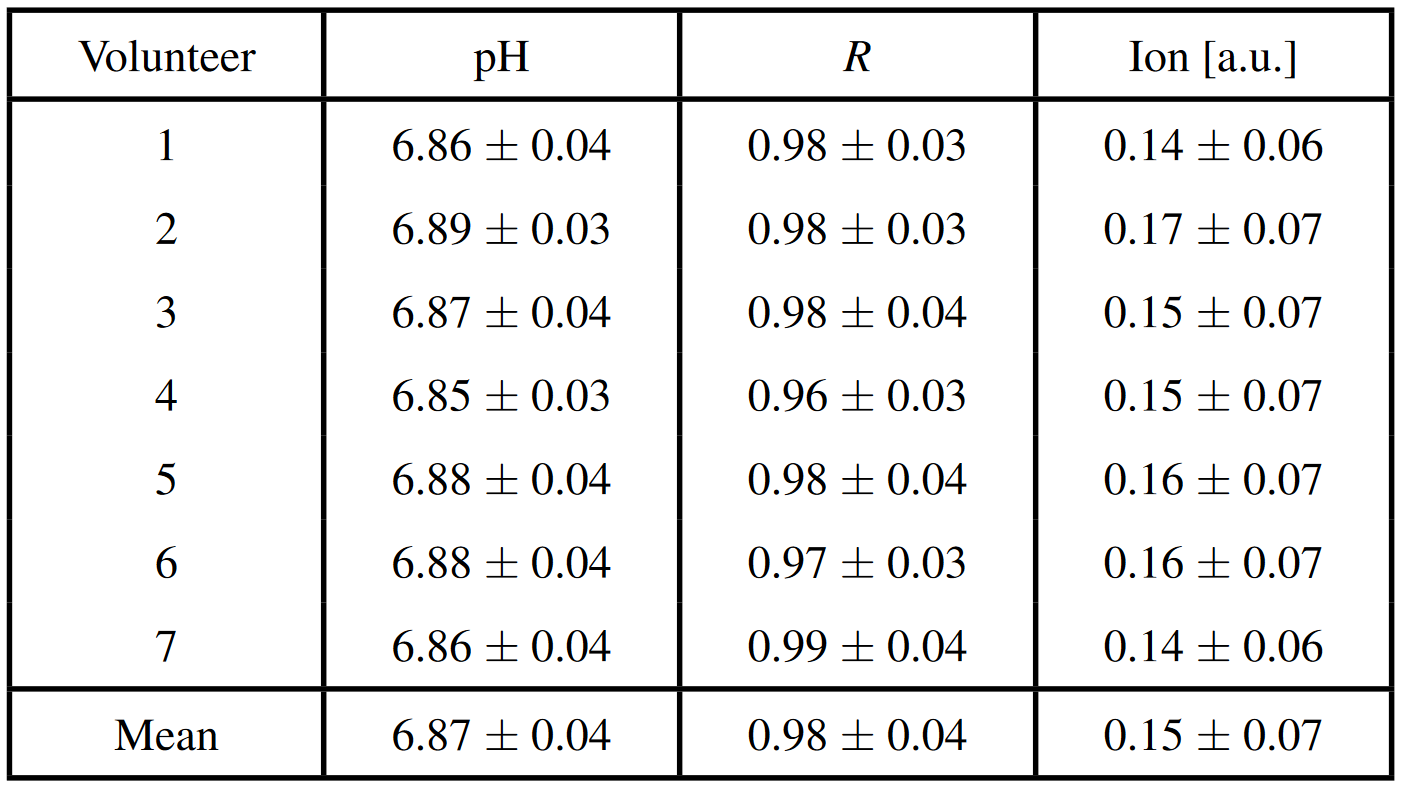
Table 1: Mean values and standard deviations of the values determined for pH, R and Ion across all voxels inside the brain tissue mask of the 31P MRSI datasets of healthy volunteers. The magnesium ion content is given as R = [Mgtot]/[ATPtot]. At the bottom, the mean value across all seven volunteers is given for each case.