3035
Boosting SNR for sodium bSSFP imaging1Department of Bioengineering, Imperial College London, London, United Kingdom, 2London Collaborative Ultra-High Field MRI System (LoCUS), King’s College London, London, United Kingdom, 3Neuroradiology, King’s College Hospital, London, United Kingdom, 4Biomedical Engineering Department, King's College London, London, United Kingdom, 5Department of Electrical and Computer Engineering, Boise State University, Boise, ID, United States
Synopsis
Keywords: Non-Proton, Non-Proton
Motivation: Sodium MRI (23Na-MRI) holds significant potential, but its clinical use is still limited due to challenges arising from low signal-to-noise ratio (SNR). Balanced steady-state free precession (bSSFP) sequences are particularly suited for 23Na-MRI, being highly SNR efficient, but there are opportunities to extract more signal components.
Goal(s): We aim to improve SNR in sodium bSSFP acquisitions and produce additional tissue contrasts.
Approach: We propose a strategy to extract additional information from a series of phase-cycled bSSFP images using different linear combinations.
Results: Three different signal components were extracted from phase-cycled bSSFP data and combined. Results showed an SNR boost in fluid.
Impact: This offers a simple way to improve the SNR in sodium MRI images, with potential applications being pathologies that alter biofluid sodium concentrations.
Introduction
Sodium MRI (23Na-MRI) provides a unique and direct window into the sodium content within human tissues, which can be used as a biomarker for disease. However, low Signal-to-Noise Ratio (SNR) is a significant challenge. The quadrupolar characteristics of sodium nuclei contribute to the complex and rapid decay of the MR signal. This compounds the SNR issue as a substantial portion of the signal dissipates within mere milliseconds1.Balanced steady-state free precession (bSSFP) sequences are rapid, volumetric imaging techniques. Additionally, bSSFP yields high SNR due to its efficient refocusing of residual magnetization from prior excitations2. However, it is sensitive to static field (B0) inhomogeneity, resulting in dark bands which obscure parts of the resulting images. Phase cycling shifts these bands systematically, and the resulting phas-cycled images can be averaged to produce a band-free image3.
In terms of the extended phase graph formalism4,5, this averaging across a phase-cycled bSSFP series corresponds to isolating the zeroth F-state (F0) signal component. However, the same phase-cycled bSSFP data could be combined using a different complex sum, which would isolate higher-order F-states with different contrasts. In this work, we investigate the inclusion of higher-order F-state signals to boost the SNR or produce alternative contrasts in 23Na-MRI.
Method
Acquisition23Na-MRI scanning was carried out on a 7T Siemens MAGNETOM Terra (Erlangen, Germany), using a 1Tx/32Rx 23Na head coil (Rapid Biomedical, Rimpar, Germany). A uniform, saline bottle phantom and a healthy volunteer were imaged using the protocol in Table 1.
Reconstruction
Three signal components (F0, F1, and F-1) were obtained by complex summation of the six phase-cycled datasets. Each signal component was adjusted by its own linear phase cycling increment ($$$\Delta\phi$$$) using the following expression:
$$data(F_n)=\sum_{\Delta\phi}(data_{\Delta\phi}e^{-i\Delta\phi n})$$
where n is the order of the F-state, and $$$\Delta\phi$$$ is in radians.
Each dataset was then reconstructed using a nonuniform-FFT 6 (conjugate-gradient SENSE), producing corresponding images for each F-state component. A composite image was then generated by combining all three F-state images via root sum of squares.
The SNR was calculated using Equation 1:
$$ SNR =\left(\frac{mean\ (ROI)}{std \ dev \ (Background)}\right ) \times 0.66 \tag {1}$$
As SNR was calculated using magnitude images, a correction factor of 0.66 for the Rayleigh distributed noise was included.
Results & Discussion
Figure 2 shows an estimated SNR comparison of the different signal components and the proposed composite image in a saline phantom. Additional signal components are present from the F-1 and F1 states (b-c), which can be combined with the F0 signal (a) to improve SNR in a saline solution (d).Figure 3 shows an estimated SNR comparison of the different signal components and the proposed composite image in the brain of a healthy volunteer. CSF has significant signal components in the F-1 and F1 states. This not only leads to the generation of different contrasts in those components, but those components can also be combined to improve fluid SNR over that obtained in the F0 image. However, this is of limited use in white matter, where the F-1 and F1 signal components have low SNR.
This approach therefore holds promise in boosting SNR in situations where the tissue of interest is primarily fluid (cerebrospinal fluid, synovial fluid, inner ear), particularly when: 1) these are inaccessible to sampling; 2) where disregulation of sodium homeostasis may be implicated in disease processes; or 3) there are benefits in depicting fluid compartments with differing sodium concentrations. Even if SNR benefits are not realised in white matter, the F1 and F-1 images can be reconstructed from the same underlying data. They can provide free additional contrasts which may be useful in qualitative or quantitative assessment.
Conclusion
In summary, our proposed approach to phase-cycled bSSFP in sodium MRI offers complementary images with enhanced SNR in fluids and a broader range of contrasts for assessing tissue properties without the need for any additional acquisitions.Acknowledgements
We acknowledge and thank the Saudia Arabia Cultural Bureau in London, The Wellcome Trust (WT201526/Z/16/Z; 220473/Z/20/Z), The Edmond J Safra Foundation, UK Dementia Research Institute, NIHR Imperial Biomedical Research Centre, and National Institutes of Health (R01EB002524) for their generous support. We would also like to thank the people who volunteered to participate in this study.
References
1. Madelin, G. & Regatte, R. R. Biomedical applications of sodium MRI in vivo. Journal of Magnetic Resonance Imaging 38, 511–529 (2013).
2. Radiol, E., Scheffler, K. & Lehnhardt, S. Principles and applications of balanced SSFP techniques. (2003) doi:10.1007/s00330-003-1957-x.
3. Zur, Y., Wood, M. L. & Neuringer, L. J. Motion-insensitive, steady-state free precession imaging. Magn Reson Med 16, 444–459 (1990).
4. Weigel, M. Extended phase graphs: Dephasing, RF pulses, and echoes - pure and simple. Journal of Magnetic Resonance Imaging 41, 266–295 (2015).
5. Hennig, J. Multiecho imaging sequences with low refocusing flip angles. Journal of Magnetic Resonance (1969) 78, 397–407 (1988).
6. Knoll, F., Schwarzl, A., ;Diwoky, C. & Sodickson DK. gpuNUFFT - An open source GPU library for 3D regridding with direct Matlab interface. p4297 Preprint at https://archive.ismrm.org/2014/4297.html (2014).
Figures
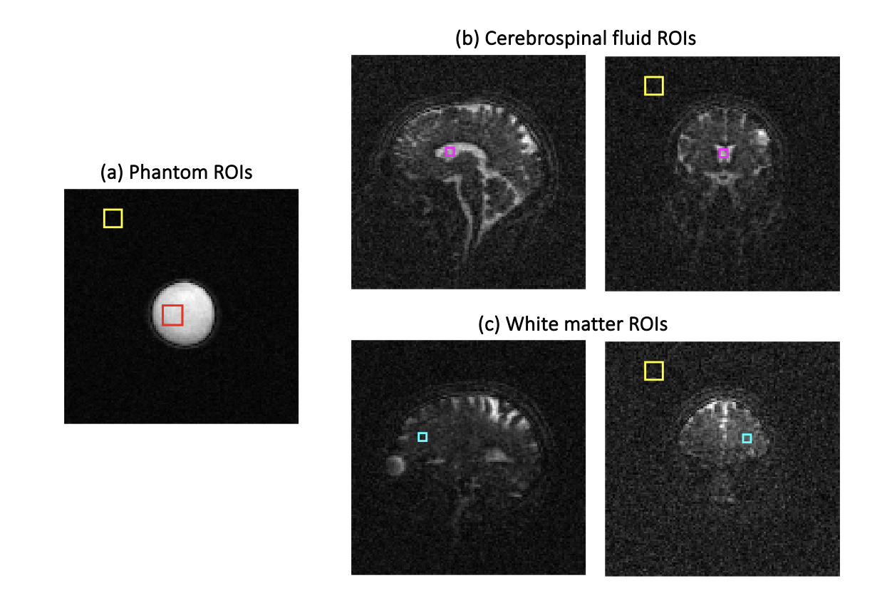
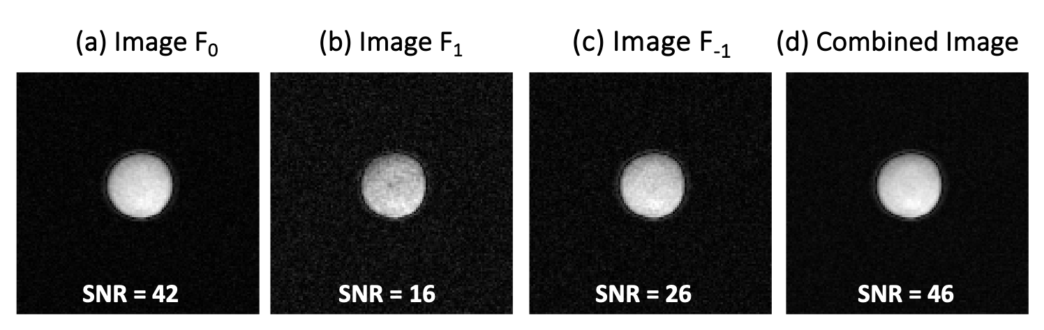
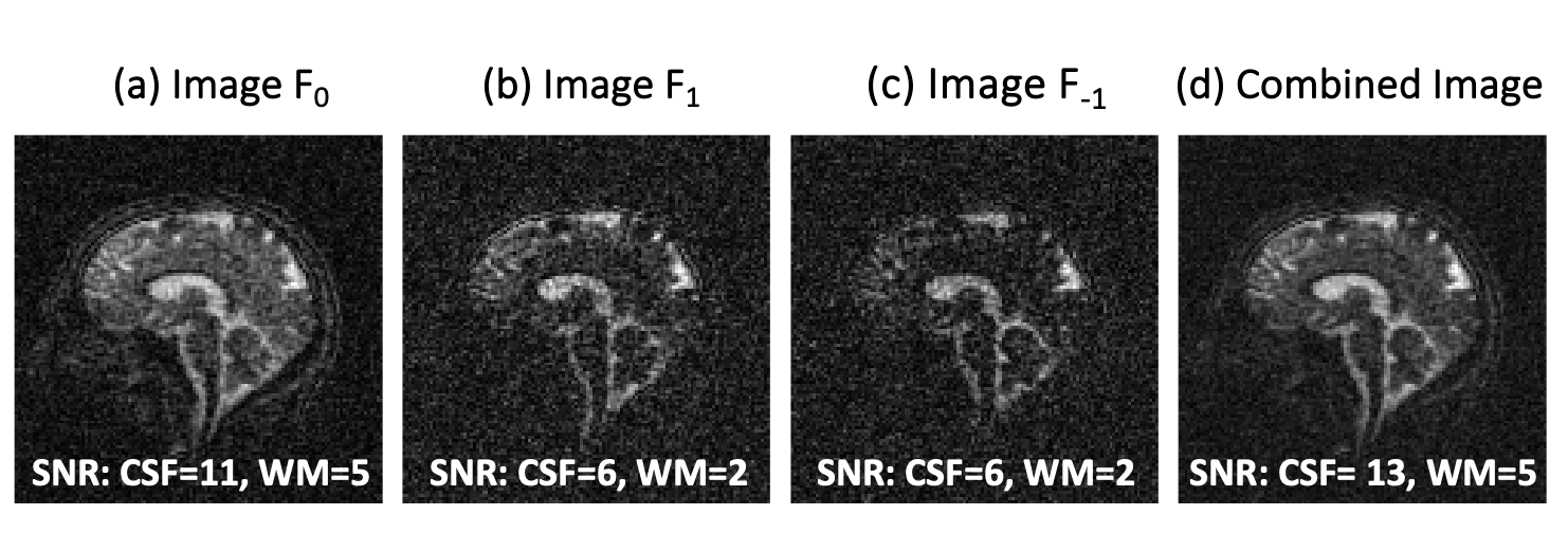
Fig.3: SNR estimates in a healthy volunteer brain for (a-c) each F-state signal component, and (d) the proposed combination. F1 and F-1 components (b and c) have a different contrast to the F0 component (a). Cerebrospinal fluid has significant signal components in the F1 and F-1 states when compared to white matter. This leads to: 1) the F1 and F-1 components (b-c) having a different contrast to the F0 component (a); and 2) improved fluid SNR in the combined image. F1 and F-1 states have low signal in white matter, however, and hence SNR boost is limited to fluid in the combined image.
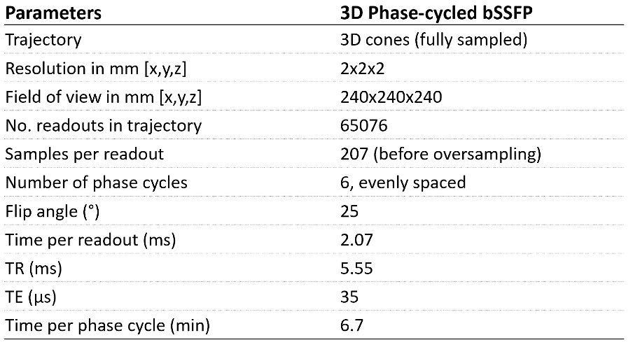
Table 1: 23Na-MRI acquisition parameters